Background: Retroperitoneal leiomyosarcomas are aggressive malignancies. Complete surgical resection with negative margins is crucial to decrease the risk of recurrence but can be risky due to vascular involvement. The aim of our study was to evaluate the different approaches to IVC and renal vein management and their impact on postoperative complications. Methods: We performed a retrospective review of patients who underwent surgery for retroperitoneal leiomyosarcoma with IVC and/or renal vein involvement at our institution from 2016 to 2024. Patients were stratified by intraoperative vascular management, including ligation only versus varying forms of vascular reconstruction. Postoperative complications, including bleeding, transfusions, the need for acute and chronic hemodialysis, and thromboembolic events, were recorded. Chi-squared tests were used to compare rates of postoperative complications by vascular management. A p-value of 0.05 was considered statistically significant. Results: We identified 60 patients at our institution who underwent surgery for leiomyosarcoma with IVC and/or renal vein involvement. Ten patients underwent IVC ligation alone due to thrombosis, thirty-six had IVC replacement, and fourteen had patch angioplasty. In the entire cohort, twenty-six patients (43.3%) experienced an adverse event after surgery. When looking at postoperative adverse events by IVC management, we did not find any statistically significant differences among rates of adverse events by group. There were also no statistically significant differences in complications following renal vein ligation versus renal vein reconstruction. Conclusions: Patients with leiomyosarcoma with IVC and/or renal vein involvement have several options for intraoperative vascular management. Our data demonstrates that there are no statistically significant differences in rates of complications among the different groups.
Malignant peripheral nerve sheath tumors (MPNSTs) are aggressive sarcomas. An obstacle to treating MPNSTs is a lack of effective systemic therapies. Although over 70% of human MPNSTs have lost or inactivated the epigenome regulator polycomb repressive complex 2 (PRC2), its activity and contribution to canine PNST progression remain unclear. This study compared canine peripheral nerve sheath tumors (PNSTs) and human MPNSTs across biological and clinical features, including PRC2 activity. Immunohistochemical analysis was performed for a human tissue microarray of 54 neurofibromas and 139 MPNSTs, and 63 canine PNSTs for H3K27me3, a repressive histone mark deposited by intact PRC2, and H3K27ac, which increases globally upon H3K27me3 loss. To understand the genomic alterations present in canine PNSTs, we analyzed tumor mutation burden, copy number alteration, and transcriptomes of eight canine PNST/normal pairs. The results suggested that H3K27me3 loss and associated gain of H3K27ac epigenetically drive human and canine tumors. These findings warrant further studies to evaluate whether these epigenetic deregulations alter similar gene signatures across species.
Abstract Background: Soft tissue sarcomas (STS) are rare and heterogenous cancers, with limited effective treatments and poor prognosis. While some patients with STS respond to immune checkpoint blockade (ICB), the majority do not benefit. This highlights an urgent need to elucidate mechanisms of response and resistance to ICB and develop novel immunotherapeutic approaches for patients with STS. We and others have shown that tumor infiltrating B cells (TIL-Bs), particularly those in tertiary lymphoid structures (TLS) are associated with response to ICB and longer survival. However, the antigen specificity, mechanisms of action, and therapeutic potential of these TIL-Bs and their B cell receptors (BCRs) remain unknown. Methods: We recently completed a phase 2 clinical trial evaluating ICB administered in the neoadjuvant setting in patients with resectable undifferentiated pleomorphic sarcoma (UPS) of the extremity/trunk or dedifferentiated liposarcoma (DDLPS) of the retroperitoneum (Roland et al. Nature Cancer 2024). Leveraging a unique biospecimen resource from this trial, we reconstructed intratumoral BCR heavy and light chain (VH and VL) repertoires using pre-treatment baseline tumor biopsies (n = 27), RNA-sequencing, and an in-house pipeline including TRUST4. We then performed single intratumoral B cell immunoglobulin sequencing using cell suspensions from 4 TLS-positive tumors to reconstruct and express 105 recombinant BCR antibodies (rBCR Abs) as IgG1. Flow cytometry (FC) was performed to characterize the binding features of a subset of 20 rBCR Abs with human DDLPS (LPS224, LPS246) and UPS cell lines (UPS186, RIS819.1). Binding was quantified as the ratio of the mean fluorescent intensity (MFI) of the antibody to the MFI of the isotype control for each cell line. Abs with an MFI ratio ≥ 1.2 were classified as binders and tumor surface binding was further assessed by live cell microscopy. Antibody-dependent cellular cytotoxicity (ADCC) assays were performed to assess the functional capacity of this cohort of rBCR Abs to mediate NK-cell dependent tumor killing. The anti-tumor efficacy of 1 lead rBCR Ab was assessed using an MCA205 immunocompetent murine STS model. Results: Of 105 rBCR Abs screened, 23 (22%) exhibited reproducible surface binding to at least one sarcoma cell line by FC. Among these, 13 (12%) bound LPS lines and 18 (17%) bound UPS lines. To date, 20 rBCR Abs have been confirmed to bind the surface of sarcoma cell lines by live cell staining of which 5 mediated tumor cell killing in vitro. One candidate rBCR Ab has been evaluated in vivo, localizing to tumor and synergized with PD-1/PD-L1 blockade to suppress tumor growth when administered intraperitoneally. Conclusion: These findings highlight the potential of rBCR Abs to serve as a novel therapeutic strategy and as tools to elucidate the roles of B cells and the BCR repertoire within the tumor immune microenvironment of patients with STS. Citation Format: Varshini Arunkumar, Manoj Chelvanambi, Joshua B. Plummer, Monika Zelazowska, Fabiana J. Veguilla, Elise Nassif Haddad, Noha M. Osman, Wei-Lien Wang, Davis Ingram, Khalida M. Wani, Angela Bhalla, Sharon M. Landers, Keila E. Torres, Jennifer A. Wargo, Bin Liu, Alexander Lazar, Neeta Somaiah, Christina L. Roland, Kevin McBride, Emily Z. Keung. Harnessing tumor-informed B cell receptors for discovery and antibody-based immunotherapy in sarcoma [abstract]. In: Proceedings of the American Association for Cancer Research Annual Meeting 2026; Part 1 (Regular Abstracts); 2026 Apr 17-22; San Diego, CA. Philadelphia (PA): AACR; Cancer Res 2026;86(7 Suppl):Abstract nr 6458.
Soft tissue sarcomas are aggressive malignancies with limited responses to immunotherapy. Strategies that induce viral mimicry and type I interferon (IFN-I) signaling may enhance tumor immunogenicity but remain poorly explored in sarcoma. Here, we show that the CDK4/6 inhibitor abemaciclib triggers viral mimicry and robust IFN-I responses in sarcoma cells, leading to tumor growth suppression and remodeling of the tumor microenvironment toward an immune-permissive state. However, sarcoma cells activate resistance mechanisms that limit IFN-induced stress. We identify lysophosphatidic acid receptor 4 (LPAR4) as a key mediator of this adaptation. LPAR4 dampens IFN signaling, reduces oxidative stress, and promotes tumor cell survival under therapeutic pressure. Genetic silencing of LPAR4 enhances IFN responses, increases reactive oxygen species, and sensitizes sarcoma cells to apoptosis, resulting in delayed tumor growth. These findings identify LPAR4 as a central regulator of IFN-driven stress adaptation and a potential therapeutic target to enhance viral mimicry-based strategies in sarcoma.
Background: Undifferentiated pleomorphic sarcoma (UPS) is the most common subtype of soft-tissue sarcoma (STS). At our institution, preoperative management typically consists of neoadjuvant chemotherapy plus radiotherapy (CT-RT) or radiotherapy (RT) alone. This study aimed to assess (1) the impact of treatment strategies on therapeutic response, and (2) the utility of multiparametric MRI (mp-MRI), qualitative and morphologic features from contrast-enhanced susceptibility-weighted imaging (CE-SWI) and dynamic contrast-enhanced perfusion-weighted imaging (PWI/DCE), for response evaluation. Methods: In this IRB-approved retrospective study, 36 UPS patients underwent presurgical mp-MRI between 02/2021 and 03/2025. Unlike our prior STS research, we intentionally excluded clinically unavailable software-based semiquantitative, quantitative, and radiomic analyses, focusing instead on morphologic and qualitative features readily accessible to clinical radiologists. Lesions were morphologically classified by CE-SWI-T2* and PWI/DCE in six patterns, respectively. PWI/DCE Time-intensity curves (TICs) types III—V, characterized by rapid arterial upstroke, were considered “aggressive,” while TIC I–II as non-aggressive. Patients with ≥90% pathology-assessed treatment effect (PATE) on the surgical specimen were designated as responders (n=20), >30 and <90% PATE as partial-responders (n=10), and ≤30% as non-responders (n=6). <90% patients were combined into a single group (partial/non-responders n=16). Results: Of 36 patients, 23 received CT-RT and 13 received RT alone. The CT-RT group demonstrated a higher responder rate (61% vs. 45%) and a higher mean PATE (81% vs. 66%; P=0.054) than the RT group. Among CT-RT responders, 86% showed a CE-SWI-complete-ring pattern (P=2.72×10⁻⁹), 86% a PWI/DCE-capsular-pattern (P=1.05×10⁻⁷), and 93% TIC-type-II (P=1.35×10⁻⁵). A predictive model combining PWI/DCE-capsular-enhancement, TIC-II, and favorable CE-SWI-patterns (“full blooming” or “complete ring”) achieved excellent performance (AUC=0.98) for distinguishing responders from non-responders, outperforming RECIST (AUC=0.52). In the RT group, all responders demonstrated TIC-II (P=0.014), and a model based solely on TIC-II achieved perfect classification (AUC=1.0). Conclusion: The CT-RT group shows a non-statistically significant higher proportion of responders and PATE-average than patients receiving RT-alone. MP-MRI-based predictive models using PWI/DCE and CE-SWI morphologic and qualitative clinically available features substantially outperform RECIST in predicting UPS pathology response. While the CT-RT model closely resembled a previously published treatment-agnostic UPS response model, pathology response in RT-alone patients was reliably predicted by the presence of presurgical TIC-II alone, indicating that optimal classification models for distinguishing responders vary by both tumor histology and treatment regimen, underscoring the importance of personalized imaging approaches.
Leiomyosarcoma is a smooth muscle-derived malignancy marked by significant clinical heterogeneity. The extent and nature of cellular heterogeneity and molecular underpinnings remain poorly understood. To address this at transcriptomic and epigenomic levels, we performed single-nucleus multiome sequencing on untreated primary leiomyosarcoma tissues. Malignant cells segregated almost exclusively into two previously unrecognized and epigenetically distinct states: a dedifferentiated, mesenchymal-like subtype (MES) and a differentiated smooth muscle-enriched subtype (SMC). Chromatin accessibility profiling revealed strong enrichment of nuclear factor I (NFI) transcription factor motifs in MES cells, whereas AP-1 family motifs-most prominently FOSL2-were selectively accessible in SMC cells. Established leiomyosarcoma cell lines faithfully recapitulated these subtypes, and targeted depletion of NFI or AP-1 factors suppressed proliferation, invasion, and in vivo tumor growth, demonstrating functional dependency on these transcriptional programs. Spatial transcriptomics across 328 tissue cores from 128 leiomyosarcomas showed that immunosuppressive macrophages preferentially cluster around MES regions, revealing a subtype-specific tumor-immune niche. Clinically, MES-dominant tumors were associated with significantly worse patient outcomes. Through an epigenetic inhibitor screen, we identify and validate SMARCA4/2 inhibition as a promising therapeutic vulnerability for MES leiomyosarcomas. Together, this work defines two epigenetically driven, transcription factor-regulated, and clinically relevant states of leiomyosarcoma, revealing mechanistic underpinnings of tumor heterogeneity and uncovering actionable therapeutic strategies.
Abstract INTRODUCTION: Malignant peripheral nerve sheath tumors (MPNSTs) are aggressive sarcomas. An obstacle to treating MPNSTs is a lack of effective systemic therapies. Although over 70% of human MPNSTs have lost or inactivated the epigenome regulator polycomb repressive complex 2 (PRC2) (DeRaedt et al., Nature, 2014), its activity and contribution to canine PNST progression remain unclear. METHODS: This study compared canine peripheral nerve sheath tumors (PNSTs) and human MPNSTs across biological and clinical features, including PRC2 activity. Immunohistochemical analysis was performed for a human tissue microarray of 54 neurofibromas and 139 MPNSTs, and 63 canine PNSTs for H3K27me3, a repressive histone mark deposited by intact PRC2, and H3K27ac, which increases globally upon H3K27me3 loss. To understand the genomic alterations present in canine PNSTs, we analyzed tumor mutation burden, copy number alteration, and transcriptomes of eight canine PNST/normal pairs. RESULTS: The results suggested that H3K27me3 loss and associated gain of H3K27ac epigenetically drive human and canine tumors. This study provided evidence that human and canine PNSTs are clinicopathologically similar and may also be similarly driven by epigenetic mechanisms. CONCLUSIONS: Further studies are warranted to evaluate whether these epigenetic deregulations alter similar gene signatures in humans and canine patients. The knowledge gained from this work advances our understanding of the molecular drivers of MPNST and informs potential therapeutics to evaluate in future clinical studies. Citation Format: Jace P. Landry, Angela D. Bhalla, Sharon M. Landers, Rossana Lazcano, Lindsay A. Parker, Tasha M. Miller, Noelle Niemi, Heather G. Lyu, Heather A. Lillemoe, Emily Z. Keung, Christopher P. Scally, Christina L. Roland, Kelly K. Hunt, John M. Slopis, Ian E. McCutcheon, Beth Boudreau, Heather Wilson-Robles, Alexander J. Lazar, Kunal Rai, Dominique J. Wiener, Brian W. Davis, Brandan Wustefeld-Janssens, Keila E. Torres. Comparative study of human and canine nerve sheath tumors in terms of morphology, prognosis, treatment, epigenetics, transcriptomics, and genomics [abstract]. In: Proceedings of the American Association for Cancer Research Annual Meeting 2026; Part 1 (Regular Abstracts); 2026 Apr 17-22; San Diego, CA. Philadelphia (PA): AACR; Cancer Res 2026;86(7 Suppl):Abstract nr 3250.
INTRODUCTION:Limb-sparing resection of extremity soft-tissue sarcoma (eSTS) often leaves complex defects that require reliable coverage. Although perforator flaps offer theoretical advantages, their reliability in eSTS reconstruction remains debated. We directly compared outcomes between perforator and traditional (i.e., non-perforator) flaps for eSTS reconstruction. METHODS:This retrospective cohort study included patients who underwent limb-sparing eSTS excision with flap reconstruction at a quaternary cancer center between 2016 and 2021. Patients were categorized into perforator and traditional flap groups. Outcomes included operative time, surgical site complications, prolonged wound care, reoperation, readmission, and need for eventual amputation. RESULTS:A total of 156 patients were included (53 perforator and 103 traditional flaps). Perforator flaps were more frequently used for tumors distal to the elbow (17% vs. 1.9%, p = 0.002) and for recurrent tumors (21% vs. 7.8%, p = 0.019). Operative time and overall complication rates were comparable between the groups. Among free tissue transfers, perforator flaps were associated with a shorter length of stay than traditional flaps (6 vs. 11 days, p = 0.003); however, this association was not maintained on multivariable analysis. CONCLUSIONS:Perforator flaps provide oncologically safe, functionally reliable reconstruction for eSTS, with similar complication rates compared to traditional flaps. These findings support their integration into the eSTS reconstructive armamentarium.
Well-differentiated and dedifferentiated liposarcoma (WDLPS and DDLPS) exhibit markedly different clinical behaviors, with DDLPS showing greater aggressiveness, higher recurrence and metastasis rates, and worse outcomes. Using single-nucleus multiome sequencing, epigenomic profiling, and spatial transcriptomics, we characterized cellular and epigenetic heterogeneity between these subtypes at single-cell and spatial resolution. We found distinct phenotypic states reflecting altered lineage differentiation and plasticity: DDLPS is dominated by early-differentiated progenitor-like cells, sclerotic WDLPS displays broader mesenchymal lineage plasticity, and adipocytic WDLPS contains abundant committed adipocytes. The DDLPS immune microenvironment was dominated by immunosuppressive macrophages, whereas WDLPS harbored more T cells and inflammatory macrophages. Notably, sclerotic WDLPS displayed intermediate cellular and molecular features, suggesting it may represent a distinct WDLPS subtype. Importantly, we identified novel gene regulatory circuits underlying each state, including FABP4/PPARG programs in adipocytic WDLPS, GLI2/TCF7L2/RBPJ/KLF7 programs in sclerotic WDLPS, and KLF7/FOSL2/SP3/GLI2/RBPJ programs in DDLPS. H3K27ac-marked enhancers were enriched near adipocytic marker genes in WDLPS and mesenchymal markers in DDLPS. Together, these findings reveal the cellular heterogeneity of tumor and immune compartments across liposarcoma subtypes and identify regulatory programs driving their differentiation states.
INTRODUCTION:Surveillance practices after resection of extremity and truncal soft-tissue sarcoma (ETSTS) vary, with guidelines recommending physical exam and imaging every 3-6 months. Local and distant recurrence risks differ based on clinicopathological features including tumor grade and histopathologic subtype. The aim of this study was to evaluate surveillance patterns after ETSTS resection at our high-volume sarcoma referral center and determine if opportunities exist to de-escalate intensity of surveillance in appropriate patients. METHODS:A retrospective review of patients with primary ETSTS who underwent resection and surveillance at our institution from 2016-2021 was performed. Patients with less than 2 years of follow-up after resection were excluded. Sarculator, a validated nomogram for soft-tissue sarcoma, was used to stratify patients into high- (< 60%) and low-risk (≥ 60%) prognostic groups based on their predicted overall survival (OS) at 10 years. RESULTS:The cohort of 296 patients included 112 high- and 184 low-risk. 5-year OS was 70.5% in the high-risk group versus 90.9% in the low-risk group (P < 0.001). Most patients did not recur during the study period (n = 206). 26 patients recurred within 6 months from their initial surgery (18 high-risk, 8 low-risk) and all were offered immediate treatment after recurrence diagnosis. Of this cohort, 84.6% were offered immediate treatment after diagnosis of recurrence. CONCLUSION:Surveillance strategies after primary resection should be tailored to patient's individual risk. Patients with low-risk clinicopathologic features have a lower chance of recurrence in the first two years and frequent surveillance visits may not impact their oncologic outcomes.
Undifferentiated pleomorphic sarcoma (UPS) is the largest subgroup of soft-tissue sarcomas. It demonstrates post-therapeutic hemosiderin deposition, granulation tissue formation, fibrosis, and calcification. Our research aims to establish the multiparametric MRI (mp-MRI) value for predicting UPS treatment response. An IRB-approved retrospective study included 33 extremity UPS patients with pre-operative mp-MRI, including diffusion-weighted imaging (DWI), contrast-enhanced susceptibility-weighted imaging (CE-SWI), and perfusion-weighted imaging with dynamic contrast-enhancement (PWI/DCE), and surgical resection between February 2021 and May 2023. Lesions were visually classified on CE-SWI into one of 6 morphology patterns. On PWI/DCE, lesions were classified into one of 6 patterns, and time-intensity curves (TICs) were classified as types I-V. Patients were categorized into three groups based on the percentage of pathology-assessed treatment effect (PATE) in the surgical specimen: Responders (> = 90
PURPOSE Undifferentiated pleomorphic sarcomas (UPSs) demonstrate therapy-induced hemosiderin deposition, granulation tissue formation, fibrosis, and calcification. We aimed to determine the treatment-assessment value of morphologic tumoral hemorrhage patterns and first- and high-order radiomic features extracted from contrast-enhanced susceptibility-weighted imaging (CE-SWI). MATERIALS AND METHODS This retrospective institutional review board–authorized study included 33 patients with extremity UPS with magnetic resonance imaging and resection performed from February 2021 to May 2023. Volumetric tumor segmentation was obtained at baseline, postsystemic chemotherapy (PC), and postradiation therapy (PRT). The pathology-assessed treatment effect (PATE) in surgical specimens separated patients into responders (R; ≥90%, n = 16), partial responders (PR; 89%-31%, n = 10), and nonresponders (NR; ≤30%, n = 7). RECIST, WHO, and volume were assessed for all time points. CE-SWI T2* morphologic patterns and 107 radiomic features were analyzed. RESULTS A Complete-Ring (CR) pattern was observed in PRT in 71.4% of R ( P = 7.71 × 10 –6 ), an Incomplete-Ring pattern in 33.3% of PR ( P = .2751), and a Globular pattern in 50% of NR ( P = .1562). The first-order radiomic analysis from the CE-SWI intensity histogram outlined the values of the 10th and 90th percentiles and their skewness. R showed a 280% increase in 10th percentile voxels ( P = .061) and a 241% increase in skewness ( P = .0449) at PC. PR/NR showed a 690% increase in the 90th percentile voxels ( P = .03) at PC. Multiple high-order radiomic texture features observed at PRT discriminated better R versus PR/NR than the first-order features. CONCLUSION CE-SWI morphologic patterns strongly correlate with PATE. The CR morphology pattern was the most frequent in R and had the highest statistical association predicting response at PRT, easily recognized by a radiologist not requiring postprocessing software. It can potentially outperform size-based metrics, such as RECIST. The first- and high-order radiomic analysis found several features separating R versus PR/NR.
Desmoid tumors are rare mesenchymal neoplasms characterized by a clonal proliferation of fibroblasts and myofibroblasts. Using the novel contrast-enhanced susceptibility-weighted imaging (CE-SWI) for characterizing desmoid tumors can enhance the separation between fibrous T2-hypointense and cellular T1-enhancing components. We aim to evaluate the effectiveness of the CE-SWI signal, volumetric, and radiomics-derived features in assessing desmoid treatment response. This IRB-approved study included 17 single-lesion extremity desmoid fibromatosis patients who underwent standard-of-care MRI, including CE-SWI, from March 2021 to February 2024. Measurements of maximum diameter, volume, and the modified Choi (m-Choi: tumor/muscle T2 ratio) were computed based on CE-SWI and T2-STIR volumetric tumor segmentations. 107 shape, first-order, and textural radiomic features were calculated. Patient response was assessed using conventional RECIST as a reference standard and compared against T2-STIR and CE-SWI volumetric, m-Choi, and radiomics features. RECIST-progression (n = 3): In two patients, CE-SWI volume detected progression 10 months earlier than T2-STIR-based RECIST. Only 33% were characterized as progression by the routine radiologic report (RRR). RECIST-stability (n = 14): 5% exhibited at least one expected first-order response/progression-related change in the mean, skewness, 10th percentile, or 90th percentile, with all four changes present in 33% of cases. In RECIST-progression, CE-SWI showed an average of 15% more voxels at the 90th percentile than T2-STIR. Volume and CE-SWI/T2-STIR shape-derived size dimensional features demonstrated the highest separation between progressive and responding patients. CE-SWI has a higher sensitivity than T2-WI in detecting the active/progressive enhancing component. Volume and Shape-derived and, to a lesser extent, textural radiomic features and m-Choi effectively distinguish between progressive and responding cases, outperforming first-order radiomics, RRR, and RECIST. Particularly, progression prediction by CE-SWI/T2-STIR-volume and response prediction by CE-SWI-m-Choi outperform and precede RRR and RECIST. The novel CE-SWI enhances tumor insight and desmoid treatment-response prediction by effectively separating responding T2-hypointense-collagenized-mature components from potentially progressive T1-shortened/enhancing T2-hyperintense-immature components.
Neurofibromatosis type 1 (NF1) is an inherited disorder that predisposes individuals to malignant peripheral nerve sheath tumors (MPNSTs), a highly aggressive sarcoma with limited treatment options and poor prognosis. This study explores the potential of targeting the interaction between Galectin-1 and Ras as a novel therapeutic strategy for MPNSTs. Through molecular docking, we identified critical residues involved in the Galectin-1 and H-Ras interaction. We developed LLS30, a compound designed to target this Ras-binding pocket on Galectin-1, and tested its efficacy. LLS30 effectively disrupted the Galectin-1/Ras interaction, causing Ras delocalization from the plasma membrane and inhibiting Ras signaling. In vitro experiments showed that LLS30 significantly decreased MPNST cell proliferation and induced apoptosis. In vivo, LLS30 demonstrated potent anti-tumor effects, reducing tumor size, inhibiting metastasis, and extending survival in animal models. Transcriptome analysis further revealed the downregulation of KRAS signaling and inhibition of pathways associated with epithelial–mesenchymal transition. These findings suggest that targeting Galectin-1 with LLS30 offers therapeutic potential for MPNSTs and could be beneficial for other cancers driven by Galectin-1 and Ras signaling.
Introduction Accurate diagnosis of lung metastasis for patients with soft tissue sarcoma (STS) can impact overall survival, but there is considerable variability in imaging utilized to detect metastasis. Chest x-ray (CXR) or computer tomography (CT) scan of the chest are the most common studies in current practice. Methods A systematic literature search was performed. Databases were searched from inception to January 3, 2024. Articles were reviewed to determine if CXR or CT chest was associated with improved overall or recurrence-free survival in patients with STS. Articles were also reviewed for data on cost-effectiveness of CXR versus CT chest. The quality of evidence was assessed by the Critical Appraisal Skills Programme. Results 259 abstracts were screened, and twenty-seven studies were selected for full-text review. Nine studies met all inclusion criteria. Seven studies included data on survival or recurrence rates and four included data on the cost of imaging modality. The seven studies all had conflicting results on the impact of CXR versus chest CT on survival and recurrence. The four studies that looked at cost found that CXR was more cost-effective, but in certain patients, screening with chest CT could be cost-effective. Conclusion:The literature defining the optimal surveillance method for lung metastases for patients with STS is limited. Most of the studies had a low quality of evidence due to study design and significant risk of bias. Randomized controlled trials are needed to further understand the best imaging modality for lung metastasis surveillance in this patient population.
11576 Background: MPNSTs are aggressive sarcomas with poor prognosis due to their high propensity for metastasis, rapid growth and limited response to standard chemotherapy (CT). While commonly associated with neurofibromatosis type 1 (NF1), they can also occur sporadically (non-NF1). This study aims to evaluate baseline characteristics and outcomes in NF1 and non-NF1 associated MPNST in order to understand CT effectiveness and set a benchmark for future therapies. Methods: A retrospective chart review was conducted at MDACC, including 258 patients diagnosed with MPNST (173 NF1 and 85 non-NF1). Data collected included demographic information, primary tumor location and size, disease stage at diagnosis (localized vs. metastatic), efficacy of CT regimens utilized, and survival data. Descriptive statistics were used to summarize patient characteristics. Chi squared tests or Fisher’s exact tests, and t-test/ANOVA were used to compare patient’s characteristics and distributions of overall survival (OS), and progression-free survival (PFS) were estimated by the Kaplan-Meier method. Results: Median age at diagnosis was 33 yrs (IQR 22-44) and 50 yrs (IQR 38-61), and median tumor size at diagnosis 7.4cm and 6.3cm for the NF1 and non-NF1 cohorts, respectively. Tumors were most commonly located in the trunk, followed by the lower extremity, with head and neck involvement more frequent in non-NF1 cases. Metastatic disease was present at diagnosis in 28.3% of NF1 (49/173) and 22.4% (19/85) of non-NF1 cases. Adriamycin with Ifosfamide (AI) was the most utilized first-line CT regimen while gemcitabine plus docetaxel was the preferred second-line regimen. 154 patients, 87 patients, and 37 patients received frontline, second-line, and third-line CT, respectively. Median PFS for front-line CT was 8.0 months (4.7, NR) for patients with metastatic disease at presentation and 11.7 months (8.94, NR) for patients who received AI. Median OS was 2.34 yrs (1.88, 3.15) vs 1.81 yrs (0.89, 3.76) for NF1 and non-NF1 cohorts (p = 0.038), respectively. 5-yr OS was 42% (33, 51) for local disease and 9% (3, 19) for metastatic disease at presentation. Additional analysis of PFS by NF1 status, lines of therapy, and regimen will be presented at the conference. Conclusions: Patients with metastatic MPNST have dismal outcomes and CT efficacy and utilization drops after frontline treatment. These findings highlight the importance of early diagnosis and tailored novel therapeutics for MPNST patients.
ATRX is one of the most frequently altered genes in sarcoma and encodes an ATP-dependent chromatin remodeler implicated in maintaining heterochromatin. However, ATRX alterations have not been leveraged for sarcoma treatment. We observed loss of ATRX protein in 14% of soft tissue leiomyosarcoma (STLMS, n =127), 53% of uterine leiomyosarcoma (ULMS, n = 95), 37% of undifferentiated pleomorphic sarcoma (UPS, n = 82), and 8% of dedifferentiated liposarcoma (DDLPS, n = 84). ATRX loss was associated with significantly worse outcomes in ULMS, UPS, and DDLPS. ATRX knockout in sarcoma cells increased proliferation in cooperation with TP53 deletion. ATRX knockout led to chromatin de-repression and enrichment of PRDM4 and NFIX transcription factor (TF) motifs. PRDM4 and NFIX knockdown in ATRX -mutant sarcoma lines resulted in reduced proliferation and invasion suggesting epistatic relationship. Consistent with the known functional relationship between PRMD4 and YAP1, we observed that ATRX/TP53 KO cells were more sensitive to the TEAD inhibitor VT103 compared to TP53 KO and ATRX WT controls. Overall, our results identify ATRX loss as a prognostic factor of worse outcomes, implicate the ATRX-PRDM4-YAP1 axis as a novel underlying mechanism, and suggest use of TEAD inhibition as a potential therapeutic strategy for ATRX -deficient sarcomas. ![Figure][1] GRAPHICAL ABSTRACT ### Competing Interest Statement Kunal Rai reports equity in Jivanu Therapeutics and Koshika Therapeutics. NIH Common Fund, https://ror.org/001d55x84, T32 CA009666, 5K12CA088084, P50CA272170, P30 CA016672 Conquer Cancer Foundation, https://ror.org/027327e78, Young Investigator Award The QuadW Foundation, https://ror.org/0353cdc64 National Leiomyosarcoma Foundation [1]: pending:yes
Introduction:The dietary habits of children and adolescents in Chile have been identified as inadequate, potentially contributing to low levels of essential vitamins and minerals, as well as the development of chronic diseases such as obesity and diabetes. However, the nutritional profile of Chilean university students and the impact of their diets on this profile remain largely unknown. This study aimed to assess the nutritional profile of Finis Terrae University students in the Santiago Metropolitan Region, Chile. Methods:A retrospective 24-h dietary survey, using the Automated Self-Administered 24-Hour Dietary Assessment Tool (ASA24®), was administered to 124 students between September and October 2024. Data on macronutrient and micronutrient consumption was analyzed using GraphPad Prism. Results:Among those surveyed, 90% reported consuming at least three meals daily (breakfast, lunch, and dinner). However, 61% consumed fewer calories than recommended, only 7% met the daily intake goals for dairy, 11% met the recommended fruit and vegetable intake, and 16% fiber intake. In contrast, 65% exceeded the upper recommended limits for sodium intake. Significant deficiencies were observed in the intake of calcium, magnesium, potassium, and vitamins A, C, D, and E. Meanwhile, the intake of copper, phosphorus, selenium, folate, niacin, riboflavin, thiamine, vitamin B6, and vitamin B12 met the recommended dietary allowances. Discussion:Although most of the students consumed three meals daily, the quality of their nutritional intake was suboptimal. These findings highlight the need for educational and nutritional programs to promote healthier eating habits and improve the quality of life of university students, emphasizing regular, balanced meals, developing skills in meal preparation to reduce reliance on ultra-processed foods, and prioritizing hydration with water.
Background and purpose: Rhabdomyosarcoma (RMS) is the most common soft-tissue sarcoma in the pediatric population and an aggressive cancer subtyped as embryonal, alveolar, pleomorphic, or not otherwise specified (NOS). It demonstrates post-therapeutic hemosiderin deposition, granulation tissue formation, fibrosis, and calcification. This pilot study aims to provide initial evidence for developing a multiparametric MRI-feature-derived (mp-MRI) predictive response model to outperform RECIST.Methods: A UT MD Anderson Cancer Center IRB-approved retrospective pilot study of 11 extremity and pelvic RMS patients with presurgical mp-MRI, including diffusion-weighted imaging (DWI), contrast-enhanced susceptibility-weighted imaging (CE-SWI), and perfusion-weighted imaging with dynamic-contrast-enhancement (PWI/DCE), with surgical resection between 02/2021-06/2024. Lesions were categorized into 6 CE-SWI and 6 PWI/DCE morphologic patterns. Time-intensity curves (TICs) were classified as types I-V. Patients were categorized by the percentage of pathology-assessed treatment effect (PATE) in the surgical specimen as responders (PATE> 90%, n = 3) and partial/non-responders (PATE < 90%, n = 8).Results: The ADC-mean for the 6 extremity RMS increased minimally from 1,425 ± 476 x 10-6 mm2/s at baseline (BL) to 1,494 ± 386 x 10-6 mm2/s at the presurgical time point (PS). The ADC-mean for the 5 pelvic RMS increased from 1,093 ± 342 x 10-6 mm2/s at BL to 1,677 ± 313 x 10-6 mm2/s at PS. All responders and partial/non-responders presented presurgical RECIST, WHO, and volume stability. At PS, 50% of responders displayed CE-SWI Complete Ring pattern (p = 0.5578), PWI/DCE Capsular pattern (p = 0.6065), and TIC Type-2 (p = 0.6065). No statistically significant differences were observed at PS in ADC or CE-SWI first- or high-order radiomics and PWI/DCE semi-quantitative parameters comparing responders vs. partial/non-responders at PS. PS ROC Analysis: The model based on the CE-SWI textural radiomic GLSZM Large-Area-High-Gray-Level-Emphasis yielded perfect classification performance (AUC = 1.0) separating responders vs. partial/non-responders, outperforming other radiomic, morphologic, and qualitative features such as ADC-Maximum-2D-Diameter-Slice (0.83), CE-SWI Complete Ring (AUC = 0.67), PWI/DCE Capsular (0.67), RECIST (0.67), and TIC type-2 (0.6).Conclusion: Our pilot study provides initial evidence supporting a model utilizing a single CE-SWI-derived high-order texture GLSZM radiomic feature that, in our small sample, obtained a perfect classification performance (AUC = 1.0), further building on the body of evidence supporting the use of mp-MRI with CE-SWI in soft-tissue sarcoma response assessment and suggesting its potential in predicting RMS response, outperforming RECIST (AUC = 0.67). These promising early results are hindered by a limited statistical power (10%) inherent to the small sample size and the retrospective nature of an exploratory pilot study, highlighting the necessity for further validation through a larger, more representative, and balanced prospective study.
 Alexander J. Lazar合作论文数Department of Anatomical Pathology, Division of Pathology-Lab Medicine, The University of Texas M.D. Anderson Cancer Center;Department of Genomic Medicine, Division of Basic Science Research, The University of Texas MD Anderson Cancer Center;Department of Translational Molecular Pathology, Division of Pathology/Lab Medicine, The University of Texas MD Anderson Cancer Center74
 Christina L. Roland合作论文数Department of Surgical Oncology, The University of Texas MD Anderson Cancer Center;Division of Surgery, The University of Texas MD Anderson Cancer Center;Department of Cancer Network, The University of Texas MD Anderson Cancer Center62
 Kelly K. Hunt合作论文数Department of Breast Surgical Oncology, Division of Surgery, The University of Texas MD Anderson Cancer Center59
 Barry W. Feig合作论文数Department of Surgical Oncology, Division of Surgery, The University of Texas MD Anderson Cancer Center48
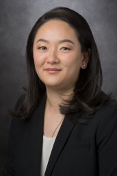 Emily Z. Keung合作论文数47
 Neeta Somaiah合作论文数42
 Wei-Lien Wang合作论文数Department of Anatomical Pathology, Division of Pathology-Lab Medicine Div, The University of Texas M. D. Anderson Cancer Center;Departments of Pathology and Translational Molecular Pathology, The University of Texas M. D. Anderson Cancer Center42
 Davis R Ingram合作论文数39
 Dina C Lev合作论文数33Christopher P Scally合作论文数32
 B. Ashleigh Guadagnolo合作论文数Division of Radiation Oncology, Department of Radiation Oncology, The University of Texas MD Anderson Cancer Center;Department of Health Services Research, The University of Texas MD Anderson Cancer Center28Janice N. Cormier合作论文数27Bishop Andrew J合作论文数25Chiang Yi-Ju合作论文数23
 Raphael E Pollock合作论文数College of Medicine, The Ohio State University;The Ohio State University Comprehensive Cancer Center21Sharon M Landers合作论文数20
 Vinod Ravi合作论文数Department of Sarcoma Medical Oncology, Division of Cancer Medicine, The University of Texas MD Anderson Cancer Center;Department of Genomic Medicine, Division of Cancer Medicine, The University of Texas MD Anderson Cancer Center20
 Ian E. McCutcheon合作论文数Division of Surgery, Department of Neurosurgery, The University of Texas MD Anderson Cancer Center16Ravin Ratan合作论文数15
 Robert S. Benjamin合作论文数Department of Sarcoma Medical Oncology, Division of Cancer Medicine, The University of Texas MD Anderson Cancer Center13Sannaa Ghadah Al合作论文数13Svetlana Bolshakov合作论文数13
 Kunal Rai合作论文数Univ Texas MD Anderson Canc Ctr, Dept Genom Med, Houston, TX 77030 USA12Lewis Valerae O合作论文数12Joseph A. Ludwig合作论文数Department of Sarcoma Medical Oncology, MD Anderson Cancer Center12
 Justin Bird合作论文数12Angela D. Bhalla合作论文数11
 Elizabeth Demicco合作论文数Department of Laboratory Medicine and Pathobiology, Temerty Faculty of Medicine, University of Toronto11Slopis John M合作论文数11
 Russell Witt合作论文数Department of Surgery, University of California at Davis Medical Center11
 Shreyaskumar R. Patel合作论文数Department of Sarcoma Medical Oncology, Division of Cancer Medicine, The University of Texas MD Anderson Cancer Center11Heather Lin合作论文数10Shulin Li合作论文数Department of Pediatrics–Research, The University of Texas MD Anderson Cancer Center, 77030, Houston, TX USA10Markus P. Ghadimi合作论文数10
 Elise Nassif Haddad合作论文数9Watson Kelsey L合作论文数9
 Lillemoe Heather A合作论文数9Xueqing Xia合作论文数9Bryan Moon合作论文数9
 Eric D Young合作论文数8Lahat Guy合作论文数8
 Jennifer Wargo合作论文数Department of Surgical Oncology, Division of Surgery, The University of Texas MD Anderson Cancer Center;Department of Genomic Medicine, Division of Cancer Medicine, The University of Texas MD Anderson Cancer Center8
 Naruhiko Ikoma合作论文数Department of Surgery, University of Texas Health Science Center at Houston;Department of Surgery, Saitama Social Insurance Hospital;Saitama Social Insurance Hospital, University of Texas Health Science Center at Houston8
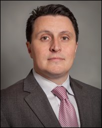 Anthony P. Conley合作论文数Department of Sarcoma Medical Oncology, Division of Cancer Medicine, The University of Texas MD Anderson Cancer Center8Caitlin D. May合作论文数7Raymond S. Traweek合作论文数7Maria A Zarzour合作论文数7Lusby Kristelle合作论文数7Lopez Gonzalo合作论文数7




