Compressed ultrafast photography (CUP) is applied to laser breakdown in argon and xenon under pressures up to 40atm to obtain 2D images of the plasma dynamics of single events with a spatial resolution of 250x100 pixels and an equivalent frame rate of 500 GHz. Light emission as a function of position and time is measured through red, green, blue, and broad-band filters. The spatially encoded and temporally sheared image normally used in CUP is now enhanced by the introduction of a constraint given by a spatially integrated and temporally sheared unencoded signal. The data yield insights into the temperature, opacity, the plasma formation process, and heat flow within the plasma and to the surrounding ambient gas. Contours of constant emission indicate that plasmas formed from sufficiently dense gas contract rather than expand despite having a temperature of a few eV. Plasmas formed from relatively low pressure gases such as 7atm argon can radiate with emissivity near unity. Modeling transport and opacity as arising from inverse Bremsstrahlung requires a degree of ionization that strongly exceeds expectations based on Saha's equation even as customarily modified to include density and screening. According to this model, both electrons and ions are strongly coupled with a plasma coefficient >1. During the first few nanoseconds after formation, Stefan-Boltzmann radiation and thermal conduction to ambient gas are too weak to explain the observed cooling rates, suggesting that transport within the plasma dominates its evolution. Yet, thermal conduction within the plasma itself is also small as indicated by the persistence of thermal inhomogeneities for far longer timescales. The fact that plasma is isolated from the surroundings makes it an excellent system for the study of the equation of state and hydrodynamics of such dense plasmas via the systems and techniques described.
Imaging the human body's morphological and angiographic information is essential for diagnosing, monitoring, and treating medical conditions. Ultrasonography performs the morphological assessment of the soft tissue based on acoustic impedance variations, whereas photoacoustic tomography (PAT) can visualize blood vessels based on intrinsic hemoglobin absorption. Three-dimensional (3D) panoramic imaging of the vasculature is generally not practical in conventional ultrasonography with limited field-of-view (FOV) probes, and PAT does not provide sufficient scattering-based soft tissue morphological contrast. Complementing each other, fast panoramic rotational ultrasound tomography (RUST) and PAT are integrated for hybrid rotational ultrasound and photoacoustic tomography (RUS-PAT), which obtains 3D ultrasound structural and PAT angiographic images of the human body quasi-simultaneously. The RUST functionality is achieved in a cost-effective manner using a single-element ultrasonic transducer for ultrasound transmission and rotating arc-shaped arrays for 3D panoramic detection. RUST is superior to conventional ultrasonography, which either has a limited FOV with a linear array or is high-cost with a hemispherical array that requires both transmission and receiving. By switching the acoustic source to a light source, the system is conveniently converted to PAT mode to acquire angiographic images in the same region. Using RUS-PAT, we have successfully imaged the human head, breast, hand, and foot with a 10 cm diameter FOV, submillimeter isotropic resolution, and 10 s imaging time for each modality. The 3D RUS-PAT is a powerful tool for high-speed, 3D, dual-contrast imaging of the human body with potential for rapid clinical translation.
Fetal alcohol spectrum disorders (FASDs) are caused by prenatal exposure to ethanol (EtOH), leading to developmental brain abnormalities. Cortical organoids derived from human-induced pluripotent stem cells provide physiologically relevant models to study such neurotoxic effects. However, accurate imaging of nuclear morphology, a key indicator of cytotoxicity and developmental impairment, remains difficult. Traditional fluorescence-based techniques rely on staining and sectioning, limiting throughput and potentially altering native structures. Herein, we present an ultraviolet photoacoustic microscopy system that targets endogenous nucleic acids at 266 nm excitation, resulting in a lateral resolution of 278 nm, which is sufficient to resolve individual nuclei in situ. The system integrates precise z-axis scanning for focal-plane alignment, enabling high-resolution depth-resolved imaging of subvolumes within 3D intact live organoids without physical sectioning. Using EtOH-treated cortical organoids as a model of FASD-associated neurotoxicity, we observed significant reductions in nuclear area, diameter, and circularity by 46.1%, 20.8%, and 6.0%, respectively, indicating structural damage consistent with apoptosis and impaired neurodevelopment. This method is the first to demonstrate label-free nuclear imaging in intact live brain organoids, providing a robust and preparation-free platform for probing disease-relevant phenotypes, accelerating drug screening, and enabling early toxicity assessments in neurodevelopmental disorder models.
Acoustic wave detection techniques like ultra-sound (US) and photoacoustic (PA) tomography are widely used in biomedical imaging but often require expensive transducer arrays and complex setups for 3D imaging. To overcome these challenges, recent research has explored using an ergodic relay (ER) with a single-element transducer. This approach improves imaging speed at low cost and reduces complexity. This study presents a dual-mode system that utilizes an ER for single-shot, 3D PA and US imaging with a single-element detector. Each ultrasonic or optical excitation pulse generates a 1D signal, which is then used to reconstruct a 3D image in US or PA mode. While the PA mode provides optical absorption-based contrast, the US mode offers complementary acoustic scattering-based contrast. We demonstrate the system’s capabilities through in vivo imaging of blood vessels and skin structure in human hands. The system is non-invasive, label-free, and ultrafast, enabling 4D imaging with simplified hardware requirements.
OBJECTIVE:Current breast imaging has limitations. Mammography uses radiation and compression; ultrasound depends on user expertise; MRI requires time and intravenous contrast. Development of novel technologies for breast imaging may be improved with patient surveys. METHODS:Breast cancer patients scheduled for breast operations or undergoing neoadjuvant therapy were enrolled in studies to evaluate photoacoustic computed tomography (PACT) at a single institution. After each imaging session, the patients were surveyed. The survey included Likert scale, multiple choice, and open-ended questions. RESULTS:Of 49 patients, 86 % completed at least one survey with 42 % completing three. Survey completion took <10 min. Features assessed specific to the imaging technology included water bath, duration, positioning, and environment. Patients overwhelmingly reported ease of PACT over mammography and MRI. Suggestions included better cushioning, improved head support, well-fitting laser safety glasses. CONCLUSION:Photoacoustic breast imaging is feasible to breast cancer patients. User feedback informs on clinical technology improvement.
Transcranial photoacoustic computed tomography (PACT) faces challenges due to skull-induced aberrations in measured pressure data. Although optimization-based image reconstruction methods have been developed to mitigate these aberrations, they require precise knowledge of skull properties that are generally unknown. Parameterized joint reconstruction (JR) methods improve computational tractability by estimating simplified skull models but only partially capture skull heterogeneity, potentially compromising modeling accuracy. Therefore, there remains a need for a reconstruction method that accounts for uncertainties in a fully heterogeneous skull model to enable accurate PACT image reconstruction in humans. In this study, a JR method for 3D transcranial PACT is developed to concurrently estimate the initial pressure distribution and CT-informed heterogeneous viscoelastic properties of the skull. The proposed approach leverages adjunct CT data and a linear relationship between CT voxel values and acoustic properties to parameterize a fully heterogeneous skull model. A gradient-free joint reconstruction method is used to estimate subject-dependent bone property parameters concurrently with the 3D initial pressure distribution. The method was systematically evaluated using realistic, full-scale 3D numerical head phantoms through in-silico studies. Results demonstrate that incorporating CT-informed heterogeneous skull modeling significantly improved image quality compared to model-based methods that assume a homogeneous skull model or a heterogeneous skull model with nominal parameter values. These findings highlight the importance of accurate skull modeling for robust transcranial PACT imaging.
Photoacoustic tomography is uniquely capable of high-resolution deep-tissue blood-oxygenation (sO2) imaging (oximetry) due to its optical absorption contrast. However, wavelength-dependent optical fluence changes within tissue, i.e., spectral coloring, have impeded the development of photoacoustic oximetry. We present the arterial prior method (APM+; + denotes intravascular fluence correction), which leverages the high arterial sO2 to locally calibrate the optical fluence within tissue to circumvent spectral coloring and reliably estimate the sO2 near the artery. In phantom experiments with ex vivo animal tissue, APM+ resulted in a median estimation error of 2.9% compared to 9.8% from the traditional linear unmixing method (LUM). In human imaging experiments of the radial artery-vein pair in eight healthy adult volunteers, the estimated venous sO2s from APM+ (median: 72.3%, interquartile range/IQR: 8.9%) were concentrated around the typical 60%-80% range in healthy individuals, whereas those from LUM (median: 75.2%, IQR: 34.4%) varied widely. When imaging the wrists of the eight subjects through ex vivo animal tissue of thicknesses up to 1.5 cm, APM+ provided more consistent estimates than LUM, indicating its robustness with depth.
From a series of optical-resolution photoacoustic microscopy imaging experiments performed using a lab-made laser diode driver, we found that although the pulse duration of the applied laser pulses was as long as 230 ns, it was still possible to acquire high-quality in vivo images with a sufficient signal-to-noise ratio, even when using a 25 MHz commercial high-frequency ultrasonic transducer. To interpret this interesting result, we performed additional experiments and consequently concluded that the slope of the rising or falling edge of an applied laser pulse may be a more critical factor than the overall pulse duration. In this paper, we present in vivo mouse ear imaging results acquired using both long and short pulses in a comparative manner as supporting evidence, along with the pulse profiles of the applied laser pulses.
Tomographic imaging modalities are described by large system matrices. To improve the temporal resolution of functional imaging in tomography, sparse spatial sampling is often employed, which degrades the system matrix and introduces artifacts in reconstructed images. Various existing techniques improve the image quality without correcting the system matrix and have limitations. Here, we compress the system matrix to improve computational efficiency (e.g., 42 times) using singular value decomposition and fast Fourier transform. Enabled by the efficiency, we propose fast sparsely sampling functional imaging by incorporating a densely sampled prior image into the system matrix, which maintains the critical linearity while mitigating artifacts. We demonstrate the methods in 3D photoacoustic computed tomography with significantly improved image quality and clarify their applicability to X-ray CT and radial-sampling MRI due to the similarities in system matrices.
Ultrasonography is a vital component of modern clinical care, with handheld probes routinely used for diagnostic imaging and procedural guidance. However, handheld ultrasound imaging is limited by factors such as the partial cross-sectional field of view, operator dependency, contact-induced distortion and lack of transmission contrast. Here we demonstrate a new system that enables whole cross-sectional ultrasound tomography of humans in both reflection and transmission modes. We generate two-dimensional images of the entire in vivo human cross-section in the abdomen and thighs with uniform in-plane resolution using a custom 512-element circular ultrasound receiver array and a rotating transmitter. Sequential scans with our system show strong agreement with clinical magnetic resonance imaging counterparts. To address unmet clinical needs, we explore two key applications. First, we observe abdominal adipose distributions in our images, enabling adipose thickness assessment without ionising radiation or mechanical deformation. Second, we demonstrate an approach for video-rate biopsy needle localization with respect to internal tissue features. These capabilities make whole cross-sectional ultrasound tomography a potential practical tool for clinical needs currently unmet by other modalities.
 Konstantin Maslov合作论文数Multimodal Imaging & Sensing Lab, Washington University in St. Louis187
 Junjie Yao (姚俊杰)合作论文数Department of Biomedical Engineering, Pratt School of Engineering, Duke University;Duke Cancer Institute, Duke University;Duke Global Health Institute, Duke University117
 Lei Li合作论文数George R. Brown School of Engineering, Rice University;California Institute of Technology67
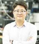 Chulhong Kim合作论文数Bio Optics and Acoustics Laboratory, Department of Creative IT Engineering, Pohang University of Science and Technology65
 Song Hu合作论文数Multimodal Imaging & Sensing Lab, McKelvey School of Engineering, Washington University in St. Louis58
 Mark Anastasio合作论文数Department of Bioengineering, The Grainger College of Engineering, University of Illinois Urbana-Champaign;Computational Imaging Science Laboratory, The Grainger College of Engineering, University of Illinois Urbana-Champaign45
 Qifa Zhou合作论文数Department of Biomedical Engineering and Ophthalmology, Viterbi School of Engineering, University of Southern California;Ginsburg Institute for Biomedical Therapeutics, University of Southern California;Department of Ophthalmology, Keck School of Medicine, University of Southern California45
 K. Kirk Shung合作论文数Department of Biomedical Engineering, University of Southern California42
 Jun Xia合作论文数Optical Imaging Laboratory, Department of Biomedical Engineering and39
 Li Chiye合作论文数39
 Alejandro Garcia-Uribe合作论文数39
 Yong Zhou合作论文数Tsinghua Univ, Tsinghua Univ.38
 Xin Cai合作论文数37Junhui Shi合作论文数36
 Geng Ku合作论文数Department of Cancer Systems Imaging, The University of Texas MD Anderson Cancer Center;Department of Mechanical Engineering, University of Kansas36
 Jun Zou合作论文数Department of Electrical and Computer Engineering, College of Engineering, Texas A&M University35
 Peng Hu (胡鹏)合作论文数School of Biomedical Engineering, ShanghaiTech University33
 Li Lin (林励)合作论文数College of Biomedical Engineering and Instrument Science, Zhejiang University33
 Ruimin Chen合作论文数Department of Biomedical Engineering, University of Southern California31
 Younan Xia (夏幼南)合作论文数Department of Materials Science and Engineering, Whiting School of Engineering, Johns Hopkins University;Department of Biomedical Engineering, Whiting School of Engineering, Johns Hopkins University30
 Xiao Xu合作论文数29
 Zhang Chi合作论文数29
 Yuecheng Shen (沈乐成)合作论文数East China Normal University29
 Joon-Mo Yang合作论文数Dept. of Computer Science and Information Engineering, Chung-Ju National University29
 Zhang Ruiying合作论文数28
 Christopher Favazza合作论文数Department of Radiology, Mayo Clinic27
 Zhu Liren合作论文数Optical Imaging Laboratory, Washington University in St. Louis27
 Dmitry Zimnyakov合作论文数26
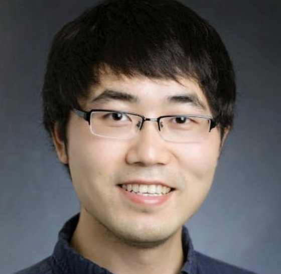 Liang Gao合作论文数Department of Bioengineering, School of Engineering, University of California, Los Angeles26
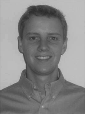 Todd N. Erpelding合作论文数26
 Puxiang Lai合作论文数Department of Biomedical Engineering, Faculty of Engineering, The Hong Kong Polytechnic University;Photonics Research Institute, The Hong Kong Polytechnic University26
 Liu Yan合作论文数Opt Imaging Lab, Washington Univ25
 Yide Zhang (张以德)合作论文数Department of Electrical, Computer & Energy Engineering, College of Engineering and Applied Science, University of Colorado Boulder25
 Jinyang Liang合作论文数Institut National de la Recherche Scientifique/Centre Énergie Matériaux Télécommunications, 1650 boulevard Lionel-Boulet, Canada24
 Zijian Guo (郭子建)合作论文数School of Chemistry and Chemical Engineering, Nanjing University;State Kay Laboratory of Coordination Chemistry, Nanjing University23Kwang Hyun Song合作论文数23
 George Stoica合作论文数21Rui Cao (曹锐)合作论文数College of Software, Taiyuan University of Technology21
 Bin Rao合作论文数Dept Biomed Engn, Washington Univ21
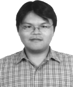 Meng-Lin Li合作论文数21
 Terence T. W. Wong (黃子維)合作论文数Department of Chemical and Biological Engineering, School of Engineering, The Hong Kong University of Science and Technology;PhoMedics20
 Manojit Pramanik合作论文数Biomedical Imaging Laboratory, Department of Electrical and Computer Engineering, College of Engineering, Iowa State University of Science and Technology;Faculty of Biomedical Engineering, Iowa State University of Science and Technology20Cheng Ma (马骋)合作论文数20
 Changhui Li (李长辉)合作论文数College of Engineering, Peking University20
 Yu S Zhang合作论文数Department of Medicine, Harvard Medical School;Harvard Stem Cell Institute;Brigham and Women's Hospital19
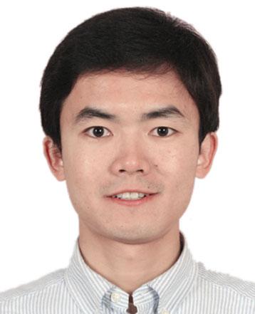 Liang Song (宋亮)合作论文数Laboratory for Biomedical Optics and Molecular Imaging, Institute of Biomedical and Health Engineering, Shenzhen Institute of Advanced Technology, Chinese Academy of Sciences;University of Chinese Academy of Sciences18
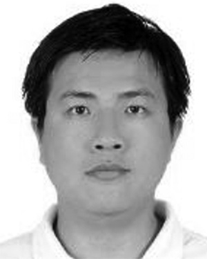 Hsin-I Wu合作论文数18Yeh Cheng-Hung合作论文数17
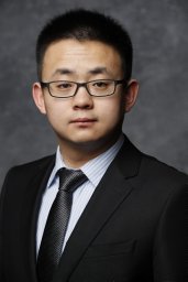 Hai Pengfei合作论文数17




