Segmental root developmental arrest (SRDA) is an uncommon developmental pattern in immature permanent teeth with apical periodontitis, in which a separated apical segment continues to develop independently of the main root. This retrospective study evaluated the frequency, clinical features, and factors associated with SRDA after regenerative endodontic procedures (REPs) or apexification. A total of 103 immature permanent teeth with apical periodontitis treated with REPs or apexification were retrospectively reviewed. Clinical records and radiographs were assessed for SRDA. Data collected included age, sex, tooth type, etiology, Nolla root developmental stage, treatment modality, timing of SRDA detection, and apical inflammation. Associations between candidate factors and SRDA were analyzed using the chi-square test or Fisher exact test, with Bonferroni correction for pairwise comparisons. SRDA was identified in 12 of 103 teeth (11.65
INTRODUCTION:Tooth morphology may predispose tooth cracking, yet evidence linking occlusal morphology to cracked molars remains limited. This study aimed to investigate the association between them, to establish reference intervals for early risk detection and management of cracked teeth. METHODS:Eighty-three cracked molars (68 patients) and 83 matched controls (69 volunteers) were analyzed. Occlusal morphologies (functional/nonfunctional cuspal inclination/height/slope) were measured using intraoral scans (Trios, 3Shape, Denmark) and analyzed using 3D digital design software. The crack initiation side (CIS) and propagation side (CPS) of crack lines were identified. Paired t-tests were used to compare the differences between groups. Multivariate logistic regression was used to assess correlations between occlusal morphology and cracked molars. RESULTS:Maxillary cracked molars exhibited steeper functional cuspal inclinations (CIS: [31.87 ± 7.72]°, CPS: [29.03 ± 7.50]°) than controls ([26.01 ± 7.01]°, [25.61 ± 7.47]°, P < .05), with 4°-6° differences. Mandibular cracked molars showed steeper inclinations only in functional cusps on CIS ([30.79 ± 7.89]° vs [24.53 ± 6.97]°, P < .05). Functional cuspal heights were 0.2-0.4 mm higher in cracked teeth than in controls for both arches (P < .05). Cracked molars exhibited flatter functional cusp slopes. Multivariate analysis identified CIS inclination (OR 1.098) and CPS height (OR 3.407) of functional cusps as maxillary risk factors; CIS functional cuspal height (OR 8.177) showed the highest risk for mandibular cracked molars. CONCLUSIONS:Within the limitations of this study, specific occlusal morphologies were correlated with cracked teeth. The identification of an excessively steep or high functional cusp in a molar warrants a thorough examination for crack lines. Morphology-guided occlusal adjustments should be considered in the treatment of cracked molars.
Background Pulpitis is a common dental disease driven by complex microbial infections, yet its microbial origins, diversity, and pathogenic mechanisms remain incompletely understood. A major clinical challenge is the absence of objective biological criteria to assess the severity and reversibility of pulpal inflammation, which is essential for decision-making in vital pulp therapy (VPT).Objective This review aims to synthesize current evidence on the microbial landscape of pulpitis and to explore microbial- and host-derived biomarkers that may enable objective assessment of inflammation severity and support precision VPT.Design We comprehensively reviewed microorganisms implicated in pulpitis and their distinct virulence mechanisms underlying inflammatory responses and tissue damage. Particular emphasis was placed on host responses of dental pulp stem cells (DPSCs) to different microbial infections. Biomarker candidates reported across multiple studies were summarized, and single-cell transcriptomic evidence was integrated to validate microbe-specific DPSC responses.Results Distinct microorganisms associated with pulpitis exhibit heterogeneous virulence strategies, inducing diverse inflammatory and degenerative processes within the dental pulp. DPSCs display infection-specific transcriptional responses, revealing molecular signatures linked to inflammation severity and tissue repair potential. Emerging biomarkers derived from both microbial factors and host responses show consistency across studies, with single-cell analyses providing high-resolution validation of these microbe-specific patterns.Conclusions Microbial- and host-derived biomarkers hold significant translational potential for stratifying pulpitis severity, informing VPT decision-making, and predicting treatment prognosis. Integrating microbial characterization with host response profiling may advance objective diagnosis and personalized management of pulpitis.
Pulpotomy, which belongs to vital pulp therapy, has become a strategy for managing pulpitis in recent decades. This minimally invasive treatment reflects the recognition of preserving healthy dental pulp and optimizing long-term patient-centered outcomes. Pulpotomy is categorized into partial pulpotomy (PP), the removal of a partial segment of the coronal pulp tissue, and full pulpotomy (FP), the removal of whole coronal pulp, which is followed by applying the biomaterials onto the remaining pulp tissue and ultimately restoring the tooth. Procedural decisions for the amount of pulp tissue removal or retention depend on the diagnostic of pulp vitality, the overall treatment plan, the patient’s general health status, and pulp inflammation reassessment during operation. This statement represents the consensus of an expert committee convened by the Society of Cariology and Endodontics, Chinese Stomatological Association. It addresses the current evidence to support the application of pulpotomy as a potential alternative to root canal treatment (RCT) on mature permanent teeth with pulpitis from a biological basis, the development of capping biomaterial, and the diagnostic considerations to evidence-based medicine. This expert statement intends to provide a clinical protocol of pulpotomy, which facilitates practitioners in choosing the optimal procedure and increasing their confidence in this rapidly evolving field.
AIM:The aim of the present study was to investigate the effects of Capnocytophaga periodontitidis (C. periodontitidis) and Porphyromonas gingivalis (P. gingivalis) on dental pulp stem cells (DPSCs) subclusters. MATERIALS AND METHODS:DPSCs were collected from healthy donors and were infected with C. periodontitidis or P. gingivalis. High-throughput single-cell RNA sequencing of infected DPSCs was performed to analyze gene expression changes, focusing on differentiation, hypoxia, and cell cycle-related pathways. RESULTS:Based on our single-cell resolution analysis, four major DPSC clusters were identified: classical-like, fibroblast-like, smooth muscle-like, and perivascular-like. C. periodontitidis infection induced differentiation into smooth muscle-like cells, while P. gingivalis promoted fibroblast (mineralized) lineages. P. gingivalis had a more significant effect on DPSCs differentiation. A specific subcluster responded uniquely to C. periodontitidis, with gene expression linked to B cell activation, muscle proliferation, and ROS pathways. CONCLUSION:DPSCs showed the homogeneity and heterogeneity when infected by these two pathogens. DPSCs differentiated into smooth muscle-related cells and mineralization-related cells when infected by C. periodontitidis and P. gingivalis, respectively. Our results will facilitate understanding and therapeutic strategies of primary periodontal disease with secondary endodontic involvement.
Apical microsurgery is accurate and minimally invasive, produces few complications, and has a success rate of more than 90%. However, due to the lack of awareness and understanding of apical microsurgery by dental general practitioners and even endodontists, many clinical problems remain to be overcome. The consensus has gathered well-known domestic experts to hold a series of special discussions and reached the consensus. This document specifies the indications, contraindications, preoperative preparations, operational procedures, complication prevention measures, and efficacy evaluation of apical microsurgery and is applicable to dentists who perform apical microsurgery after systematic training.
Enamel demineralization, the formation of white spot lesions, is a common issue in clinical orthodontic treatment. The appearance of white spot lesions not only affects the texture and health of dental hard tissues but also impacts the health and aesthetics of teeth after orthodontic treatment. The prevention, diagnosis, and treatment of white spot lesions that occur throughout the orthodontic treatment process involve multiple dental specialties. This expert consensus will focus on providing guiding opinions on the management and prevention of white spot lesions during orthodontic treatment, advocating for proactive prevention, early detection, timely treatment, scientific follow-up, and multidisciplinary management of white spot lesions throughout the orthodontic process, thereby maintaining the dental health of patients during orthodontic treatment.
Intentional tooth replantation (ITR) is an advanced treatment modality and the procedure of last resort for preserving teeth with inaccessible endodontic or resorptive lesions. ITR is defined as the deliberate extraction of a tooth; evaluation of the root surface, endodontic manipulation, and repair; and placement of the tooth back into its original socket. Case reports, case series, cohort studies, and randomized controlled trials have demonstrated the efficacy of ITR in the retention of natural teeth that are untreatable or difficult to manage with root canal treatment or endodontic microsurgery. However, variations in clinical protocols for ITR exist due to the empirical nature of the original protocols and rapid advancements in the field of oral biology and dental materials. This heterogeneity in protocols may cause confusion among dental practitioners; therefore, guidelines and considerations for ITR should be explicated. This expert consensus discusses the biological foundation of ITR, the available clinical protocols and current status of ITR in treating teeth with refractory apical periodontitis or anatomical aberration, and the main complications of this treatment, aiming to refine the clinical management of ITR in accordance with the progress of basic research and clinical studies; the findings suggest that ITR may become a more consistent evidence-based option in dental treatment.
Background Infection control is important in root canal treatment. Effective cleaning and shaping are challenging due to complex anatomy, particularly in the isthmus—narrow connections between canals that can harbor bacteria. Conventional needle irrigation (CNI) is inadequate in this region, prompting the use of passive ultrasonic irrigation (PUI) and high-frequency acoustic instruments like EDDY. This study evaluates the cleaning effects of four irrigation protocols using 3D-printed isthmus models. Methods Sixty digital root canal models with isthmuses in the coronal, middle, and apical thirds were designed using Ansys 19.0 and 3D printer (20 specimens per isthmus location). Specimens were prepared to 30#, 0.04 without irrigation. Debris accumulation in the isthmus was photographed and analyzed using Image J to calculate the initial debris area (S1). Specimens were then irrigated using CNI, low-frequency sonic irrigation (EndoActivator, EA; Dentsply, Charlotte, NC, USA), PUI, or high-frequency sonic irrigation (EDDY), followed by re-imaging to calculate remaining debris area (S2). Debris reduction percentage was determined using the formula: (S1–S2)/S1 × 100%. Results Debris reduction varied with isthmus position. In the coronal third, EDDY achieved the highest debris reduction (86.18 ± 2.25%), followed by PUI, EA, and CNI, with significant differences among groups (P < 0.05). The same trend was observed in the middle third, with EDDY showing the highest efficacy (73.96 ± 6.75%). In the apical third, debris reduction was lower overall, with no significant difference between EDDY and PUI, but both outperformed EA and CNI. Discussion Our results showed that EDDY demonstrated superior debris removal in the coronal and middle thirds, but all irrigation protocols showed limited efficacy in the apical third.
The overall health condition of patients significantly affects the diagnosis, treatment, and prognosis of endodontic diseases. A systemic consideration of the patient's overall health along with oral conditions holds the utmost importance in determining the necessity and feasibility of endodontic therapy, as well as selecting appropriate therapeutic approaches. This expert consensus is a collaborative effort by specialists from endodontics and clinical physicians across the nation based on the current clinical evidence, aiming to provide general guidance on clinical procedures, improve patient safety and enhance clinical outcomes of endodontic therapy in patients with compromised overall health.
Chemical cleaning and disinfection are crucial steps for eliminating infection in root canal treatment. However, irrigant selection or irrigation procedures are far from clear. The vapor lock effect in the apical region has yet to be solved, impeding irrigation efficacy and resulting in residual infections and compromised treatment outcomes. Additionally, ambiguous clinical indications for root canal medication and non-standardized dressing protocols must be clarified. Inappropriate intracanal medication may present side effects and jeopardize the therapeutic outcomes. Indeed, clinicians have been aware of these concerns for years. Based on the current evidence of studies, this article reviews the properties of various irrigants and intracanal medicaments and elucidates their effectiveness and interactions. The evolution of different kinetic irrigation methods, their effects, limitations, the paradigm shift, current indications, and effective operational procedures regarding intracanal medication are also discussed. This expert consensus aims to establish the clinical operation guidelines for root canal irrigation and a position statement on intracanal medication, thus facilitating a better understanding of infection control, standardizing clinical practice, and ultimately improving the success of endodontic therapy.
Isosteviol has been investigated with a cardio-treating effect on cardiomyocyte hypertrophy. However, its hydrophilicity suppressed its efficient cellular uptake for targeting the heart. Exosomes were seen as delivery vesicles with excellent histocompatibility; thus, an isosteviol-loaded exosome (Istv-exo) was prepared for cardiopulmonary delivery in this study. For in vivo evaluation, the Istv-exo and isosteviol saline solution was used for inhalation or intragastric administration on Sprague–Dawley rats for an assay of dynamic distribution. The higher biodistribution of Istv-exo in the lung and heart has been confirmed by the dynamic content curve. Istv-exo/isosteviol solution was administered by inhalation/intragastrically to Myh6-/- mice, a cardiomyocyte hypertrophy model, to explore the therapeutic effect. The heart function of the mice was measured using echocardiography and in situ imaging was used to analyze the morphology of the heart. The higher level of left ventricular internal dimension, ejection fraction, and cardiac output in echocardiography indicated that Istv-exo enhanced improvement of myocardial function. The attenuation of cardiomyocyte hypertrophy in mice treated with inhalation of Istv-exo determined that it contributed to the reversal of pathological hypertrophy phenotypes. Wheat germ agglutinin staining confirmed that cardiomyocyte hypertrophy was reversed to varying degrees in the Istv-exo, intragastrical isosteviol, and positive control groups. Hematoxylin and eosin staining exhibited the enlarging left ventricular and thinning ventricular wall of the treated mice. Drastically less collagen fiber was observed in the positive control group and Istv-exo group. The inhalation administration of isosteviol-loaded exosomes may be better in therapy for cardiomyocyte hypertrophy than intragastric usage of isosteviol.
The diterpenoid isosteviol is one of the stevia derivatives that have reported various activities. In mice inhaling isosteviol, OVA- and ACh-induced bronchial asthma showed a dose-dependent improvement of a decline in the expression of inflammatory cytokines and the infiltration of basophils. Compared with inhalation of budesonide 0.6 mg/kg, sensitized mice inhaling 1.0 mg/kg dosage of isosteviol performed a higher tolerate against the ACh stimulation. In behavior tests, the 1.0 mg/kg isosteviol nebulization reached almost 80
Endo-periodontal lesions (EPLs) involve both the periodontium and pulp tissue and have complicated etiologies and pathogenic mechanisms, including unique anatomical and microbiological characteristics and multiple contributing factors. This etiological complexity leads to difficulties in determining patient prognosis, posing great challenges in clinical practice. Furthermore, EPL-affected teeth require multidisciplinary therapy, including periodontal therapy, endodontic therapy and others, but there is still much debate about the appropriate timing of periodontal therapy and root canal therapy. By compiling the most recent findings on the etiology, pathogenesis, clinical characteristics, diagnosis, therapy, and prognosis of EPL-affected teeth, this consensus sought to support clinicians in making the best possible treatment decisions based on both biological and clinical evidence.
Dens invaginatus may be associated with peri-invagination lesions and vital pulp concurrently. This case report examines the successful preservation of vital pulp and minimally invasive treatment of invagination for Oehlers type IIIA dens invaginatus with an extensive peri-invagination lesion. A healthy 19-year-old man presented with occasional swelling of the left maxillary anterior region. Pulp vitality tests revealed vital and healthy tooth pulp. CBCT indicated Oehlers type IIIA dens invaginatus with an invagination parallel to the pulp cavity. The diagnosis was type IIIA dens invaginatus with a peri-invagination lesion. The treatment plans involved preservation of the vital pulp and minimally invasive treatment of the invagination. A 5-year follow-up revealed that both healing of the peri-invagination lesion and preservation of the vital pulp had been successful. Pulp vitality can be preserved in type IIIA dens invaginatus associated with a peri-invagination lesion through minimally invasive treatment of the invagination.
Endodontic diseases are a kind of chronic infectious oral disease. Common endodontic treatment concepts are based on the removal of inflamed or necrotic pulp tissue and the replacement by gutta-percha. However, it is very essential for endodontic treatment to debride the root canal system and prevent the root canal system from bacterial reinfection after root canal therapy (RCT). Recent research, encompassing bacterial etiology and advanced imaging techniques, contributes to our understanding of the root canal system's anatomy intricacies and the technique sensitivity of RCT. Success in RCT hinges on factors like patients, infection severity, root canal anatomy, and treatment techniques. Therefore, improving disease management is a key issue to combat endodontic diseases and cure periapical lesions. The clinical difficulty assessment system of RCT is established based on patient conditions, tooth conditions, root canal configuration, and root canal needing retreatment, and emphasizes pre-treatment risk assessment for optimal outcomes. The findings suggest that the presence of risk factors may correlate with the challenge of achieving the high standard required for RCT. These insights contribute not only to improve education but also aid practitioners in treatment planning and referral decision-making within the field of endodontics.
IntroductionMicrobial pathogens invade various human organs, including the oral cavity. Candida albicans (C.a) and Streptococcus mutans (S.m) served respectively as representative oral pathogenic fungi and bacteria to stimulate dental pulp stem cells (DPSCs) and to screen the DPSC subcluster that specifically responded to fungal infection.MethodsDPSCs were obtained from the impacted third molars of six healthy subjects. Then, cells were mixed and divided into three samples, two of which were stimulated with C.a and S.m, respectively; the third sample was exposed to cell medium only (Ctrl). Single-cell mRNA sequencing analysis of treated DPSCs was performed.ResultsDPSCs were composed of four major clusters of which one, DPSC.7, exhibited unique changes compared to those of other subclusters. The DPSC.7 cell percentage of the C.a sample was twice those of the Ctrl and S.m samples. DPSC.7 cells expressed genes associated with the response to reactive oxygen species (ROS) response. DPSC.7 subgroup cells established characteristic aggregation under the stimulation of different pathogens in UMAP. The MAPK/ERK1/2 and NF-κB pathways were up-regulated, DUSP1/5/6 expressions were suppressed, FOS synthesis was activated, the immune-related pathway was induced, and the levels of cytokines, including IL-6 and CCL2, were up-regulated in DPSC.7 cells when stimulated with C.a.ConclusionsOur study analyzed the cellular and molecular properties of DPSCs infected by oral fungi and bacteria with single-cell RNA sequencing. A subcluster of DPSCs responded specifically to infections with different pathogens, activating the MAPK and NF-κB pathways to induce immune responses via the ROS pathway. This suggests novel treatment strategies for fungal infections.
Conception of public health was firstly put forward by American professor Winslow. Ensuring and promoting the health of general population is the key connotation for the definition of public health. Oral disease has become a public health problem. Caries which preventable and curable is the most common oral disease and the etiology is also clear. Oral health comprehensive intervention program for children in central and western regions was set up in 2008 by Chinese government. The program included sealing on the first permanent molar and oral health education towards primary school children covering mid-west area. This was the first oral health program invested by government and managed by Chinese Stomatological Association. Six years later, the program was popularized to the whole nation, and renamed as national oral health comprehensive intervention program for children in China. The program had made deep impact on development of oral health service in China. The study tries to analyze the challenges of oral health service through reviewing the background, content, organization and effectiveness of the program, aiming to provide suggestions on policy, financing, system, ability and technology for the future development.
The dental operative microscope has been widely employed in the field of dentistry, particularly in endodontics and operative dentistry, resulting in significant advancements in the effectiveness of root canal therapy, endodontic surgery, and dental restoration. However, the improper use of this microscope continues to be common in clinical settings, primarily due to operators’ insufficient understanding and proficiency in both the features and established operating procedures of this equipment. In October 2019, Professor Jingping Liang, Vice Chairman of the Society of Cariology and Endodontology, Chinese Stomatological Association, organized a consensus meeting with Chinese experts in endodontics and operative dentistry. The objective of this meeting was to establish a standard operation procedure for the dental operative microscope. Subsequently, a consensus was reached and officially issued. Over the span of about four years, the content of this consensus has been further developed and improved through practical experience.
Digital guided therapy (DGT) has been advocated as a contemporary computer-aided technique for treating endodontic diseases in recent decades. The concept of DGT for endodontic diseases is categorized into static guided endodontics (SGE), necessitating a meticulously designed template, and dynamic guided endodontics (DGE), which utilizes an optical triangulation tracking system. Based on cone-beam computed tomography (CBCT) images superimposed with or without oral scan (OS) data, a virtual template is crafted through software and subsequently translated into a 3-dimensional (3D) printing for SGE, while the system guides the drilling path with a real-time navigation in DGE. DGT was reported to resolve a series of challenging endodontic cases, including teeth with pulp obliteration, teeth with anatomical abnormalities, teeth requiring retreatment, posterior teeth needing endodontic microsurgery, and tooth autotransplantation. Case reports and basic researches all demonstrate that DGT stand as a precise, time-saving, and minimally invasive approach in contrast to conventional freehand method. This expert consensus mainly introduces the case selection, general workflow, evaluation, and impact factor of DGT, which could provide an alternative working strategy in endodontic treatment.
 Xiaoying Zou (邹晓英)合作论文数Peking University Hospital23
 Xuejun Gao (高学军)合作论文数Peking University Hospital of Stomatology20
 Meihua Cui (崔梅花)合作论文数Peking University Space Center Hospital17
 Yuhong Liang (梁宇红)合作论文数Peking University Hospital of Stomatology12
 Xi Wei (韦曦)合作论文数Hospital of Stomatology, SunYat-sen University11
 Junqi Ling (凌均棨)合作论文数Hospital of Stomatology, Sun Yat-sen University10
 JiaDe Wang (王嘉德)合作论文数9
 Xudong Bao (包旭东)合作论文数Peking University School of Stomatology9
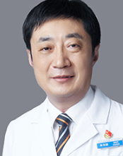 Fanghong Mu (牟方宏)合作论文数Aerospace Center Hospital9
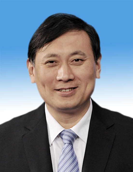 Benxiang Hou (侯本祥)合作论文数Beijing Stomatological Hospital, Capital Medical University9
 Jin Zhao (赵今)合作论文数新疆医科大学附属第一医院口腔科9
 Zhi Chen (陈智)合作论文数Hospital of Stomatology Wuhan University;School of Stomatology, Wuhan University9Wenxia Chen合作论文数8Yi Fu (傅怡)合作论文数8
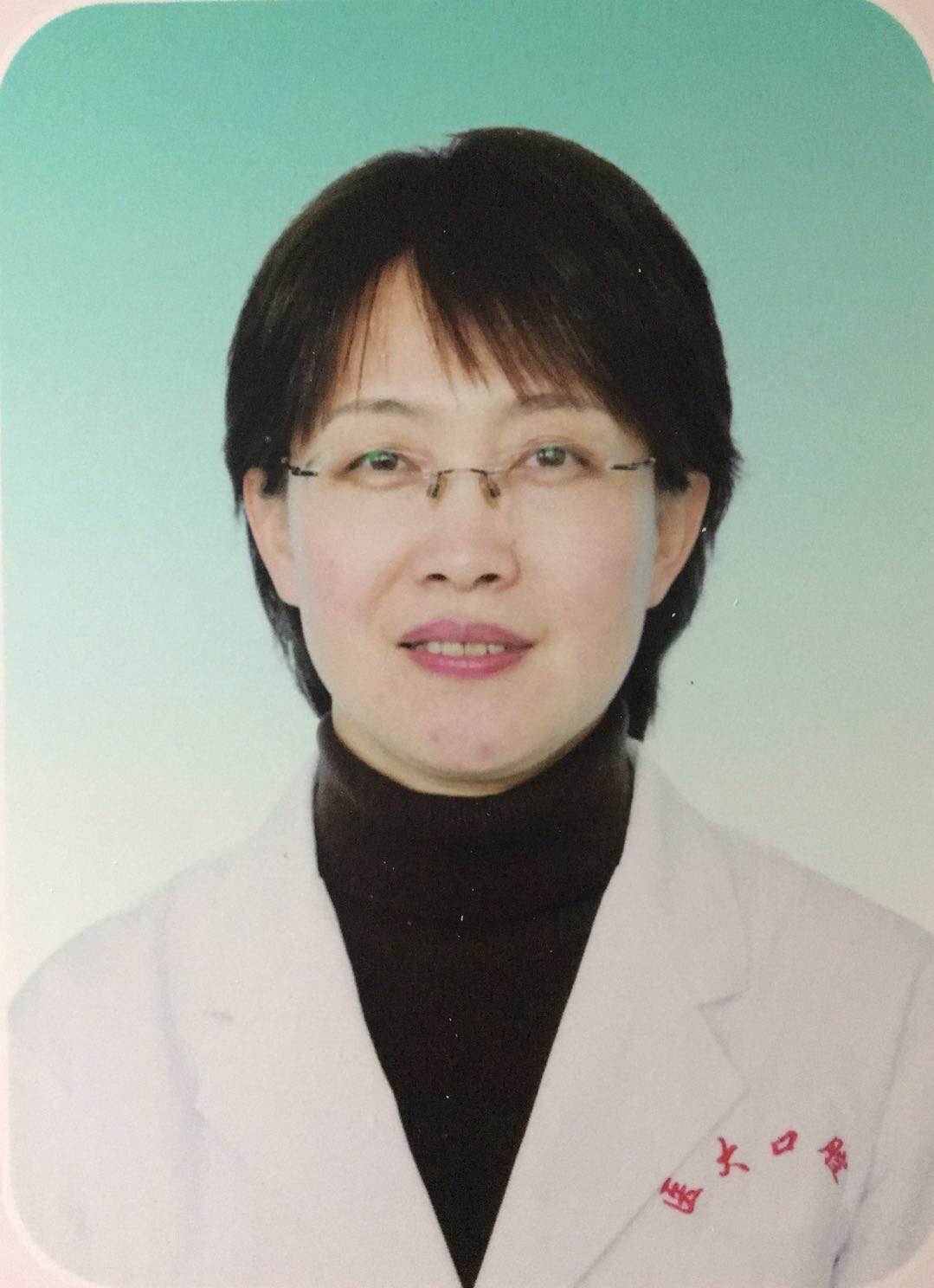 Lihong Qiu (仇丽鸿)合作论文数China Medical University8
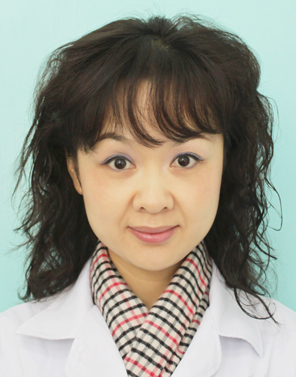 Deqin Yang (杨德琴)合作论文数College of Stomatology, Chongqing Medical University8
 Xiaoyan Wang (王晓燕)合作论文数Hospital of Stomatology, Peking University8
 Qing Yu (余擎)合作论文数第四军医大学口腔医院牙体牙髓病科7
 Guibin Yang (杨桂彬)合作论文数Aerospace Center Hospital, Peking University7Wen Tan (谭文)合作论文数Guangdong Zhongsheng Pharmaceutical Co., Ltd.;Rundu Pharma7
 Zhengwei Huang (黄正蔚)合作论文数Shanghai Ninth People's Hospital, School of Medicine, Shanghai Jiao Tong University7Changhong Ke合作论文数6ShuLi Deng (邓淑丽)合作论文数6
 Lin Feng (冯琳)合作论文数Peking University School of Stomatology6
 Jiyao Li (李继遥)合作论文数West China Hospital of Stomatology, Sichuan University6
 Dingming Huang (黄定明)合作论文数West China Hospital of Stomatology, Sichuan University6
 Jingping Liang (梁景平)合作论文数National Clinical Research Center for Oral Diseases, Shanghai Ninth People's Hospital, School of Medicine, Shanghai Jiao Tong University6Qi Zhang合作论文数5Huan Hou合作论文数5Chaohong Huang合作论文数5
 Zhengqiang Yuan合作论文数5YouPeng Yang (杨友鹏)合作论文数5
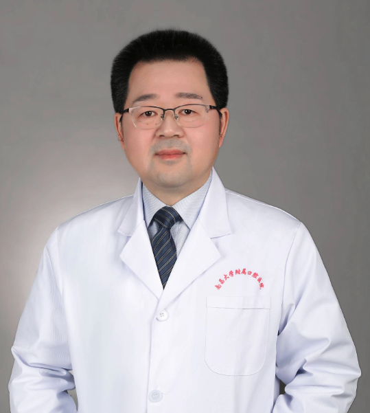 Jian Yang (杨健)合作论文数Affiliated Stomatological Hospital of Nanchang University5
 Xiaoli Xie (谢晓莉)合作论文数Hunan Xiangya Stomatological Hospital, Central South University5Kui Su合作论文数5
 Yihuai Pan (潘乙怀)合作论文数Department of Endodontics, School & Hospital of Stomatology, Wenzhou Medical University;Alberta Institute, Wenzhou Medical University5
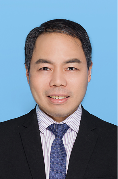 Jinhua Yu (于金华)合作论文数Endodontic Department, School of Stomatology, Nanjing Medical University;Institute of Stomatology, School of Stomatology, Nanjing Medical University5
 Jie Zhang (张杰)合作论文数Hospital of Stomatology, Peking University5Huijie Guo (郭惠杰)合作论文数5
 Xuedong Zhou (周学东)合作论文数West China School of Stomatology, Sichuan University5
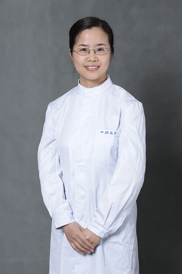 Liuyan Meng (孟柳燕)合作论文数School of Stomatology, Wuhan University;Hospital of Stomatology, Wuhan University5
 Xin Xu (徐欣)合作论文数West China Hospital of Stomatology, Sichuan University4Tiansong Xu合作论文数4Jianwu Sun合作论文数4Shuyi Li合作论文数4Xiao-bo CHEN (陈晓播)合作论文数4Qian Yuan合作论文数4Yuan Li (袁理)合作论文数4




