Objective: We have previously demonstrated a negative correlation between electroencephalographic (EEG) alpha functional connectivity (FC) and small-world organization in healthy individuals, suggesting a compensatory mechanism that maintains efficient information processing. It remains unclear whether this relationship persists in major depressive disorder (MDD), a condition associated with altered brain network dynamics. We aim to investigate whether the FC–SW correlation observed in healthy subjects is present in MDD patients. Methods: Eyes-closed EEG (30 channels, 6 min) was recorded from 37 unmedicated MDD patients. Alpha (8–12 Hz) FC was assessed with magnitude-squared coherence (MSC) and the imaginary part of coherency (ICOH). Small-worldness (SW) was computed across network densities ranging from 10
Introduction: Surface electromyography (sEMG) can be used to quantify Parkinson’s disease (PD) motor symptoms. However, traditional sEMG acquisition setups are impractical for ambulatory assessment or continuous real-world monitoring. Wearable sEMG devices could extend the use of this measurement technique to a wider clinical setting. The present study compares a wearable textile-based sEMG sleeve with a conventional sEMG system and investigates the feasibility of the wearable sleeve for characterizing upper-limb bradykinesia in PD.Methods: sEMG signals from both forearm flexor and extensor muscles were recorded in 16 healthy participants using a wearable textile sleeve and traditional Ag/AgCl electrodes during the MDS-UPDRS item 3.5—hand movements task. The burst length and averaged rectified EMG were compared between the methods using Cohen’s d, Bland–Altman analysis, and the within-session coefficient of variation (CV%). The same procedure was used with the textile electrodes to assess bradykinesia severity in 19 PD patients treated with deep brain stimulation (DBS). Measurements were performed in stimulation ON and OFF states. Commonly used sEMG features were extracted from 62 recordings during the hand movements task. The association between bradykinesia severity and sEMG-derived features was evaluated using a linear mixed-effects model (LMM) to account for repeated measures and inter-subject variability.Results: Muscle excitation burst durations were slightly longer when measured with the wearable sEMG sleeve, whereas the average rectified EMG was comparable between systems. Bland–Altman analysis demonstrated good agreement between methods. Within-session repeatability was similar for average rectified EMG, while excitation burst duration showed greater variability in both muscle groups with the textile sleeve. The final LMM examining PD bradykinesia included Willison amplitude measured from forearm extensors (β = -0.50; 95% CI [-0.78, -0.22]) and root mean square from flexors (β = -0.46; 95% CI [-0.73, -0.20]) and extensors (β = -0.34; 95% CI [-0.64, -0.03]), explaining R²ₘ = 0.71 and R²𝚌 = 0.85 of the variance. Inclusion of the DBS therapy state was associated with a mean improvement of 1.51 points in bradykinesia severity.Conclusions: Textile sleeve-based sEMG electrodes provide muscle excitation measures and reproducibility comparable to those of conventional sEMG systems and demonstrate sufficient robustness for bradykinesia assessment in individuals with PD. Further studies are warranted to validate and further characterize wearable textile-based sEMG devices, as such measurement systems may offer benefits for individuals with PD as well as healthcare providers.
Higuchi Fractal Dimension (HFD) is a widely used nonlinear metric with extensive application in biological signal analysis, including electroencephalography (EEG) signal processing. HFD relies on a single free parameter, kmax, which is the maximum scale in samples, used to assess the signal's self-similarity. While fractal dimension estimation is independent of sampling frequency (Fs) in ideally fractal signals, EEG signals do not exhibit ideal fractality, also making the HFD estimates sensitive to both parameter kmax and sampling frequency. As a result, HFD results reported across studies are difficult to compare with different parameter settings. In this work, we emphasize the need to account for the sampling frequency and present the HFD results in terms of the maximum time interval, tmax = kmax/Fs, rather than kmax, thereby facilitating comparisons across studies. Additionally, we demonstrate that the choice of maximum time interval (tmax) determines the frequency range that HFD focuses on, improving the interpretation of the results and enabling a more informed parameter selection.
Electroencephalography (EEG) is a cost-effective, noninvasive method with high temporal resolution that enables detailed assessment of neuronal activity. Detrended fluctuation analysis (DFA), a nonlinear technique for quantifying long-range temporal correlations in time-series data, has found application in EEG research across various contexts. This study investigates the temporal stability of DFA over the course of one year, during which 12 monthly EEG recordings were collected from each of nine healthy male participants. Our findings demonstrate excellent within-subject reliability, with intraclass correlation coefficients (ICCs) ranging from 0.985 to 0.997 across 30 EEG channels. These high ICCs indicate that interindividual variability exceeds intraindividual variability, supporting DFA’s reliability for long-term neural monitoring. Despite considerable differences between individuals, DFA remained consistent within subjects, highlighting its potential as a subject-specific biomarker for neurological disorders such as epilepsy, depression, and Alzheimer's disease. These findings underscore the importance of accounting for individual variability in EEG measures when developing tools for early diagnosis and clinical monitoring.Clinical Relevance— Reliable biomarkers for neuronal activity must demonstrate consistent temporal stability. This study shows that DFA offers excellent stability within individuals, supported by ICC analyses, suggesting its potential as a subject-specific biomarker for early detection of disorders characterized by altered EEG dynamics.
The clinical applicability of electroencephalography (EEG) relies on the reliability and temporal stability of its measures. While the reliability of linear EEG measures is well established, the long-term stability of both linear and nonlinear measures at the individual level, as well as interindividual variability, remains underexplored. This study evaluated the one-year stability of EEG absolute band powers (theta, alpha, beta, and gamma) and nonlinear measures (Higuchi's fractal dimension, Lempel-Ziv complexity, detrended fluctuation analysis, and in-phase Matrix Profile) across 12 monthly EEG recordings in nine healthy males aged 26-49. Intraclass correlation coefficients (ICCs) indicated excellent reliability across all measures, although beta power showed slightly reduced ICCs in temporal regions and gamma power demonstrated lower reliability in peripheral sites. At the individual level, nonlinear measures showed greater temporal stability than EEG band powers. Although a few individuals, particularly in band power measures, exhibited annual fluctuations comparable to or exceeding interindividual variability, most participants demonstrated consistent EEG profiles over time. These findings support the use of nonlinear EEG measures in longitudinal research and indicate their potential for developing personalized EEG-based neural biomarkers. They also highlight the importance of estimating expected individual variability when designing individualized monitoring approaches, as high reliability at the group level does not preclude substantial within-subject variability in some cases.
Background: Wearable sensors are mainly used in Parkinson’s disease (PD) to assess motor symptoms and to aid clinicians in patient management. Inertial measurement units that simultaneously register accelerometric and gyroscope signals have been one of the most studied and practicable methods. The heterogeneity of described methods and clinical settings studied can discourage wearable device use and highlight the need for standardization. This study compares previously proposed accelerometry and gyroscope signal features for tremor assessment measured at the wrist. Methods: An inertial measurement unit registered accelerometry and gyroscope signals at the wrist from 18 PD patients treated with deep brain stimulation (DBS). Measurements were made in DBS on and off states. Signal features for both accelerometry and gyroscope were calculated—mean linear acceleration, mean angular velocity, root mean square, maximal amplitude and power of the 3–7 Hz frequency band. The outcome features were log-transformed and correlated to the Movement Disorder Society Unified Parkinson’s Disease Rating Scale (MDS-UPDRS) item 3.17 using linear regression. Intraclass correlation coefficient (ICC) values were calculated for the signal features. Results: A total of 108 tremor episodes were investigated. All signal features exhibited a strong correlation with the MDS-UPDRS tremor amplitude scale. Tremor ratings showed a stronger correlation with accelerometry (r = 0.964–0.970) than with gyroscope-derived features (r = 0.942–0.956). The best-performing feature was the mean linear acceleration (r = 0.970, R2 = 0.940), which also showed high reliability (ICC = 0.921). Conclusions: Different accelerometry and gyroscope signal features are viable in characterizing rest tremor at the wrist. Simpler accelerometry signal features can be preferred in conducting the MDS-UPDRS item 3.17 examination in PD patients with DBS using a wrist-worn inertial measurement unit. Future research to expand the validity and usefulness of wearable technologies in PD is warranted.
The current study aims to evaluate the temporal stability of Higuchi’s Fractal Dimension (HFD) in the electroencephalographic (EEG) signals of healthy individuals. HFD depends on a single tuning parameter, kmax, which indicates the time scale range in which fractality is analyzed. A kmax value that provides stable HFD values for healthy individuals is expected to allow easier detection of deviations in brain activity associated with neurological disorders. To identify the stable region, EEG signals from 15 healthy participants, recorded twice over a three-year period, were analyzed. HFD temporal stability at different kmax values was assessed using the intraclass correlation coefficient between sessions. We hypothesized that HFD stability would be highest at lower kmax values, as a smaller range of time scales of EEG signals are likely to provide clearer representation of the fractal nature. The results support our hypothesis, demonstrating that HFD stability is highest within the range of 6 < kmax < 14. This finding enhances the reliability of the HFD measure and its potential as a valuable tool to support clinical decisions.Clinical Relevance— The identified stable region of HFD in healthy individuals enhances the method’s overall reliability, thereby improving also its potential to yield consistent results in detecting deviations associated with neurological disorders.
Chronic kidney disease is associated with cognitive impairment although the underlying mechanisms are still not fully understood. Characterization and efficient monitoring of the cognitive impact of kidney disease and ensuing therapies are critical for the accurate clinical management of patients. A vast array of imaging modalities, biomarkers, and sensors have shown relevance for the assessment of cognitive impairment. Knowing the potential and limitations of these paraclinical techniques is a necessary condition to improve the understanding of this phenomenon and to design monitoring protocols and guidelines applicable to this clinical population. The goal of this review is to provide an overview of current imaging modalities and biomarker sources available to the community, for the benefit of the research and clinical community.
Functional connectivity and small-worldness are widely used techniques to study brain network organization, and previous research has shown a negative correlation between them in the alpha frequency band. This study examines how specific electroencephalography (EEG) alpha-band connections contribute to the balance between functional connectivity and small-worldness. EEG was recorded from 80 healthy subjects in a resting-state, eyes-closed condition. Functional connectivity was estimated using magnitude-squared coherence (MSC), imaginary part of coherency (ICOH), and synchronization likelihood (SL), and small-worldness was calculated for each of these measures. The strongest negative correlation between functional connectivity and small-worldness was found in frontal-occipital and frontal connections. Given that these regions have been linked to the default mode network (DMN), the observed correlation may reflect DMN activity. These findings highlight a balance between functional network structure and connectivity and provide insight into how resting-state networks maintain functional organization. Future research should investigate how this balance shifts during cognitive tasks and in neurological disorders.Clinical Relevance— The current study is a step in understanding the physiology of a healthy brain networks, potentially aiding in the early detection and prevention of brain disorders
Glucose metabolism is an important factor in human physiology and the main source of energy for the human brain. Low blood glucose concentration (hypoglycemia) will cause several neurological and cardiological symptoms. High concentration (hyperglycemia) is an indicator of a higher risk to diabetes. The current study aims to investigate the correlation between electroencephalographic signal (EEG) functional connectivity and fasting blood glucose concentration in healthy people with normal blood glucose concentration. The present study was carried out on a group of forty-four healthy volunteers. The resting-state eyes-closed 30-channel EEG was recorded for 6 minutes and blood samples were collected from the subjects on the same morning. To describe the functional connectivity, magnitude-squared coherence (MSC) was calculated between all channels in delta, theta, alpha, beta and gamma EEG frequency bands. The negative correlation between MSC and fasting blood glucose concentration was statistically significant in delta and gamma bands. The results of the study suggest that the variations in normal blood glucose concentration can affect brain functioning.Clinical Relevance— This is a step in the development of novel biomarkers for monitoring neurological and cognitive function, potentially aiding in the early detection and prevention of metabolic and neurocognitive disorders.
This study aims to investigate the association between the natural level of blood biomarkers and electroencephalographic (EEG) markers. Resting EEG theta, alpha (ABP), beta, and gamma frequency band powers were selected as linear EEG markers indicating the level of EEG power, and Higuchi’s fractal dimension (HFD) as a nonlinear EEG complexity marker reflecting brain temporal dynamics. The impact of seven different blood biomarkers, i.e., glucose, protein, lipoprotein, HDL, LDL, C-reactive protein, and cystatin C, was investigated. The study was performed on a group of 52 healthy participants. The results of the current study show that one linear EEG marker, ABP, is correlated with protein. The nonlinear EEG marker (HFD) is correlated with protein, lipoprotein, C-reactive protein, and cystatin C. A positive correlation with linear EEG power markers and a negative correlation with the nonlinear complexity marker dominate in all brain areas. The results demonstrate that EEG complexity is more sensitive to the natural level of blood biomarkers than the level of EEG power. The reported novel findings demonstrate that the EEG markers of healthy people are influenced by the natural levels of their blood biomarkers related to their everyday dietary habits. This knowledge is useful in the interpretation of EEG signals and contributes to obtaining information about people quality of life and well-being.
Depression is currently one of the most complicated public health problems with the rising number of patients, increasing partly due to pandemics, but also due to increased existential insecurities and complicated aetiology of disease. Besides the tsunami of mental health issues, there are limitations imposed by ambiguous clinical rules of assessment of the symptoms and obsolete and inefficient standard therapy approaches. Here we are summarizing the neuroimaging results pointing out the actual complexity of the disease and novel attempts to detect depression that are evidence-based, mostly related to electrophysiology. It is repeatedly shown that the complexity of resting-state EEG recorded in patients suffering from depression is increased compared to healthy controls. We are discussing here how that can be interpreted and what we can learn about future effective therapies. Also, there is evidence that novel options of treatment, like different modalities of electromagnetic stimulation, are successful just because they are capable of decreasing that aberrated complexity. And complexity measures extracted from electrophysiological signals of depression patients can serve as excellent features for further machine learning models in order to automatize detection. In addition, after initial detection and even selection of responders for further therapy route, it is possible to monitor the therapeutic flow for one person, which leads us to possible tailored treatment for patients suffering from depression.
The correlation between electroencephalographic (EEG) and electrocardiographic (ECG) signals can provide important information about cardiovascular regulation. The current study aims to investigate the dependence of the correlation between EEG and heart rate variability (HRV) on cardiovascular indicators. The signals of a group of 30 subjects were divided into two groups of 15 subjects according to the lower and higher indicators of blood pressure and cholesterol. Relative EEG frequency band powers were calculated in theta, alpha, and beta frequency bands. From power spectral analysis of HRV, low frequency (LF) power, high frequency (HF) power and LF/HF were calculated. In the current study, the correlation between EEG and HRV is detected by two HRV features, LF and HF, in the theta band, and also by two features, HF and LF/HF, in the alpha band. The correlation by one HRV feature, LF/HF, is revealed in the beta band. The effect of cardiovascular features is reflected by LF and HF features in the theta band and by HF in the alpha band. The novel finding that even a small increase in cardiovascular features (blood pressure and cholesterol) can affect cardio-neuronal regulation is important and needs further investigation.
Background and Objective: Major depressive disorder (MDD) is the leading cause of disability worldwide. Reliable detection of MDD is the basis for early and successful intervention in treating the disorder and preventing disability. We introduce a novel feature extraction method, the in-phase matrix profile (pMP), which is specifically adapted for electroencephalographic (EEG) signals. Methods: The pMP characterizes general self-similarity of an EEG signal. The method extracts overlapping one-second-long subsegments from an EEG signal segment, calculates Euclidean distances between all possible subsegment pairs, and subsequently uses the distance values, where subsegments are most in phase, to calculate pMP. The method was applied to the resting-state eyes-closed EEG data of an MDD group and age- and gender-matched healthy controls (66 subjects). Higuchi's fractal dimension (HFD) values were calculated for the same groups for comparison. Results: Both pMP and HFD values were higher in MDD. The pMP successfully distinguished MDD and control group in all 30 EEG channels. In contrast, HFD resulted in statistically significant group distinguishability in 13 (43%) channels located mainly in the central region of the head. The highest classification accuracy for pMP was 73% and for HFD 67%. Conclusion: The present article shows that pMP outperforms HFD in detecting MDD and is a promising method for future MDD studies. Significance: The pMP is a sensitive parameter-free method for detecting MDD that can be used in future studies and is a potential method to reach clinical use for diagnosing MDD.
The decreased environment temperature can cause vasoconstriction and drops the skin perfusion, which lowers finger photoplethysmographic (PPG) signal amplitude and signal to noise ratio. The forehead is relatively insulated from external temperature changes due to the presence of the skull and the scalp. Therefore, this site is less affected by temperature compared to other peripheral PPG signal registration sites. The aim of this study was to characterize the changes in the forehead PPG and second derivative of PPG (SDPPG) waveform parameters related to arterial stiffness for the acute mental stress assessment. The PPG signals were recorded from 42 subjects during eyes open and arithmetic stress test. The signals from 34 subjects (18 females and 16 males) were included to the post-processing and analysis of PPG waveform arterial stiffness related parameters. The changes in the forehead PPG waveform parameters PPGb, PPGAI, Snorm, and SDPPG waveform parameters b/a, c/a, d/a, e/a, and AGI were statistically significantly different (p < 0.05) between eyes open and stress test states. The calculated parameters indicated the increase in arterial stiffness due to the induced stress. However, c/a changed in the opposite direction than expected. In summary, the results indicate that the changes in the forehead PPG and SDPPG parameters could be used similarly to the finger PPG signal for the stress assessment. Nevertheless, further studies are needed.
Mental stress can lead to different health problems or may increase the risk of accidents, especially in cases of high personal responsibility (pilots, policemen, military specialists etc.). Therefore, there is a need for an objective assessment of mental stress in everyday life to prevent serious mental disorders and accidents. The aim of this study was to find out whether, with the help of adding the secondary stress caused by the serial sevens test, the frontal EEG theta band power can differentiate the change in primary stress causing fatigue. The results demonstrate that in the case of increased stress, the added cognitive load does not get as many resources as at baseline. The results suggest that frontal EEG theta band power combined with the cognitive test could be a potential tool to determine the prevalence of mental stress.
EDITORIAL article Front. Digit. Health, 09 June 2023Sec. Digital Mental Health Volume 5 - 2023 | https://doi.org/10.3389/fdgth.2023.1224999
Mental disorders have an increasing tendency and represent the main burden of disease to society today. A wide variety of electroencephalographic (EEG) markers have been successfully used to assess different symptoms of mental disorders. Different EEG markers have demonstrated similar classification accuracy, raising a question of their independence. The current study is aimed to investigate the hypotheses that different EEG markers reveal partly the same EEG features reflecting brain functioning and therefore provide overlapping information. The assessment of the correlations between EEG signal frequency band power, dynamics, and functional connectivity markers demonstrates that a statistically significant correlation is evident in 37 of 66 (56%) comparisons performed between 12 markers of different natures. A significant correlation between the majority of the markers supports the similarity of information in the markers. The results of the performed study confirm the hypotheses that different EEG markers reflect partly the same features in brain functioning. Higuchi's fractal dimension has demonstrated a significant correlation with the 82% of other markers and is suggested to reveal a wide spectrum of various brain disorders. This marker is preferable in the early detection of symptoms of mental disorders.
PURPOSE:The current paper is aimed to discuss the principles and criteria for health protection to radiofrequency electromagnetic field (RF EMF) considering both thermal and non-thermal mechanisms to evaluate the reasonable level for the limits relevant to control the level of RF EMF for the general public in the living environment. The study combines the conclusions of analyses published in recent reviews on RF EMF effects and the data from RF EMF measurements in different countries to select the possible criteria and to derive proposals for the health protection limits on the level of RF EMF following the ALARA principle - as low as reasonably achievable. CONCLUSIONS:Consideration of not only energetic but also coherent qualities of RF EMF leads to two different models for determining the impact of non-ionizing radiation on human health. The thermal model, based on absorption of electromagnetic energy, has a threshold limiting the heating of tissues. The non-thermal model, based on the ability of coherent electric fields to introduce biological effects at constant temperature, has no threshold. Therefore, the impact of RF EMF on human health cannot be excluded but can be minimized by limiting the level of the radiation. The limits can be selected based on indirect criteria. The minimal level of RF EMF that has caused a biological effect is about 2 V/m. The level of long-term broadcast radiation is 6 V/m and the people can be assumed to be adapted to that level without observable health problems. The level of RF EMF measured during last years does not exceed 5 V/m and the level is decreasing with newer generations of telecommunication technology. Limiting the level of RF EMF to the peak value of 6 V/m hopefully reduces the health risk to a minimal level people are adapted to and does not restrict the further development of telecommunication technology.
Mental disorders, especially depression, have become a rising problem in modern society. The development of methods and markers for the early detection of mental disorders is an actual problem. Psychological questionnaires are the only tools for evaluating the symptoms of mental disorders in clinical practice today. The electroencephalography (EEG) based non-invasive and cost-effective method seems feasible for the early detection of depression in occupational and family medicine centers and personal monitoring. The reliability of the EEG markers in the early detection of depression assumes their high temporal stability and correlation with the scores of depression questionnaires. The study was been performed on 17 healthy people over three years. Two hypotheses have been evaluated in the current study: first, the temporal stability of EEG markers is close to the stability of the scores of depression questionnaires, and second, EEG markers and depression questionnaires’ scores are not correlated in healthy people. The results of the performed study support both hypotheses: the temporal stability of EEG markers is high and close to the stability of depression questionnaires scores and the correlation between the EEG markers and depression questionnaires scores is not detected in healthy people. The results of the current study contribute to the interpretation of results in depression EEG studies and to the feasibility of EEG markers in the detection of depression.
 Hiie Hinrikus合作论文数73
 Jaanus Lass合作论文数71
 Laura Päeske合作论文数24Viiu Tuulik合作论文数Department of Biomedical Engineering;Tallinn University of Technology;Department of Biomedical Engineering, Tallinn University of Technology20Anna Suhhova合作论文数18Tuuli Uudeberg合作论文数9
 Toomas Pold合作论文数9
 Deniss Karai合作论文数Department of Biomedical Engineering, Tallinn University of Technology9
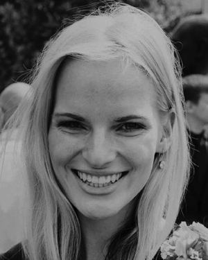 K. Kalev合作论文数8
 Ruth Tomson合作论文数8Ülle Võhma合作论文数7
 Jaan Kalda合作论文数Institute of Cybernetics;Tallinn Technical University;Institute of Cybernetics, Tallinn Technical University6
 Kaire Aadamsoo合作论文数5L. Orgo合作论文数5M. Säkki合作论文数Institute of Cybernetics, Tallinn University of Technology5Kristjan Pilt合作论文数Dept Hlth Technol, Tallinn Univ Technol4
 Ivo Fridolin合作论文数Department of Biomedical Engineering;Tallinn University of Technology;Department of Biomedical Engineering, Tallinn University of Technology4Marietta Gavriljuk合作论文数3Jaan Raik合作论文数Tallinn Technical University3
 Priit Roosipuu合作论文数2Egils Avots合作论文数2
 Tarmo Koppel合作论文数School of Business and Governance, Tallinn University of Technology2
 Milena B. Cukic合作论文数Biomedical Center, Torlak Institute2
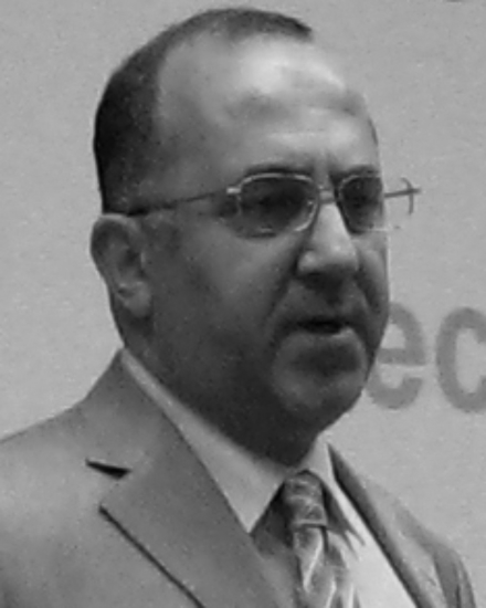 Andreas A. Ioannides合作论文数Laboratory for Human Brain Dynamics2Justina Kurganaite合作论文数1
 Ana Carina Ferreira合作论文数1Hong Xu合作论文数1
 Sol Carriazo合作论文数Instituto de Investigacion Sanitaria Fundacion Jimenez Diaz, Universidad Autonoma de Madrid;RICORS2040 (Redes de Investigación Cooperativa Orientadas a Resultados en Salud), Madrid, Spain.;Instituto de Investigacion Sanitaria Fundacion Jimenez Diaz, Universidad Autonoma de Madrid1
 Carmen Antonia Mocanu合作论文数Dept Internal Med & Nephrol, Carol Davila Univ Med & Pharm1Ardo Allik合作论文数1Marion Pépin合作论文数1vadim pesonen合作论文数1
 Konstantinos Giannakou合作论文数Dept Hlth Sci, European Univ Cyprus1
 Mickaël Bobot合作论文数Centre de Néphrologie et Transplantation Rénale, Hôpital de la Conception1
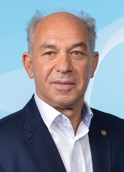 Goce Spasovski合作论文数Department of Nephrology, Saints Cyril and Methodius University of Skopje1
 Danka Savic合作论文数Vinca Institute1Dmitri Mihhailov合作论文数1Klimkowicz-Mrowiec Aleksandra合作论文数1
 Bumblyte Inga Arūne合作论文数Clinic of Nephrology, Kaunas University of Medicine1
 Vazelov Evgueniy S合作论文数Dialysis Clinic, A leksandrovska University Hospital1Pilar Delgado合作论文数1
 Giuseppe Paolisso合作论文数Dipartimento di Scienze Mediche e Chirurgiche Avanzate, Università degli Studi della Campania Luigi Vanvitelli;Azienda Universitaria Policlinico Vanvitelli, Università degli Studi della Campania Luigi Vanvitelli1
 Hans Orru合作论文数University of Tartu1
 Gholamreza Anbarjafari (Shahab)合作论文数Intelligent Computer Vision Research Lab, Technology Institute of the University of Tartu1Michelangela Barbieri合作论文数1
 Sophie Liabeuf合作论文数Clinical Pharmacology Department, CHU AMIENS - Hopital Sud1Maksim Gorev合作论文数Tallinn University of Technology1
 Wlodzimierz Klonowski合作论文数Institute of Biocybernetics and Biomedical Engineering;Polish Academy of Sciences;Institute of Biocybernetics and Biomedical Engineering, Polish Academy of Sciences1




