Background:Eosinophilic colitis (EC) is a rare eosinophilic gastrointestinal disorder with increasing frequency in recent years. Although normal eosinophil density varies substantially by colonic segment, several proposed eosinophil thresholds have been used in the literature to support the diagnosis in the appropriate clinical context. However, some patients with clinical features suggestive of eosinophilic colitis (EC) do not demonstrate increased tissue eosinophil counts on histopathologic examination. Objective:To compare eosinophil-derived cell-free granules based on expression of their surface markers between two groups of patients with symptoms consistent with EC: those with increased tissue eosinophils and those without tissue eosinophilia. Methods:This study included 10 pediatric patients with histologically confirmed EC and 13 patients with clinical suspicion of EC. Eosinophil-derived cell-free granules were evaluated in colonic tissue samples using immunohistochemistry (IHC), with antibodies against MBP and CCR3. Results:Eosinophil-derived cell-free granules were detected in all specimens from patients with suspected EC and in the majority of specimens from patients with confirmed EC. Degranulating eosinophils were identified in all specimens from both groups, although the proportion of degranulating eosinophils varied among patients. Furthermore, the abundance of granules was significantly associated with the percentage of degranulating eosinophils. Conclusion:Our findings indicate that IHC can be used to detect eosinophil-derived granules in colonic tissue. The presence of cell-free eosinophil granules with varying abundance, along with evidence of eosinophil degranulation in all specimens from patients with suspected EC, may provide supportive histopathological features that contribute to improving the diagnostic assessment of EC.
Combined immunodeficiency (CID) involves profound defects in B and T lymphocyte development and function. This study examined clinical and immunological phenotypes of CID patients with and without pulmonary manifestations. This retrospective multicenter study included 53 CID patients diagnosed between 2009 and 2022 with available thoracic computed tomography scans. Patients were categorized based on pulmonary manifestations presence. Demographic, clinical, and laboratory characteristics were compared using conservative statistical thresholds (P < 0.01). All laboratory parameters were interpreted using age-adjusted pediatric reference ranges. Among 53 patients (56.6
Abstract Serum free light chains (sFLCs) have recently been introduced as a diagnostic biomarker in common variable immunodeficiency (CVID) patients. Most patients with CVID have undetectable or lower levels of sFLCs compared to people with other types of immune system deficiencies with hypogammaglobulinemia, except for patients with agammaglobulinemia (AGG). Decreased production of kappa (κ) and lambda (λ) light chains over immunoglobulins (Ig) suggests a malfunction in early B cell development or plasma cell dysfunction may cause CVID. In this cross-sectional study, immunoturbidimetry was used to measure sFLCs in 70 immunodeficiency patients, including 39 patients with CVID, 31 patients with other immunodeficiencies with hypogammaglobulinemia such as combined immunodeficiency (CID), AGG, and other primary immunodeficiencies (PIDs), and 20 healthy controls. The levels of both κ and λ chains were significantly higher in patients with CID than patients with CVID and AGG patients (p < 0.05). The median (Q1–Q3) κ level in patients with other primary immunodeficiency disorders (PIDs) was significantly higher than that observed in patients with AGG and CVID (p < 0.01). Similarly, the median (Q1–Q3) λ level was higher in patients with other PIDs compared with those with AGG and CVID (p < 0.05).ROC analysis of κ and λ disclosed an area under the curve (AUC) for κ was 0.98 (95% CI: 0.96–0.99, p < 0.0001) and for λ was 0.95 (95% CI: 0.89–0.99, p < 0.0001) in patients with CVID. The sFLCs test can help distinguish between CVID and other immunodeficiency patients with hypogammaglobulinemia and the diagnosis of hypogammaglobulinemia.
Combined immunodeficiencies (CIDs) represent a rare group of inherited immune disorders in which defects in T- and B-lymphocyte function lead to recurrent infections, immune dysregulation, and an increased tendency toward autoimmune and rheumatologic complications. A retrospective cross-sectional analysis was performed on 150 patients with CID, diagnosed according to the European Society for Immunodeficiencies (ESID) criteria and followed at the Children’s Medical Center and Mofid Children’s Hospital (Tehran, Iran) between 2009 and 2020 Clinical records, immunologic evaluations, and rheumatologic findings were reviewed, with particular attention to autoantibody detection and disease frequency. Among 150 patients, 42 (28%) exhibited rheumatologic manifestations, with a higher frequency in females. Undifferentiated rheumatoid arthritis, undifferentiated juvenile idiopathic arthritis, and Kawasaki disease were the predominant conditions. Although lower lymphocyte counts and immunoglobulin levels were observed among non-rheumatologic patients, the differences were not statistically significant. Impairments in T-cell–mediated immunity and antibody synthesis among individuals with CID hinder the recognition of autoantibody-associated rheumatologic disorders and lead to delay diagnosis. Moreover, these conditions often present atypically in immunocompromised hosts; therefore, a vigilant clinical approach is essential for early identification and management.
BACKGROUND:Bartter syndrome (BS) is a salt-losing renal tubulopathy classically characterised by hypokalaemic metabolic alkalosis and hyperreninaemic hyperaldosteronism. METHODS:We investigated the genetic cause of a Bartter-like phenotype in an adolescent patient with progressive nephrocalcinosis, hypercalciuria, polyuria, metabolic alkalosis, hypokalaemia, significantly elevated urine chloride, failure to thrive, and a salt-losing tubulopathy. Additionally, the patient presented with hypergammaglobulinaemia, abnormal cerebral white matter signal changes, skin autoinflammation, and mild intellectual disability. RESULTS:No pathogenic variants were detected in known BS-related genes, and all recessive BS genes were outside regions of homozygosity (ROH) in this patient from a consanguineous family. Instead, exome sequencing and homozygosity mapping identified a homozygous splicing variant, c.2702-2A>G, in the epidermal growth factor receptor (EGFR) gene within an ~28 Mb ROH on chromosome 7p. RNA-Seq and RT-PCR analysis of the patient's RNA confirmed the pathogenicity of this variant, demonstrating aberrant splicing resulting in an in-frame retention of 27 nucleotides from intron 22 of EGFR. Immunofluorescence analysis of the proband's skin revealed a reduced EGFR protein level, rather than a complete absence, supporting a hypomorphic effect and likely explaining compatibility with survival into adolescence. Whereas previously reported EGFR variants have been associated with severe neonatal epithelial inflammation, bowel disease, and early mortality, our findings demonstrate that a hypomorphic variant can be compatible with survival into the second decade of life. CONCLUSION:These findings support an association between a syndromic Bartter-like salt-losing tubulopathy with epithelial autoinflammation and a homozygous splice-altering EGFR pathogenic variant, thereby expanding the phenotypic spectrum of EGFR-associated disorders.
At present, a national consensus on hematopoietic stem cell transplantation (HSCT) for patients with inborn errors of immunity (IEI) is lacking. This consensus is written based on a combination of scientific literature and comments from the expert panel of Iranian immunologists. We formed a panel of clinical immunologists at a meeting titled “Second Meeting on the Diagnosis of IEI by IEI Experts” to receive their comments in this field. All authors reviewed and agreed on the current consensus. This consensus guideline provides recommendations on donor selection, stem cell source, conditioning regimen, mobilization and collection, stem cell infusion, engraftment and chimerism assessment, and post-trans- plant care for patients with IEI. The current recommendations reflect Iranian practice and do not necessarily represent global preferences
We previously reported inherited retinoic acid-related orphan receptor γ T (RORγT) deficiency in seven patients from three ancestries (Chilean, Palestinian, and Saudi Arabian) with mycobacterial disease and chronic mucocutaneous candidiasis (CMC). We report here five additional patients from different ancestries (Afghan, Indian, Iranian, Japanese, and Sri Lankan), each homozygous for a new loss-of-function RORC variant. All but one patient-the exception receiving early prophylaxis-developed mycobacterial disease due to a near-complete depletion of innate-like adaptive T cells, including mucosa-associated invariant T and invariant natural killer T cells, low counts of adaptive TH1* and CD8+ T cells, and impaired Mycobacterium-induced IFN-γ production by the remaining cells of these subsets, NK cells, conventional CD4+ T, Vδ1, and Vδ2 γδT cells. Most patients also displayed CMC due to their low counts of TH17 and TH1* cells. One patient died from disseminated Bacille Calmette-Guérin vaccine infection, but, unexpectedly, all the other patients are still alive and clinically stable at ages of 2 to 20 years. RORγT is essential for protective immunity against mycobacteria and Candida in humans.
The Article Abstract is not available.
BACKGROUND:Inborn errors of immunity (IEI) include immunodeficiencies affecting cellular and humoral immunity. OBJECTIVES:We aimed to compare the effectiveness of the LTT and carboxyfluorescein succinimidyl ester (CFSE) assays in assessing lymphocyte proliferation in IEI patients. METHODS:We utilized radioactive [3H]-thymidine and non-radioactive CFSE to measure lymphocyte proliferation in three distinct groups: syndromic CID (SyCID), non-syndromic combined immunodeficiency (N-SyCID), and primary antibody deficiency (PAD). RESULTS:LTT identified 8 cases of abnormal lymphocyte proliferation among all patients, whereas CFSE detected 23 cases. In the N-SyCID group, LTT identified more abnormalities than CFSE, whereas, in the SyCID and PAD groups, CFSE detected more defects. Two patients with ataxia telangiectasia and CVID had positive results on both LTT and CFSE tests, and a specific ORAI1 gene mutation resulted in differing test outcomes. CONCLUSION:It was found that the CFSE method is a reliable and practical choice for measuring mitogenic T-cell responses in unclassified IEI patients for confirmation of immunologic diagnosis.
BackgroundThe majority of monogenic inborn errors of immunity presenting as actinopathies were reported originally from the Middle East and North Africa (MENA) countries indicating a high prevalence of these entities in the region. However, their prognosis is unclear due to rarity and lack of comprehensive treatment outcomes.MethodsWe evaluated clinical, immunological, and genetic abnormalities associated with 15 genetic entities of actinopathies. Based on the function of mutant genes in actin-regulatory pathways, patients were classified into CDC42- and RAC2-related subcategories.ResultsA total of 503 individuals (29.5% females) from 17 countries were considered with a median age of 120 months. Although most patients presented initially with allergic phenotypes (37.7%), the most prevalent manifestations throughout the lifespan were infection in respiratory tracts (72.2%). Primary clinical diagnosis was mainly combined immunodeficiencies (48.3%) and the majority of cases were molecularly assigned to the CDC42 pathway (64.8%). The most common genetic defects were reported within the DOCK8 (n = 209) followed by the WAS (n = 94) and the CARMIL2 (n = 15) genes. Hematopoietic stem cell transplantation (HSCT) was conducted on 24.0% of patients, which significantly improved survival in patients with defects in WAS, DOCK8 and DOCK2. Overall mortality was 23.0%, mainly due to sepsis and malignancy.ConclusionPatients with defects in RAC2-associated regulators of actin usually present with late-onset symptoms due to normal immune profiles, but a higher rate of EBV and HPV infections, autoimmune cytopenia, asthma, and lymphoproliferation compared to defects in the CDC42 pathway. The severity of mutations in patients of the CDC42 group helps to estimate the prognosis of the disease and prioritization of HSCT.
Currently, a national consensus or guideline for diagnosing and managing patients suspected of having chronic granulomatous disease (CGD) is lacking. This consensus is written based on a combination of scien- tific literature and comments from the expert panel of Iranian immunologists. A group of clinical immunol- ogists reviewed the current consensus, presented their comments at a meeting titled “First Meeting on the Diagnosis of Inborn Errors of Immunity (IEI) by IEI Experts” and agreed on this consensus. This consensus guideline provides recommendations on the diagnosis, antimicrobial prophylaxis, management of clinical manifestations, administration of interferon gamma (IFN-γ) and hematopoietic stem cell transplantation (HSCT) for patients with CGD.
Stromal interaction molecule 1 (STIM1) is a transmembrane protein located in the endoplasmic and sarcoplasmic reticulum, where it plays a crucial role in activating calcium release-activated calcium (CRAC) channels. It functions as a calcium (Ca2⁺) sensor within the endoplasmic reticulum (ER), triggering CRAC channel opening and allowing calcium entry—mechanisms essential for maintaining intracellular calcium homeostasis. Mutations in the STIM1 gene that impair calcium signaling can disrupt both T cell and muscle cell function, leading to combined immunodeficiency and congenital myopathy. Here, we describe a 9-year-old boy with these clinical features, who was found to carry a previously undescribed mutation in the STIM1 gene. The patient presented with recurrent pneumonia, blood-streaked diarrhea, eczema, muscle weakness, and failure to thrive. Whole exome sequencing identified a novel homozygous missense variant in STIM1 (c.584T > C | p.Leu195Pro), considered likely pathogenic. This classification was supported by high Combined Annotation Dependent Depletion (CADD) and Rare Exome Variant Ensemble Learner (REVEL) scores of 29.8 and 0.89, respectively. Homozygosity of the mutation was confirmed using PCR-Sanger sequencing. This case highlights a novel homozygous STIM1 variant in a child with combined immunodeficiency and congenital myopathy. The clinical presentation is consistent with previously reported phenotypes associated with STIM1 deficiency.
In recent years, many studies have been conducted on the possible link between rheumatologic diseases and inborn errors of immunity. Rheumatologic diseases may occur as manifestations of an underlying immunodeficiency disorder, and may appear before the more-common infectious manifestations more typically seen in immunodeficiency disorders. In this study, we have attempted to study such symptoms and uncover their relationship with inborn errors of immunity. In this retrospective descriptive-analytical study, 381 cases of IEIs in children that were referred to Mofid Children’s Hospital clinic between 2015 and 2019 were evaluated for eligibility to be enrolled in the study. Of these patients, 20 that had confirmed rheumatologic diagnoses were entered into the study. Patients’ demographic and medical data, including age at disease onset, age at diagnosis and type of diagnosed rheumatologic and immunodeficiency disorders, parental consanguinity rate, and relevant laboratory findings were retrieved for study and analyzed. Among 20 eligible patients, half of which were female and half were male, the average age at disease onset, average age at diagnosis of the underlying immunodeficiency disease and average age at diagnosis of the rheumatologic disease were 2.98 ± 1.56, 5.26 ± 3.45 and 3.58 ± 2.97, respectively. JIA made up 10 of the observed rheumatic diseases (“the JIA group”); the remaining 10 patients included SLE (3), FMF (2), juvenile dermatomyositis (2), MCTD (1), GPA (1) and reactive arthritis (1) (“the non-JIA group”). As for the underlying immunodeficiency disorders, CID was seen in 8 patients, followed by CVID (5), XLA (4), SIgAD (2) and CGD (1). The average age at onset of the disease and the average age at diagnosis of the rheumatologic disease were significantly lower in the JIA group than in the non-JIA group (p < 0.05). A plethora of rheumatologic manifestations may be observed in patients with IEIs; such manifestations should be actively sought out and treated in IEI patients. Not applicable.
At present, a national consensus or guideline for diagnosing and managing patients suspected of havingpredominantly antibody deficiencies (PADs) is lacking. This consensus is written based on a combinationof scientific literature and comments from the expert panel of Iranian immunologists. A group of clinicalimmunologists reviewed the current consensus, presented their comments at a meeting titled 'First Meetingon the Diagnosis of Inborn Errors of Immunity (IEI) by IEI Experts,' and agreed on this consensus. Thisconsensus guideline provides recommendations on the diagnosis, antimicrobial prophylaxis, managementof clinical manifestations, and immunoglobulin replacement therapy (IgRT) for patients with PAD.
It can sometimes be very difficult to control the manifestations of autoimmunity and lymphoproliferation in patients with primary immunodeficiency diseases, and there is no adequate response to first-line treatments. Rituximab (RTX), as a second-line treatment, is efficacious and well-tolerated for the management of these clinical manifestations. This retrospective study was conducted to analyze the clinical, immunological, and genetic findings together with the response rate to RTX therapy in subjects with inborn errors of immunity (IEI) and autoimmune or autoinflammatory manifestations. In this study, 23 individuals with IEI and autoimmune or lymphoproliferation manifestations who received RTX between April 2008 and 2021 were evaluated. Fifteen out of the 23 patients were female. The median age of cases was 12 years. The moderate and severe adverse reactions, including fever, diarrhea, and anaphylaxis shock, were manifested during RTX infusion in 5 patients. In total, 86.9% of patients responded to rituximab (complete response: n=14, partial response: n=6) while three failed to respond. The median response time to RTX treatment was 50 days. All patients were given monthly intravenous immunoglobulin (IVIG) therapy. Pneumonia and candidiasis occurred in one patient a week after receiving the second injection of RTX. Eight patients expired during follow-up. In conclusion, the response rate of RTX could be improved through administering monthly IVIG for hypogammaglobulinemia treatment following RTX infusion. Early use of rituximab leads to a better response rate in comparison with late use of rituximab in multitreated refractory patients. The efficient cumulative dose of rituximab remains undefined.
At present, a national consensus or guideline for diagnosing and managing patients suspected of having se- vere congenital neutropenia (SCN) is lacking. This consensus is written based on a combination of scientific literature and comments from the expert panel of Iranian immunologists. A group of clinical immunolo- gists reviewed the current consensus, presented their comments at a meeting titled “First Meeting on the Diagnosis of Inborn Errors of Immunity (IEI) by IEI Experts” and agreed on this consensus. This consensus guideline provides recommendations on the diagnosis, antimicrobial prophylaxis, management of clinical manifestations, administration of granulocyte colony-stimulating factor (G-CSF) and hematopoietic stem cell transplantation (HSCT) for patients with SCN.
Background: The gastrointestinal (GI) tract can be affected by immunodeficiency disorders. This study aimed to evaluate GI manifestations in children with immunodeficiency. Methods: This cross-sectional study retrospectively evaluated immunodeficient children. Demographics, immunodeficiency disorders, clinical signs and symptoms, upper endoscopy and colonoscopy findings, histopathologic and imaging findings, and laboratory test results were extracted from the patients' medical records. Participants were selected from medical records at Mofid Children's Hospital, Tehran, Iran, from 2011 to 2022, a tertiary care hospital for pediatrics with immunodeficiency disorders. Results: Of the 43 children with immunodeficiency evaluated in this study (mean age: 3.89 ± 4.32 years), 25 (58.1%) were male, and 18 (41.8%) were female. The most common immunodeficiency disorder was combined immunodeficiency (CID) in 11 patients (25.6%), followed by severe combined immunodeficiency (SCID) in 6 (14%). The most frequent clinical manifestations were diarrhea in 24 patients (55.8%) and vomiting in 21 (48.8%). Erythema of the esophagus (3/14), stomach (5/14), and duodenum (3/14) was the most common finding of upper endoscopy. Nodularity of the colon (4/11) and erythema of the rectum (4/11) were the most frequently observed findings in colonoscopy. The most frequently observed histopathologic findings were chronic esophagitis, chronic gastritis, chronic inflammation in the duodenum, and increased/enlarged lymphoid follicles in the colon and rectum. Abdominal computed tomography (CT) and ultrasound (US) revealed splenomegaly and hepatomegaly as the most frequent findings (4.6% and 20.9%, respectively). Conclusions: The GI manifestations were common in children with immunodeficiency, with diarrhea and vomiting as the predominant symptoms.
Purpose:Eosinophilic esophagitis (EoE) is the most well-known eosinophilic gastrointestinal disorder (EGID) characterized by the presence of a high number eosinophils within the esophageal epithelium and the clinical signs. Biopsies of patients with suspected EoE may not show a high number of eosinophils, however the presence of granules may help with the diagnosis. This study aims to evaluate the presence of cell-free eosinophil granules in the esophageal tissue of patients with suspected and confirmed EoE to accelerate the diagnosis and treatment of patients with low eosinophil count. Methods:Fifteen patients with confirmed EoE and 15 suspected of EoE were included in this study. Patients' esophageal tissue biopsies were stained using immunohistochemistry (IHC) to identify eosinophils and their cell-free granules. For testing, anti-major basic protein (MBP) and anti-chemokine receptor type 3 (CCR3) were used as primary antibodies and a double-staining kit containing secondary antibodies conjugated to the enzyme and related chromogens were used. Results:Cell-free granules with different degrees were observed in 53.3% and 93.3% of suspected and confirmed EoE samples, respectively. Furthermore, in esophageal biopsy of 73.3% of patients with suspected and 93.3% of patients with a definitive diagnosis of EoE, basal layer hyperplasia (BLH) was recognized. Conclusion:The results of the present study showed that IHC can be applied to detect cell-free eosinophil granules in esophageal tissue. Observation of granules and basal cell hyperplasia in biopsies of suspected EoE patients whose eosinophil count is below the threshold can be valuable findings to make a definitive diagnosis for these patients.
Hymenoptera venom allergy (HVA) is a significant cause of anaphylaxis, with a broad spectrum of symptoms. Accurate diagnosis is critical for selecting appropriate venom immunotherapy (VIT), yet multiple sensitizations complicate the identification of the culprit Hymenoptera. Component-resolved diagnostics (CRD) and the basophil activation test (BAT) are emerging approaches that may enhance the precision of HVA diagnosis. This study aimed to compare the outcomes of the BAT and CRD tests in patients with reaction to more than one type of Hymenoptera in intradermal test. Following 12 patient’s report-based determination of the Hymenoptera types, Intradermal testing with venom extracts from honey bees, paper wasps, and yellow jackets was performed. CRD was conducted using recombinant allergens for each venom, and BAT was performed to assess basophil activation using venom extracts. The intradermal test indicated 10 triple-positive reactions (83.3%) and 2 double-positive reactions (16.7%). In addition, 7 (58.4%) individuals were triple-positive and 5 (41.6%) were double-positive in the CRD test. The BAT had 6 (50.0%) double-positive individuals and 3 (25.0%) single-positive patients. The diagnostic complexity in patients with multi-sensitivity to Hymenoptera venoms is highlighted by the results. While CRD provides a more specific identification of allergenic components, valuable information is offered by the BAT, especially when the clinical diagnosis is uncertain. As a result, relying just on one test seems to have limited diagnostic capabilities and it is more efficient to use many diagnostic tests in order to get a thorough evaluation.
Background:IgG4-related disease may initially present with pulmonary pseudotumor, making the diagnosis challenging particularly in patients prone to granulomatous inflammation. Here, we describe the first case of chronic granulomatous disease with associated lung IgG4RD. Case Presentation:An 8.5-year-old male was hospitalized two years ago with exertional dyspnea, mild cough, chest pain, and nocturnal sweating and was found to have a tumor-like mass in the right lung. The histopathologic findings were consistent with extensive peripheral fibrosis and infiltration of mixed inflammatory cells without any evidence of acid-fast bacilli/fungal elements. Treatment with prednisolone resulted in considerable symptom resolution. Following the discontinuation of prednisolone by the patient, symptoms recurred, gradually exacerbated, and he developed anorexia and weight loss. The next chest spiral computed tomography (CT) scan showed a larger mass in the right lung, right lung collapse, and mediastinal metastasis. The abdominal ultrasound and CT scan were normal. In laboratory evaluation, low counts of B and T cells, normal natural killer cells, high levels of IgG4, and high inflammatory markers were detected. The nitro blue tetrazolium test was zero in two consecutive evaluations. In virtue of high IgG4 level, organ-specific mass, notable tissue fibrosis, and mixed inflammatory cell infiltrate, he was diagnosed with concurrent CGD and IgG4RD, but progressed to respiratory failure and died despite the reinstitution of steroid therapy. Conclusion:The overlap between inborn errors of immunity and IgG4RD is not common. Further studies to investigate IgG subsets among IEI patients can help elucidate clinicopathological correlations between these two immune-mediated disorders.
 Zahra Chavoshzadeh合作论文数Mofid Children Hospital, Shaheed Beheshti University65
 Mahboubeh Mansouri合作论文数Department of Allergy and Clinical Immunology of Children ' s;Medical Center;Allergy Research Institute;Tehran University of Medical Sciences;Allergy Research Institute, Tehran University of Medical Sciences32
 Delara Babaie合作论文数28
 Samin Sharafian合作论文数Mofid Children's Hospital, Shahid Beheshti University of Medical Sciences22
 Eslami Narges合作论文数22
 Sepideh Darougar合作论文数21
 Nima Rezaei合作论文数Universal Scientific Education and Research Network;Karolinska Institutet;Department of Immunology, School of Medicine, Tehran University of Medical Sciences21
 Mahnaz Jamee合作论文数Research Institute for Children's Health, Shahid Beheshti University of Medical Sciences19
 Mohammad Nabavi合作论文数Semnan University of Medical Sciences19
 Fallahpour Morteza合作论文数Rasool e Akram Hospital, Iran University of Medical Sciences17
 Saba Arshi合作论文数Rasoul Akram Hospital;Iran University of Medical Sciences;Rasoul Akram Hospital, Iran University of Medical Sciences16
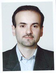 Mahdaviani Seyed Alireza合作论文数NRITLD, Shahid Beheshti Univ Med Sci15
 Mohammad Hassan Bemanian合作论文数Department of Pediatrics;Shahid Sadoughi Hospital;Shahid Sadoughi University of Medical Science;Shahid Sadoughi Hospital, Shahid Sadoughi University of Medical Science15
 Shahnaz Armin合作论文数14Zahra Pourpak合作论文数Allergy Research Institute;Tehran University of Medical Sciences;Allergy Research Institute, Tehran University of Medical Sciences14
 Sima Shokri合作论文数Rasool Akram Hosp, Iran Univ Med Sci13
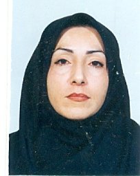 Mahnaz Sadeghi-Shabestari合作论文数Dept Immunol & Allergy, Tabriz Univ Med Sci13
 Reza Yazdani合作论文数13
 Abdollah Karimi合作论文数Pediatric Infections Research Center, Labbafinejad Hospital;Shahid Beheshti University;Labbafinejad Hospital and Pediatric Infections Research Center, Shahid Beheshti University13Alireza Shafiei合作论文数12
 Samaneh Delavari合作论文数Children's Medical Center, Tehran University of Medical Science12
 Tavakol Marzieh合作论文数Children's Medical Center, Tehran University of Medical Science12
 Asghar Aghamohammadi合作论文数Research Center for Immunodeficiencies, Tehran University of Medical Sciences12
 Hassan Abolhassani合作论文数Department of Medical Biochemistry and Biophysics, Karolinska Institutet;Pan Hammarström Laboratory, Karolinska Institutet;Research Center for Immunodeficiencies, Tehran University of Medical Sciences12
 Mazdak Fallahi合作论文数Mofid Children's Hospital, Shahid Beheshti University of Medical Sciences11Shahrzad Fallah合作论文数Mofid Children's Hospital, Shahid Beheshti University of Medical Sciences11
 Taher Cheraghi合作论文数Children’s Medical Center, Tehran University of Medical Sciences11
 Mitra Khalili合作论文数10
 Anahita Razaghian合作论文数10
 Masoud Movahedi合作论文数Tehran University of Medical Sciences10
 Behniafard Nasrin合作论文数Children's Medical Center, Tehran University of Medical Sciences9
 Tooba Momen合作论文数9Molatefi Rasool合作论文数9
 Dara Naghi合作论文数Faculty of Medicine, Shahid Beheshti University of Medical Sciences9Enayat Javad合作论文数9Roya Sherkat合作论文数Isfahan University of Medical Sciences9Ahmad Vosughimotlagh合作论文数Tehran University of Medical Sciences8
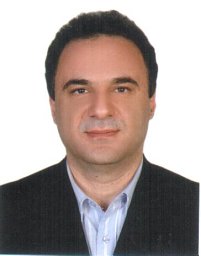 Farid Imanzadeh合作论文数8
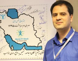 Gholamreza Azizi合作论文数Imam Hassan Mojtaba Hospital, Alborz University of Medical Sciences8
 Esmaeilzadeh Hossein合作论文数Rasool e Akram Hospital, Iran University of Medical Sciences8
 Iraj Mohammadzadeh合作论文数School of Medicine, Babol University of Medical Sciences8Behzad Shakerian合作论文数7
 F. Mousavinasab合作论文数School of Medicine, Shahid Beheshti University of Medical Sciences7
 Fereshte Salami合作论文数Children's Medical Center, Tehran University of Medical Science7
 Alyasin Soheila合作论文数Allergy Research Center, Shiraz University of Medical Sciences7
 Khoddami Maliheh合作论文数Mofid Hospital, Shaheed Beheshti University of Medical Sciences7
 Ahanchian Hamid合作论文数Faculty of Medicine, Mashhad University of Medical Sciences7
 Shiari Reza合作论文数Mofid Children's Hospital, Shahid Beheshti University7Shamsian Bibi Shahin合作论文数7




