This review was written to be included in the Special Collection 'Therapy Ultrasound: Medicine's Swiss Army Knife?' The purpose of this review is to provide basic presentation and interpretation of the fundamentals of hyperthermia biology, as it pertains to uses of therapeutic ultrasound. The fundamentals are presented but in the setting of a translational interpretation and a view toward the future. Subjects that require future research and development are highlighted. The effects of hyperthermia are time and temperature dependent. Because intra-tumoral temperatures are non-uniform in tumors, one has to account for differential biologic effects in different parts of a tumor that occur simultaneously during and after hyperthermia.
Supplementary Figures S1-S3; Tables S1, S2 from Gene Expression Profiles of Multiple Breast Cancer Phenotypes and Response to Neoadjuvant Chemotherapy
GO term analysis from Estrogen-Related Receptor α Is Critical for the Growth of Estrogen Receptor–Negative Breast Cancer
Supplementary Figures 1-2 from Systemic Overexpression of Angiopoietin-2 Promotes Tumor Microvessel Regression and Inhibits Angiogenesis and Tumor Growth
AVI file - 34.5MB, Real-time intravascular release of doxorubicin from Dox-TSL and subsequent redistribution of drug in B16 melanoma tumor tissue. The video shows drug (red) delivered over the first 20min, at 20x magnification. The eNOS-GFPtg mouse model was used in this experiment, so the blood vessels are green. (Download before playing in IE8)
Supplemental Figure 3: Effects of radiation and MnTnBuOE-2-PyP5+ on tumor cells. (A) LN-18 and (B) LN-229 glioblastoma cell lines treated with or without 50 nM MnTnBuOE-2-PyP5+ in clonogenic survival assays prior to various doses of radiation.
Supplemental Figure 1: Western analysis of myelin within the corpus callosum. Signal intensity of myelin and actin were quantitated by Image J, averaged within groups, then normalized to the saline group .
Supplementary Movie 6 from The Pervasive Presence of Fluctuating Oxygenation in Tumors
Supplementary Movie 9 from The Pervasive Presence of Fluctuating Oxygenation in Tumors
Background Chaperon-mediated autophagy (CMA) has taken on a new emphasis in cancer biology. However, the roles of CMA in hypoxic tumours are poorly understood. We investigated the anti-tumour effects of the natural product ManA through the activation of CMA in tumour progression under hypoxia. Methods The effect of ManA on CMA activation was assessed in mouse xenograft models and cells. The gene expressions of HIF-1α, HSP90AA1, and transcription factor EB (TFEB) were analysed using The Cancer Genome Atlas (TCGA) datasets to assess the clinical relevance of CMA. Results ManA activates photoswitchable CMA reporter activity and inhibits Hsp90 chaperone function by disrupting the Hsp90/F 1 F 0 -ATP synthase complex. Hsp90 inhibition enhances the interaction between CMA substrates and LAMP-2A and TFEB nuclear localisation, suggesting CMA activation by ManA. ManA-activated CMA retards tumour growth and displays cooperative anti-tumour activity with anti-PD-1 antibody. TCGA datasets show that a combined expression of HSP90AA1 High /HIF1A High or TFEB Low /HIF1A High is strongly correlated with poor prognosis in patients with lung cancer. Conclusions ManA-induced CMA activation by modulating Hsp90 under hypoxia induces HIF-1α degradation and reduces tumour growth. Thus, inducing CMA activity by targeting Hsp90 may be a promising therapeutic strategy against hypoxic tumours.
Supplementary Figure 1 from Tumor Necrosis Factor-α Is a Potent Endogenous Mutagen that Promotes Cellular Transformation
Inflammatory breast cancer (IBC), an understudied and lethal breast cancer, is often misdiagnosed due to its unique presentation of diffuse tumor cell clusters in the skin and dermal lymphatics. Here, we describe a window chamber technique in combination with a novel transgenic mouse model that has red fluorescent lymphatics (ProxTom RFP Nu/Nu) to simulate IBC clinicopathological hallmarks. Various breast cancer cells stably transfected to express green or red fluorescent reporters were transplanted into mice bearing dorsal skinfold window chambers. Intravital fluorescence microscopy and the in vivo imaging system (IVIS) were used to serially quantify local tumor growth, motility, length density of lymph and blood vessels, and degree of tumor cell lymphatic invasion over 0–140 h. This short-term, longitudinal imaging time frame in studying transient or dynamic events of diffuse and collectively migrating tumor cells in the local environment and quantitative analysis of the tumor area, motility, and vessel characteristics can be expanded to investigate other cancer cell types exhibiting lymphovascular invasion, a key step in metastatic dissemination. It was found that these models were able to effectively track tumor cluster migration and dissemination, which is a hallmark of IBC clinically, and was recapitulated in these mouse models.
Supplementary Movie 5 from The Pervasive Presence of Fluctuating Oxygenation in Tumors
Advances in energy balance and cancer research to date have largely occurred in siloed work in rodents or patients. However, substantial benefit can be derived from parallel studies in which animal models inform the design of clinical and population studies or in which clinical observations become the basis for animal studies. The conference Translating Energy Balance from Bench to Communities: Application of Parallel Animal-Human Studies in Cancer, held in July 2021, convened investigators from basic, translational/clinical, and population science research to share knowledge, examples of successful parallel studies, and strong research to move the field of energy balance and cancer toward practice changes. This review summarizes key topics discussed to advance research on the role of energy balance, including physical activity, body composition, and dietary intake, on cancer development, cancer outcomes, and healthy survivorship.
Supplemental Figure 2: Neurocognitive analysis of mice by Morris watermaze (A and B), novel object test (C and D), and fear conditioning (E and F). Tests were performed as described (29). (G and H) Distance and velocity during the novel object training and test periods and overall averages.
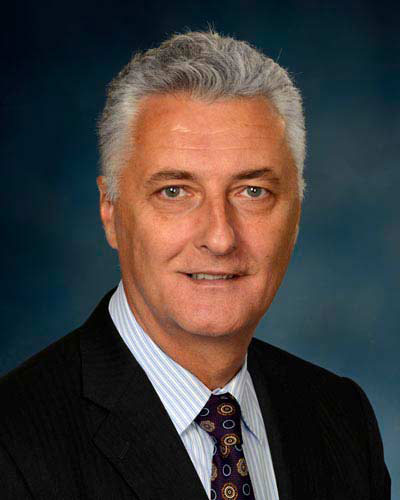 Zeljko Vujaskovic合作论文数Division of Translational Radiation Sciences, Department of Radiation Oncology, School of Medicine, University of Maryland;Greenebaum Cancer Center, University of Maryland;Normal Tissue Laboratory, Department of Radiation Oncology, Duke University Medical Center85
 Gregory M. Palmer合作论文数Department of Radiation Oncology, Duke University School of Medicine;Duke Cancer Institute, Duke University School of Medicine;Zenalux Biomedical, Inc56
 David Manfield Brizel合作论文数Clinical Science Departments, School of Medicine, Duke University54
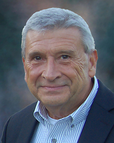 Donald E Thrall合作论文数School of Veterinary Medicine, Ross University49
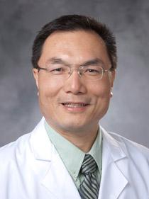 Chuan-Yuan Li合作论文数Department of Dermatology, Duke University School of Medicine;Duke Cancer Institute;Duke Regeneration Center47
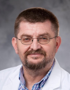 Ivan Spasojevic合作论文数Department of Medicine, School of Medicine, Duke University School of Medicine45
 Ines Batinic-Haberle合作论文数Department of Radiation Oncology, School of Medicine, Duke University44
 Thies Schroeder合作论文数Institute of Physiology and Pathophysiology, Johannes Gutenberg University Mainz43
 Timothy W. Secomb合作论文数Department of Biomedical Engineering, College of Medicine, University of Arizona;Department of Physiology, College of Medicine, University of Arizona42
 Rod D. Braun合作论文数42Tv Samulski合作论文数41
 Kimberly Lynn Blackwell合作论文数Department of Medicine, Duke University37Zahid N. Rabbani合作论文数37
 Benjamin Viglianti合作论文数Division of Nuclear Medicine, Medical School, University of Michigan35Rl Page合作论文数33
 Kathleen Ashcraft合作论文数32Paul R. Stauffer合作论文数31
 Leonard Richard Prosnitz合作论文数School of Medicine, Duke University30Mark Oldham合作论文数30Pierre Sonveaux合作论文数30
 Oana I. Craciunescu合作论文数Radiation Oncology, Duke University Medical Center30Jr Oleson合作论文数29
 David Needham合作论文数Department of Mechanical Engineering and Materials Science, Duke University28Daohai Yu合作论文数26Benjamin J. Moeller合作论文数26
 Shiva Das合作论文数Department of Radiation Oncology, School of Medicine, University of North Carolina26Yiting Cao合作论文数Shanghai Jiao Tong University, Shanghai Jiaotong Univ.25
 Mary-Keara Boss合作论文数25
 Nimmi Ramanujam合作论文数Department of Biomedical Engineering, Pratt School of Engineering, Duke University;Department of Pharmacology & Cancer Biology, Duke University23
 Ashutosh Chilkoti合作论文数Department of Biomedical Engineering, Pratt School of Engineering, Duke University23
 Charles Steven Greenberg合作论文数22
 Andrew Fontanella合作论文数22
 Ellen Jones合作论文数Department of Radiation Oncology, School of Medicine, University of North Carolina21Rachel Richardson合作论文数20Siqing Shan合作论文数20
 Chelsea Landon合作论文数Department of Pathology, Duke University School of Medicine20
 Bruce Klitzman合作论文数Department of Cell Biology, Duke University School of Medicine20Mitchell S. Anscher合作论文数20
 John P. Kirkpatrick合作论文数Department of Radiation Oncology, Duke University School of Medicine19
 Fan Yuan合作论文数Department of Biomedical Engineering, Pratt School of Engineering, Duke University19
 Gary L. Rosner合作论文数Division of Biostatististics and Bioinformatics, Department of Oncology, Johns Hopkins University;Sidney Kimmel Comprehensive Cancer Center, Johns Hopkins University19
 Lee Jones合作论文数Department of Medicine, Memorial Sloan Kettering Cancer Center19
 Hanna Gabi合作论文数18Matthew R. Dreher合作论文数18ZA Haroon合作论文数17
 Jen-Tsan Ashley Chi合作论文数Department of Molecular Genetics and Microbiology, Duke University School of Medicine17
 Olivier Feron合作论文数Faculty of Pharmacy and Biomedical Sciences, Universite Catholique de Louvain;Faculty of Medicine, Universite Catholique de Louvain17James R. MacFall合作论文数Department of Radiology, Duke University Medical Center17Diane R. Fels合作论文数17




