BACKGROUND:The surgical pathology report contains vital information guiding postoperative care; however, it often lacks critical details regarding final resection margin status, leaving downstream providers vulnerable to misinterpretation. We sought to quantify the interpretability of our institution's oral cancer surgical pathology reports. METHODS:Head and neck surgeons, pathologists, medical and radiation oncologists retrospectively reviewed 149 pathology reports. Reviewers classified final margin status in a two-tiered process: first, as "ambiguous" or "unambiguous." Then, unambiguous margin status was classified into negative, positive, or at risk (< 5 mm). RESULTS:Rates of margin status agreement varied significantly across reviewers. Reported ambiguous margin status ranged from 15.4% to 40.9%. Roughly two-thirds of pathology reports were deemed ambiguous by at least one reviewer. CONCLUSION:Significant margin status ambiguity and low interrater agreement highlight the limitations of current surgical pathology reporting. Standardized reporting practices are needed to improve clarity, enhance multidisciplinary communication, and optimize postoperative care.
INTRODUCTION:Isolated soft tissue deposits (ISTDs) are believed to be tumor emboli from lymphatic drainage pathways, discontinuous from primary tumors and lacking lymph node architecture. While associated with poor prognosis in various malignancies, characterization of ISTDs in papillary thyroid carcinoma (PTC) is limited. METHODS:This single-center cohort study examined 11 cases of PTC with ISTDs. Multi-platform next-generation sequencing analyzing driver mutations and tumor suppressor loss of heterozygosity (LOH) was performed on primary tumors, lymph nodes, and ISTDs. Clinical and histopathologic features were compared to a 3:1 matched control cohort. RESULTS:Genomic alterations were identical across paired tumors, lymph nodes, and ISTDs. LOH was present in 78% of cases with driver mutations. Compared to controls, ISTD cases demonstrated increased rates of high lymph node burden (p < 0.01), extranodal extension (p = 0.012), and lymphovascular invasion (p < 0.01). CONCLUSIONS:Associated with aggressive disease features, the molecular profiles of ISTDs mirror primary tumors and lymph node metastases.
BACKGROUND:Squamous cell carcinoma (SCC) of the dorsal tongue accounts for only 2%-5% of tongue cancers; midline dorsal tongue cancers account for less than 1% of these tumors. Basaloid SCC (BSCC) is usually associated with more aggressive features than conventional SCC. There are fewer than 30 reported cases of lingual BSCC. METHODS:A 76-year-old woman presented with irregular, exophytic, rugated, and furrowed changes of the midline dorsal tongue, which was initially misdiagnosed as mucoepidermoid carcinoma on an outside institution biopsy. Other than mild linear enhancement of the oropharyngeal tongue, MRI was unremarkable. A midline partial glossectomy with primary closure was performed with the aid of intraoperative frozen section analysis. RESULTS:Final pathology was rediagnosed as pT2 invasive BSCC, with depth of invasion of 2.43 mm, worst pattern of invasion of WPOI-3, and negative final margins. The decision was made to proceed with surveillance without any adjuvant therapy. CONCLUSIONS:This is the first reported case of BSCC of the midline dorsal tongue. This case report highlights the diagnostic challenges of BSCC and underscores the importance of comprehensive evaluation of all histological features using immunohistochemistry studies, as BSCC can mimic other pathologic entities but may confer a different prognosis. Early and definitive diagnosis of this disease has implications for management decisions and prognosis.
BACKGROUND:Tumor tissue-modified viral (TTMV) human papillomavirus (HPV) DNA testing has high diagnostic and surveillance performance in HPV-associated oropharyngeal squamous cell carcinoma, but its utility in HPV-associated non-oropharyngeal (non-OP) head and neck squamous cell carcinoma (HNSCC) is not well defined. METHODS:We performed a retrospective cohort study of patients with biopsy-confirmed HPV-positive non-OP HNSCC who underwent TTMV-HPV DNA testing at a single tertiary academic center from April 2020 to January 2025. Diagnostic performance was assessed for pretreatment testing, and surveillance performance was assessed per test and per patient. RESULTS:The diagnostic cohort included 19 patients and demonstrated 80% sensitivity and 100% specificity. The surveillance cohort included 15 patients who underwent 32 tests. Per-test surveillance sensitivity was 87%, specificity 100%, positive predictive value 100%, and negative predictive value 89%. Per-patient surveillance sensitivity was 86%, specificity 100%, positive predictive value 100%, and negative predictive value 89%. In two patients, positive surveillance tests preceded clinical or radiographic recurrence by 18 and 110 days. CONCLUSIONS:TTMV-HPV DNA testing demonstrated strong diagnostic and surveillance performance in HPV-associated non-OP HNSCC and may be a useful adjunct in posttreatment surveillance.
BACKGROUND:The American Thyroid Association's most recent clinical practice guidelines include margin status as a key factor to estimate risk of recurrence. Unlike most thyroid cancers, the successful resection of invasive pT4 thyroid cancers necessitates the use of frozen section analysis (FSA). There is no standardized method for intraoperative surgical margin assessment. We developed a novel intraoperative workflow and software platform, MarginView3DTM (MV3D), to enhance the precision of FSA and improve surgical pathology reporting in head and neck cancer. Here, we demonstrate its use in invasive thyroid cancer. METHODS:We describe the MV3DTM surgical pathology reporting software and detail our surgical workflow, which incorporates 3D scanning technology and standardized "timeouts" to facilitate intraoperative communication and pathologic documentation. This approach was utilized in five invasive pT4 thyroid cancer cases. RESULTS:MV3DTM was used to guide intraoperative margin assessment, communication, and pathologic documentation in four tracheal resections and one sternal mass resection. A final surgical pathology report was generated for each case, integrating 3D scans, annotated radiographs, and audiovisual summaries into a dynamic interface for multidisciplinary use. We highlight case 5, where MV3DTM facilitated bidirectional communication between the surgeon and pathologist to guide precise harvesting of supplemental margins. The surgeon and pathologist collaborated in real time to address at-risk areas requiring immediate re-resection. An R0 resection was achieved, and all relevant details were captured within MV3DTM. CONCLUSIONS:We have utilized our novel approach extensively in head and neck cancer. Here, we highlight it as a tool for the management of invasive pT4 thyroid cancer. As a brief summary of new and innovative research, this study introduces MV3DTM as a novel and scalable approach to intraoperative margin assessment and documentation in advanced thyroid cancer, with the potential to inform future practice in surgical oncology.
BACKGROUND:Due to their shared embryologic origin, the association between ectopic parathyroid glands and the thymus is well recognized. Abnormal implantation of thymic tissue during embryological migration to the mediastinum can lead to ectopic cervical thymus. METHODS:A 33-year-old woman with persistent primary hyperparathyroidism (89 pg/mL) was referred following failed parathyroidectomy. Preoperative 4D computed topography identified soft tissue in the right carotid sheath corresponding to an area of increased uptake on sestamibi imaging. Revision parathyroidectomy was performed. RESULTS:Intraoperatively, thymic tissue and an associated parathyroid adenoma were found in the right carotid sheath extending superiorly toward the hyoid bone. Post-excision parathyroid hormone levels fell within normal limits (24.2 pg/mL). CONCLUSIONS:We describe a novel location for an ectopic parathyroid adenoma within ectopic cervical thymic tissue outside of the thyroid bed and mediastinum. This highlights embryologic relationships that may aid in diagnostic and therapeutic management, particularly in revision or non-localizing cases.
Contemporary cancer management relies on the precise transfer of information regarding disease and treatment details between a range of providers and facilities. The time of surgery is uniquely responsible for many ambiguities in this oncological information flow, as intraoperative communication and documentation are often unregimented and imprecise. Downstream providers such as radiation oncologists, medical oncologists, and radiologists rely on surgical pathology reports for planning postoperative treatment and surveillance. However, traditional pathology reports lack critical details about the actions taken during surgery and do not make the oncologic clearance status explicit. We identified agents of change to improve information transfer and designed an improved surgical workflow and pathology report that make use of a novel pathologic reporting software to address gaps in the oncologic care timeline. Our updated workflow results in a dynamic final pathology report that integrates annotated 3D scans of the surgical specimen and extirpative defect, unequivocal reconciliation of at-risk and supplemental margins, highlighted preoperative radiographs, and brief narrative summaries by the surgeon and pathologist. These tangible changes aim to improve clarity and continuity in oncologic care.
Context Supraphysiologic T4 doses are used in intermediate- and high-risk patients with differentiated thyroid cancer (IR/HR-DTC) to suppress tumor progression by TSH. However, preclinical data suggest that T4 can also act as a growth stimulus for cancer, but there is no clinical evidence supporting this claim. Objective We analyzed the association between free T4 (FT4) and progression-free survival (PFS) in patients with IR/HR-DTC. Methods This longitudinal cohort study, approved by multi-institutional review board, included patients with IR/HR-DTC treated uniformly with total thyroidectomy, radioiodine, and TSH suppression therapy, with at least 3 TSH and FT4 values available. Association between FT4 and PFS at landmarks 6, 12, and 18 months was assessed by Kaplan-Meier survival curves, whereas competing risks were assessed through Cox proportional hazards model. Results From 739 screened patients, 382 met the inclusion criteria and were characterized by a median age of 46 (34-59) years, 64.1% women, and treated with a median radioiodine dosage of 159 (110-410) mCi. During follow up of 7.1 (3.4-12.7) years, 34.6% experienced disease progression. Elevated FT4, observed in 29.3% of patients, was not associated with worse PFS (hazard ratio [HR], 0.9; CI, 0.54-1.5; P = .69), whereas age (HR, 1.02; CI, 1.004-1.04; P = .01), tumor size (HR, 1.15; CI, 1.04-1.28; P = .01) and metastases to the lateral neck lymph nodes (HR, 2.9; CI, 1.7-4.74; P < .001), bones (HR, 4.87; CI, 1.79-13.3; P = .002), and brain (HR, 5.56; CI; 2.54-12.2; P < .001) were associated with shorter PFS. Conclusion Contrary to preclinical evidence, elevated FT4 levels do not affect PFS in patients with IR/HR-DTC.
Surgical pathology reports are crucial to the delivery of personalized and coordinated cancer management. However, traditional reports often lack clarity and comprehensiveness, which can lead to misinterpretation or suboptimal therapeutic planning. To address these issues, we developed MarginView3DTM (MV3D), a novel surgical pathology reporting software. This software integrates real-time three-dimensional (3D) visualizations of surgical specimens, surgical defects, anatomic models, annotated radiographs, and audiovisual narrative summaries from surgeons and pathologists to enhance the care of patients with head and neck cancer. We detail the internally developed MV3D surgical pathology reporting platform created by a multidisciplinary team incorporating open-source platforms, commercial 3D scanners, and medical illustrators. This software was piloted in 26 head and neck cancer cases over 6 months. The software facilitates comprehensive margin mapping and pathology reporting in a range of procedures: palatomaxillectomy (n = 5), parotidectomy (n = 5), mandibulectomy (n = 5), oral cavity soft tissue resection (n = 5), laryngectomy/laryngopharyngectomy (n = 4), thyroidectomy (n = 1), and facial cutaneous malignancy resection (n = 1). Notably, 24 out of 26 surgeries achieved well-documented negative final margin statuses. The median number of inadequate margins (“margins at-risk”) per case was 1.5 (range 0–6). The median number of supplemental tissue samples harvested per case was two (range 0–13). MV3D enhances surgical pathology documentation by consolidating precise anatomic orientation, clear margin reconciliation, and narrative summaries offered by the surgeon and pathologist into one platform to aid in the treatment of head and neck cancer. MV3D has the potential to improve margin reporting, care coordination, adjuvant treatment planning, and personalized cancer management.
SAGE-Journals-Accessible-Video-Player 10.1177/23299738251380151.M1 sj-vid-1-vde-10.1177_23299738251380151
The successful surgical management of head and neck cancer relies on intraoperative communication between surgical and pathological teams to achieve oncologic clearance. Precise documentation of the actions taken and pathological information obtained during surgery is crucial for planning adjuvant treatment and to meaningfully interpret surveillance imaging. Achieving oncologic clearance of the cancer has significant implications with respect to reducing the risk of recurrence, reducing the need for multimodality adjuvant therapy, and improving quality of life. The surgical pathology report guides postoperative care, and yet it falls short of conveying the comprehensive body of information obtained at the time of surgery. We report the use of 3D scanning technology, principles of standardization, and software developments to improve the current surgical workflow and final pathology report. The approach described here integrates dynamic three-dimensional (3D) visuals, a series of intraoperative timeouts, annotated radiographs, and a novel surgical pathology reporting software to improve both intraoperative communication as well as postoperative understanding by medical and radiation oncologists. This unique methodology addresses shortcomings in the current and stagnant standard of care for pathologic documentation, paves the way for significant innovations in surgical pathology reporting, and holds the promise of improving patient outcomes.
BACKGROUND:Although head and neck cancer (HNC) in pregnant patients is rare, the incidence of oral cavity cancer has increased in younger women of childbearing age. We detail the complexities of two cases of HNC during pregnancy requiring surgical resection with free flap reconstruction, followed by general guidelines for HNC care of this population. METHODS:We report two cases of head and neck cancer management in the pregnant patient. RESULTS:Both patients underwent similar free flap reconstruction for oral cavity cancer. One was able to avoid adjuvant and deliver a healthy child at term, while the second required early termination followed by chemotherapy and radiation. Both remain disease free. CONCLUSIONS:Multidisciplinary management is crucial in care for the pregnant HNC patient. Goals include cumulative radiation dose exposures below 100 mGy, operating in the second trimester when the risk is lowest to the fetus and mother, and avoiding chemotherapy or radiation in the first trimester when possible.
OBJECTIVE:Dermal metastasis from head and neck squamous cell carcinoma (HNSCC) is a rare and poorly understood manifestation of advanced disease, historically linked to poor prognosis. This systematic review aims to better characterize the clinical associations, treatment paradigms, and survival outcomes associated with this entity. DATA SOURCES:PubMed, Embase, and Scopus databases were systematically searched through May 1, 2025. REVIEW METHODS:The protocol was registered on PROSPERO, and the systematic review was reported in accordance with PRISMA guidelines. Patient-level data on demographics, disease characteristics, treatments, and survival outcomes were extracted from cases, and pooled analyses were conducted using logistic regression, Kaplan-Meier survival estimates, and Cox proportional hazards regression. RESULTS:Seventy-eight studies comprising 279 cases were included. Most patients were male (83%). The majority had advanced-stage primary disease (87%) originating in the oral cavity (42%), larynx (21%), and oropharynx (18%). Dermal metastases most often affected the neck (57%) or trunk (23%). Median survival following diagnosis was 3.5 months. The receipt of treatment was independently associated with a 45% lower hazard of death compared with no treatment (HR: 0.55; 95% CI: 0.32-0.96; p = 0.036), although no individual modality conferred a significant survival advantage. Primary tumor characteristics, anatomical location of dermal lesions, and the presence of locoregional or distant metastatic disease were not predictive of survival. CONCLUSIONS:Dermal metastasis from HNSCC signifies aggressive disease with limited survival. These findings underscore the importance of individualized patient counseling and the need for further research into effective therapeutic strategies.
Background Paragangliomas are rare neuroendocrine tumors derived from autonomic paraganglia of neural crest origin. While typically found at predictable sites in the head and neck, ectopic paragangliomas in unusual locations are exceptionally rare and diagnostically challenging.Methods We describe a novel presentation of an ectopic paraganglioma found as a discrete soft tissue mass in the neck.Results A 45-year-old female with a pathogenic mutation in the SDHB gene was diagnosed with an isolated ectopic paraganglioma in left level III. Intraoperatively, the soft tissue mass was anatomically separate from the carotid sheath and all thyroid and laryngeal neurovascular structures. Histopathology revealed the absence of lymph node architecture, excluding the diagnosis of metastasis.Conclusions This case represents a previously unreported location of an ectopic head and neck paraganglioma, unrelated to known neurovascular structures or nodal metastasis, highlighting key diagnostic and management considerations-especially in the context of hereditary disease.
PURPOSE:This study aimed to establish reference values for laryngeal adductor reflex latency and amplitude under general anesthesia using a noninvasive technique with commercially available electromyographic endotracheal tubes. METHODS:This retrospective observational study included 380 patients undergoing head and neck surgery. The inclusion criteria were recording the laryngeal adductor reflex from the nonsurgical side and the absence of pathology in the recurrent laryngeal nerve. Measurements were taken before incision and surgical completion. RESULTS:The R1 component medians for the latency and amplitude were 20 ms and 448 µV for the right and 22 ms and 425 µV for the left LARs, respectively. There were significant differences in the R1 latencies between the sides. Quantile regression models revealed that 95th percentile amplitudes exceeded 400 µV, whereas 5% of our data set surpassed 600 µV. The R2 component was present in 26% of patients initially but decreased to 8% at surgical conclusion. The percentage difference between the median R1 value at the end of the surgery, compared with the opening value, was up to 4% for latency and 10% for amplitude. CONCLUSIONS:The R1 component of the laryngeal adductor reflex remains a reliable tool for intraoperative neuromonitoring. This is the largest study to provide reference values for laryngeal adductor reflex, aiding future diagnostic applications in head and neck surgeries.
Tracheoesophageal puncture (TEP) with voice prosthesis (VP) placement is commonly used to restore voice in laryngectomy patients. The conventional procedure utilizes a rigid esophagoscope to open and visualize the pharyngeal inlet. However, this approach is challenging in patients with postradiation changes, reduced neck extension, or trismus. Here, we demonstrate a modified technique involving flexible endoscopy and endotracheal tube placement to reestablish the TEP tract in a patient with a challenging anatomic profile. This step-by-step video demonstrates retrieval of a dislodged prosthesis and TEP/VP placement under general anesthesia, which results in effective voice restoration. This modified technique can safely and effectively restore voice in patients with difficult exposure.
Background: Radiofrequency and other ablative treatments for thyroid nodules and thyroid malignancies are increasingly being adopted into clinical practice. The safety of these procedures with respect to the recurrent laryngeal nerve (RLN) is not well-characterized. Summary: Our current understanding of RLN injury with thermal ablation procedures is nascent. Review of surgical literature and practices offers a framework for voice and laryngeal evaluation and reveals important features of RLN injury. At present, current strategies to mitigate and manage RLN injury in thyroid ablation procedures are offered by physicians of varying specialties and are empirical. An in-depth understanding of lessons learned from surgical management of the thyroid and RLN should optimize the safety of thermal ablation procedures. Critically examining commonly recommended ablation techniques through a surgical framework may provide practical insights that can be used to minimize the risk of RLN injury. Conclusions: Further work is needed to characterize the risk of thermal injury to the RLN in ablation procedures. Research should address the safety and efficacy of hydrodissection and cold irrigation techniques.
Objective: The final surgical pathology report follows the patient throughout their cancer journey. For locoregionally advanced cancers, lack of surgeon-pathologist communication can lead to understaging, adversely impacting management. Our study aims to improve the accuracy of staging grossly invasive thyroid cancer by introducing an anatomic checklist, enhancing surgeon-pathologist communication. Methods: We studied 35 consecutive patients with either gross extrathyroidal or extranodal extension, 29 of whom underwent primary resections requiring AJCC staging. Surgeon A initially only dictated an operative report. Surgeon B transmitted an anatomic checklist to the pathologist in addition to the standard operative note. Final pathology reports were reviewed for AJCC staging accuracy. Surgeon A transitioned to submission of an anatomic checklist for his final six cases. Results: 13 of the 14 final pathology reports without a checklist were understaged. All 15 cases with a surgeon completed anatomic checklist were accurately staged. There was a statistically significant improvement in the accuracy of staging reported in the final pathology reports when an anatomic checklist was submitted as compared to when it was not (P < 0.01, Fisher exact test, two-tailed). All final pathology reports for recurrent cases without a checklist failed to define the anatomic parts that were resected. The time to complete the checklist was less than 90 s. Conclusion: A surgeon-completed anatomic checklist allows pathologists to more accurately stage grossly invasive thyroid cancers. This rapidly completed form eliminates the need for pathologists to analyze the operative note and facilitates both risk of recurrence and AJCC stage determination.
OBJECTIVE:Poorly-differentiated thyroid cancer (PDTC) is a highly aggressive malignancy which is recently defined and understudied in the radiologic literature. Necrosis is a key histopathologic criterion for the diagnosis of PDTC. We illustrate the current difficulty in accurate identification of histopathologic necrosis on preoperative imaging.METHODS:A series of seven patients with the final diagnosis of PDTC from our institution were identified. Multimodality preoperative imaging was analyzed by two head and neck radiologists. Final pathology reports were queried confirming histopathologic evidence of necrosis.RESULTS:Patients presented with a wide range of preoperative imaging features. A consistent imaging appearance confirming necrosis was not identified. All patients were subsequently upstaged to PDTC following final pathological analysis.CONCLUSION:A lack of definitive evidence of necrosis on preoperative imaging does not exclude the possibility of PDTC. We demonstrate the need for further research to establish a clear methodology for the preoperative diagnosis of PDTC.
 Daniel Buchbinder合作论文数60
 Adam S Jacobson合作论文数57
 Azita Khorsandi合作论文数55
 Hugh F. Biller合作论文数54Margaret Brandwein-Weber合作论文数43
 Mark S Persky合作论文数43Eric M. Genden合作论文数37H Weinberg合作论文数36
 Kenneth S. Hu合作论文数Department of Otolaryngology—Head and Neck Surgery, Grossman School of Medicine, New York University35B. Culliney合作论文数30Monica H. Xing合作论文数28William Lawson合作论文数26
 Waleed F. Mourad合作论文数Department of Radiation Oncology, University of Mississippi Medical Center26Harrison Louis B合作论文数25
 Tran Thuy合作论文数School of Medicine, Stanford University23
 Raymond Chai合作论文数23Likhterov Ilya合作论文数22Louis B. Harrison合作论文数22Lauren E Yue合作论文数THANC (Thyroid, Head and Neck Cancer) Foundation, Icahn School of Medicine at Mount Sinai, Mount Sinai Beth Israel, New York, New York.20
 Sami Moubayed合作论文数Department of Surgery, Université de Montréal Hospital Center20Bruce M. Wenig合作论文数20
 Jason B Clain合作论文数19
 Cathy Lynn Lazarus合作论文数19Margaret S Brandwein合作论文数19
 Henry Kangming Su合作论文数18Scherl Sophie合作论文数18Wen X. Zhang合作论文数17John R Sims合作论文数16
 Neil Mundi合作论文数Department of Otolaryngology Head &Neck Surgery, Western University;Department of Otolaryngology Head & Neck Surgery, Western University16Danielle A Kapustin合作论文数15
 Laura Dos Reis合作论文数15Peter M. Som合作论文数15Carlin Vickery合作论文数15Ammar Matloob合作论文数14
 Mykayla L Sandler合作论文数THANC Thyroid Head & Neck Canc Fdn14Kayvon F Sharif合作论文数14
 Eric Dowling合作论文数13
 R Michael Tuttle合作论文数David H. Koch Center for Cancer Care at Memorial Sloan Kettering Cancer Center13
 Saral Mehra合作论文数Yale Department of Surgery, Yale School of Medicine, Yale University13Shourbaji Rania A合作论文数13
 Eliza Dewey合作论文数13Stimson P. Schantz合作论文数13
 Devin Okay合作论文数12Quinn F O'Malley合作论文数12
 Lisa A. Orloff合作论文数Otolaryngology (Head and Neck Surgery) Department, Stanford University12
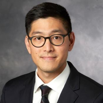 Fred M Baik合作论文数Otolaryngology (Head and Neck Surgery) Department, Stanford University12
 Gregory W. Randolph合作论文数Department of Otolaryngology Head and Neck Surgery, Harvard Medical School, Harvard University;Division of Thyroid and Parathyroid Endocrine Surgical Service, Massachusetts Eye and Ear Infirmary;Massachusetts General Hospital12Bruce Culliney合作论文数11Samuel J Rubin合作论文数11




