Submaximal aerobic exercise is an evidence-informed strategy for concussion management. However, its impact on the concussed pediatric brain remains poorly understood. A seminal adult study reported stability of default mode network (DMN) functional connectivity before and immediately after aerobic exercise in adults with mild traumatic brain injury. Comparable data do not exist in children, despite known developmental neurophysiological differences between children and adults. This study examined DMN network stability before and after submaximal aerobic exercise in pediatric sport-related concussion. In a controlled cohort design, 18 concussed participants (within 4 weeks of injury; 15.2 ± 1.8 years; 33% female) and 18 age- and sex-matched controls (14.5 ± 2.1 years; 50% female) completed resting-state functional magnetic resonance imaging scans pre- and post-exercise. Exercise intensity was set to 85% of the individualized symptom-limited heart rate achieved on a Buffalo Concussion Treadmill Test performed 24-48 h prior. Functional connectivity was assessed across four DMN regions of interest (posterior cingulate cortex, medial prefrontal cortex, left lateral parietal cortex, right lateral parietal cortex). Graph theory analyses conceptualized each region as a network node. Region of interest-based analyses demonstrated reduced pre- to post-exercise correlations across DMN pairs in both groups. Mean percent change was -25.8% (±12.7%) in concussion and -15.0% (±7.2%) in controls. However, no statistically significant within- or between-group correlational differences were observed, consistent with adult findings. In contrast, graph theory revealed significant post-exercise reductions in network efficiency (β = -0.17, p = 0.015), cost (β = -0.18, p = 0.014), and degree centrality (β = -0.53, p = 0.001) exclusively in the concussion group, driven primarily by the right lateral parietal cortex. While correlational analyses suggest DMN stability similar to adults, graph metrics indicate reduced network connectedness following exercise in pediatric concussion. These findings underscore the need to move beyond symptom-based frameworks and examine exercise-related neurophysiological responses in youth concussion.
With a focus on physical activity and physiological variables this scoping review synthesizes recent trends in machine learning for glycemic prediction in individuals with Type 1 diabetes. A structured PRISMA-ScR search (2010–2025) identified 41 studies which resulted in three dominant application areas: (1) Multi-horizon prediction of glycemia and physical activity detection, driven mainly by recurrent neural networks (RNN)-most commonly long short-term memory (LSTM)-with evidence that incorporating energy expenditure improves model performance; (2) prediction of exercise-induced dysglycemia and nocturnal hypoglycemia, which share overlapping temporal horizons, indicating potential for unified forecasting models; and (3) translation of prediction models into bolus-optimization strategies, though real-world validation is limited. The review identifies two critical gaps: (1) The handling of physiological drift and model decay as a result of physiological training or detraining; (2) Menstrual cycle integration and its use as a feature remains unexplored, while multiple studies have demonstrated the decrease of insulin sensitivity in the late luteal phase of the cycle.
There is an urgent need for interventions which reduce dementia risk in aging adults. People with subjective cognitive decline (SCD) have a greater risk of developing dementia compared to age-matched cognitively normal individuals. Impaired cerebral blood flow (CBF) and cerebral glucose hypometabolism are leading mechanisms underlying dementia that could be ideal interventional targets for this population. Recent research, including our own work, has shown that oral consumption of ketone monoesters (KME) can improve CBF and cerebral metabolism, which in turn can improve cognition. The purpose of this study is to investigate the hypothesis that a 14-day KME supplementation intervention in middle-to-older adults with SCD will increase CBF, brain functional connectivity, and cognitive performance in comparison to placebo. A total of 34 middle-to-older adults (50
Background GERAS DANcing for Cognition and Exercise (DANCE) was developed with rehabilitation and geriatric medicine expertise for older adults (age 60 +) looking to improve brain health or mobility. This trial aimed to assess the feasibility, acceptability, and safety of delivering virtual GERAS DANCE to older adults in a home-based setting. A single-center, prospective, parallel-group randomized feasibility trial was conducted to assess the feasibility of virtual GERAS DANCE. Fifty older adults were randomized to the virtual GERAS DANCE intervention group or a control group receiving usual care. The progressive dance curriculum was live-streamed with videoconference by a certified GERAS DANCE instructor in 1-h sessions held twice weekly for 6 weeks. Participants used their personal tablets, desktop computers, and laptops. Feasibility was evaluated based on predefined criteria, including process measures (e.g., recruitment and retention rates), outcome measures, resource utilization, and the acceptability of the intervention to participants. One hundred ninety three of 206 individuals met the eligibility criteria, indicating that the inclusion criteria were well-defined and suitable for the target population. The enrollment-to-screening ratio was 25:103, with recruitment completed in 8 weeks. Fifty older adults were randomized, and 46 completed baseline assessments (mean age = 75.02(5.89) years, range 63–92, 92
Youth mental health-related problems and disorders have garnered increased attention due to global prevalence estimates that have, in some cases, increased following the COVID-19 pandemic. Various methodologies have been proposed to leverage artificial intelligence (AI) for detecting mental health problems in the general population; however, research specifically focused on AI methods for youth remains limited. Shortcomings in modern AI include limited training data modalities (i.e., types of input data used for model training), reliance on offline training, and the use of static models. This scoping review provides an overview of evidence that uses AI methods applied to youth mental health (YMH) and provides an assessment of the current state of research that integrates multimodal AI (i.e., models that incorporate multiple data modalities) and/or online learning (i.e., incremental or continual model training from streaming data) for the diagnosis, monitoring, and treatment of YMH-related problems. The findings indicate that research in AI applied to YMH is limited in the areas of multimodal AI and online learning. The number of studies in this field is steadily growing. Studies incorporating online learning demonstrate that this approach enhances model performance and adaptability, which is crucial for developing translational models capable of addressing real-world challenges effectively. Despite these advances, key challenges remain, including the availability and long-term validity of multimodal data, the lack of participant-related information in certain databases and studies, the ethical and logistical difficulties of collecting data from minors, and the computational costs of training robust AI models.
PURPOSE:Diffusion MRI is widely used to characterize tissue microstructure, but standardization remains challenging, particularly for advanced models or regions with crossing fibers. Phantoms provide controlled environments to assess measurement repeatability independent of biological variability. This study evaluated the repeatability of higher-order diffusion tensor metrics using a novel anisotropic diffusion phantom designed to mimic white matter tract geometry. METHODS:The phantom, containing linear, crossing (30°, 45°, 90°), and bifurcating synthetic fiber bundles, was scanned seven times using a GE Healthcare 3.0 T MRI system. Four acquisition protocols were evaluated: 30-direction DTI (b = 1000s/mm2), 60 and 90-direction High Angular Resolution Diffusion Imaging (HARDI; b = 1300s/mm2), and 30-direction Diffusion Kurtosis Imaging (DKI; b = 250, 500, 750, 1000, 1500, 2000, 2500, 3000 s/mm2). Repeatability was quantified using coefficient of variation (CoV) and intraclass correlation coefficient (ICC) for scalar diffusion metrics across six regions of interest. Fiber orientation distribution functions (fODFs) were analyzed to assess crossing fiber resolution accuracy. RESULTS:DTI-derived metrics demonstrated excellent repeatability, with fractional anisotropy (FA) CoV < 10% and mean, axial, and radial diffusivities < 3%. DKI-derived metrics exhibited greater variability, though kurtosis FA remained stable (CoV ∼7%). Generalized FA showed improved reliability with increased angular resolution (ICC = 0.8445 for 90-direction HARDI). fODFs accurately resolved crossing fibers at 90° (RMSE = 3.49°) and 45° (RMSE = 8.92°) but failed at 30° separation. CONCLUSION:The phantom provides reliable repeatability for standard DTI metrics and demonstrates utility for quality assurance of advanced diffusion models with high angular resolution protocols.
Diffusion tensor imaging (DTI) has emerged as a powerful neuroimaging modality for investigating white matter microstructure and its alterations following brain injury. This review presents a comprehensive overview of DTI, encompassing its physical principles, mathematical modeling of diffusion tensors, and known limitations of the technique. We explore key methodological considerations, including acquisition protocols, preprocessing pipelines, vendor-related variability, atlas registration, and the role of diffusion phantoms in calibration. With the rise of big data in medical imaging, we highlight the influence of large-scale, multisite datasets and open-source neuroimaging repositories in advancing DTI research. A central focus is placed on the application of DTI in mild traumatic brain injury, a condition that often eludes detection in conventional imaging settings. We evaluate emerging computational strategies, including Z-score analysis, principal component analysis, random forests, and generative adversarial networks-that improve the sensitivity, specificity, and interpretability of DTI metrics in both clinical and research settings. By bridging methodological rigor with translational insight, this review underscores the evolving potential of DTI as a neuroimaging biomarker for brain injury assessment.
To provide a thorough comparison of the SNR between sodium MRI k-space sampling schemes in the brain within clinically feasible time constraints (∼10 min) at 3 T. Density-adapted radial (DA-3DPR), constant-amplitude radial, Cartesian, FLORET, rotated spiral, and 3D cones trajectories were designed with parameters optimized for brain tissue SNR. The sequences were acquired in both a phantom and 13 healthy participants (age = 28.7 ± 3.4, M:F = 7:6). SNR was measured and corrected for point-spread function (PSF) volume and scan duration for a less-biased assessment. CSF-to-brain-tissue contrast and CNR were also measured. The data were linearly modeled, and ANOVA was used to determine if the sampling scheme contributed to the variance with the obtained metrics. The sampling schemes contributed significantly to the variance (p < 0.001) for all metrics. The DA-3DPR sampling scheme provided the highest SNR in both the phantom and the participants. The Cartesian sampling scheme had the highest absolute contrast, but the largest CNR was shared between the DA-3DPR, 3D cones, and FLORET sampling schemes. When considering the PSF and the requirement for a clinically feasible scan time, a 15 ms read-out DA-3DPR trajectory provides the highest SNR at 3 T, without losing any desired contrast.
The aim of this study was to evaluate the contributions of age, sex, and MRI vendor to variance in Diffusion Tensor Imaging (DTI) metrics, with a focus on understanding the impact of these factors in large-scale healthy brain datasets. A dataset of 2,700 DTI scans from healthy controls across multiple sites and MRI vendors was analyzed. The DTI scalar metrics fractional anisotropy (FA) and mean diffusivity (MD) were processed and the influence of age, sex, vendor, and brain atlas selection were determined. A statistical analysis was conducted and revealed significant (p<0.05) age-related differences in DTI metrics, with older participants showing reduced FA and increased MD, in line with known microstructural changes. Sex differences were observed, with females exhibiting slightly higher FA and lower MD in certain brain regions. Vendor variability was also noted, with all three MRI vendors showing significant differences in FA with Siemens machines typically exhibiting higher FA values and GE machines lower FA values (i.e. FA Siemens > FA Philips > FAGE GE ). Atlas selection also highlighted some specific ROI behaviour (e.g. tapetum of the corpus callosum) as one of the most significant regions of interest (ROIs) in the JHU-Tracts atlas that demonstrated a large amount of deterioration with age, particularly in females. These findings emphasize the need to account for biological factors such as age and sex, as well as technical factors like ROI selection and MRI vendor, when interpreting DTI data. The results demonstrate the potential of large-scale, multi-vendor datasets to uncover meaningful biological trends, while also addressing the challenges of scanner-specific variability. Although previous work has shown sex and age differences, this is the first large scale DTI analysis that has included age, sex, and MRI vendor as sources of variance in one model.
Context:To understand energy balance, whole-room indirect calorimetry (WRIC) allows for accurate measurement of energy expenditure (EE). Objective:To examine the relationship between cold-induced resting EE and brown adipose tissue (BAT) activity measured by magnetic resonance imaging (MRI) and to evaluate WRIC system (WRICS) performance and feasibility of use in children and adults. Methods:The WRICS was equipped with a Promethion High-Definition Room Calorimetry System. Technical validation utilized nitrogen (N2) and carbon dioxide (CO2) gas infusions. Healthy adults (n = 21) and children aged 8-17 years (n = 17) attended two 4-hour WRIC visits (one week apart) and one MRI visit. Resting EE at 25 °C (REE25) was compared between visits and to REE at 18 °C (REE18). Recruitment and completion rates were examined. BAT activity was assessed by MRI as the decline in supraclavicular proton density fat fraction during 18 °C cold exposure. Results:Gas infusion testing confirmed high accuracy (respiratory exchange ratio [RER] = 0.99; 95% CI 0.991-0.996). Study completion rates were high (adults: 20/21; children: 17/18). REE25 over a 10-minute period was consistent between visits (adults: 1.68 ± 0.462 vs 1.66 ± 0.301 kcal/min, P = 0.77; children: 1.50 ± 0.358 vs 1.58 ± 0.348 kcal/min, P = .25). Cold exposure increased fasting EE by 0.21 kcal/min (adults) and 0.14 kcal/min (children). BAT activity was correlated with REE18 in adults (r = 0.49, P = .04). Conclusion:WRICS use was feasible in adults and children. Changes in EE during cold (ie, cold-induced thermogenesis) were measurable and related to BAT activity, supporting the usefulness of this system in the assessment of EE in response to interventions in adults and children.
Abstract Context Accurate assessment of excess body fat and its cardiometabolic risk is essential in clinical and epidemiologic research. Body mass index (BMI), although widely used, does not capture visceral or ectopic fat. Objective To evaluate how anthropometric measures—BMI, percent body fat, waist circumference (WC), and waist-to-hip ratio (WHR)—relate to MRI-measured visceral adipose tissue (VAT) and hepatic fat fraction (HFF), overall and by sex. Design Cross-sectional analysis within the Canadian Alliance for Healthy Heart and Minds (CAHHM) cohort. Setting Community-based, pan-Canadian prospective study. Patients or Other Participants 6,683 apparently healthy adults (mean age 57±9 years; 3,665 females) with baseline anthropometric and MRI measures; analyses adjusted for study center. Intervention(s) Not applicable. Main Outcome Measure(s) MRI-derived VAT and HFF. Associations were estimated using linear regression and mixed models, stratified by sex, ethnicity, age, and BMI category. Results . Mean BMI was 26.7 kg/m2, WC 88.3 cm, VAT 71 mL, and HFF 5.7%. Females had lower VAT and HFF than males. Correlations with VAT ranged from 0.35–0.77 and with HFF from 0.26–0.47. Each 10-cm higher WC was associated with 21.0 mL higher VAT and 2.1% higher HFF; each 5-kg/m2 higher BMI predicted 24.7 mL and 2.8% higher values. When modeled jointly, WC remained strongly predictive, while BMI contributed modestly. Associations were smaller in females. VAT and HFF increased across WC tertiles within BMI categories. Conclusions . WC is a robust surrogate for visceral and hepatic fat across BMI categories, supporting its use when MRI is not feasible.
Abstract Clinical adoption of new biomedical techniques depends on establishing reference values against which individual patients can be compared. In resting-state functional MRI (rsfMRI), most biomarker research has relied on the case-control paradigm, whose underlying assumptions are often invalid as diseases are frequently heterogeneous, limiting biomarker generalizability. Normative modeling offers a complementary alternative by characterizing individual deviations against a reference population. However, in rsfMRI, normative modeling has been applied almost exclusively to functional connectivity, with limited attention to age trajectories and sex effects. We address these gaps by developing a spatial normative model of four rsfMRI metrics that capture complementary features of the blood-oxygen-level-dependent (BOLD) signal across age and sex. Five publicly available datasets were aggregated to form a sample of 1,978 participants aged 10-30 years. Four metrics were computed for each of 110 grey matter regions: amplitude of low-frequency fluctuations (ALFF), fractional amplitude of low-frequency fluctuations (fALFF), regional homogeneity (ReHo), and Hurst exponent. A machine-learning model based on hierarchical Bayesian regression with a non-Gaussian likelihood was fitted per metric, modeling non-linear age effects, sex, and multi-site acquisition. Models were well calibrated across all four metrics, with fALFF showing the strongest predictive performance and Hurst exponent the weakest. Normative trajectories varied across brain regions for each metric, but on average, the median of each distribution remained bounded across regions, while the spread was more regionally variable. All four metrics showed predominantly negative slopes with age, indicating a decrease in each metric over the age window. This work provides a normative reference across four rsfMRI metrics that capture distinct features of the BOLD signal, complementing the case-control paradigm and supporting individual-level inference.
We investigated the potential of diffusion tensor imaging (DTI) in identifying alterations in cerebellar white matter (WM) in concussion patients. We hypothesized that DTI, combined with a Z-score analysis approach, could provide personalized assessment of cerebellar changes in these patients. Fifty-one mTBI patients (25 females, age: 40.5 ± 11.7 years and 26 males, age: 45.0 ± 14.1 years) and 2700 healthy age/sex/vendor-matched controls were analyzed in this study. DTI data analysis focused on scalar metrics fractional anisotropy (FA) and mean diffusivity (MD). A Z-score approach was used to identify subject-specific deviations, while statistical models (linear regression, random forests, hierarchical clustering) compared the sensitivity of Z-scores and the Post-Concussion Symptom Scale (PCSS) in capturing individualized differences. Significant cerebellar DTI abnormalities were found in the concussion group. Z-score analysis showed higher sensitivity to cerebellar regions associated with balance and sleep. Statistical models revealed distinct patterns by age, sex, region, and symptoms, underscoring the value of personalized analysis and the subjectivity that remains in existing assessments like the PCSS. The combination of DTI and Z-score methods provides a promising framework for personalized detection of cerebellar changes in mTBI, potentially improving diagnostic accuracy and enabling more targeted interventions.
Background: There has been rapid growth in the field of deep learning with convolutional neural networks (CNNs) for imaging-related tasks. More recently, vision transformers (ViTs) have shown competitive performance to CNNs, while also uniquely possessing a novel 'attention mechanism'. ViTs may replace CNNs; however, more work is required to show if transformer architecture, specifically the new attention mechanism, is more receptive to salient information from input images. This issue is becoming increasingly critical as the use of machine learning continues to expand in healthcare. Thus, we proposed to assess whether the attention that ViTs receive is misplaced. Methods: The attention heads of ViT and the important pixels for a ResNet50 model were compared to radiologist annotations to determine appropriateness of each model's attention mechanisms in classifying two datasets. We used the VinDr-CXR dataset of 18,000 chest X-rays and the Mini-DDSM dataset of 10,000 mammograms to classify healthy, benign, and malignant tumours. The attention heads were examined through attention rollout and the ResNet50 model using the occlusion method. Models were evaluated with accuracy and level of agreement calculated with a pixel-wise logical XNOR operator. Results: The VinDr-CXR ResNet50 and the ViT models had test accuracy of 70.4% and 77.4%, respectively. The Mini-DDSM ResNet50 and ViT models had test accuracy of 90.26% and 95.53%, respectively. Agreement was higher for transformer models at 94.72% and 96.96% compared to 88.07% and 94.85% for the CNN models. Conclusions: We show that our attention is not misplaced as the transformer approach shows overall better performance and agreement compared to CNNs.
We evaluated a quality control (QC) phantom designed to mimic diffusion characteristics and white matter fiber tracts in the brain. We hypothesized that acquisition of diffusion tensor imaging (DTI) data on different vendors and over multiple repeated measures would not contribute to significant variability in calculated diffusion tensor scalar metrics such as fractional anisotropy (FA) and mean diffusivity (MD). The DTI QC phantom was scanned using a 32-direction DTI sequence on General Electric (GE), Siemens, and Philips 3 Tesla scanners. Motion probing gradients (MPGs) were investigated as a source of variance in our statistical design, and data were acquired on GE and Siemens scanners using GE, Siemens, and Philips vendor MPGs for 32 directions. In total, 8 repeated scans were made for each GE/Siemens combination of vendor and MPGs with 8 repeated scans on a Philips machine using its stock DTI sequence. Data were analyzed using 2-way ANOVAs to investigate repeat scan and vendor variances and 3-way ANOVAs with repeat, MPG, and vendor as factors. No statistical differences (i.e., P > 0.05) were found in any DTI scalar metrics (FA, MD) or for any factor, suggesting system constancy across imaging platforms and the specified phantom’s reliability and reproducibility across vendors and conditions. A DTI QC phantom demonstrates that DTI measurements maintain their consistency across different MRI systems and can contribute to a standard that is more reliable for quantitative MRI analyses.
Visceral adipose tissue (VAT) and hepatic fat (HF) contribute to multiple health risks, including diabetes, hypertension, cardiovascular disease, cognitive decline, and cancer. The objective of this study is to determine whether VAT and HF are associated with carotid atherosclerosis beyond traditional cardiovascular risk factors. Participants in the Canadian Alliance of Healthy Hearts and Minds (CAHHM) cohort study (n = 6760; average age= 57.1; 54.9
The purpose was to assess the agreement in measures of acute knee cartilage thickness and composition change after loading in clinical knee osteoarthritis (OA) between two magnetic resonance imaging (MRI) acquisition approaches: (1) single sequence approach using quantitative double-echo in steady-state (qDESS), which allows simultaneous morphological and compositional scanning, versus (2) multi-sequence approach that captures morphology (fast spoiled gradient recalled (FSPGR) or qDESS) and composition (multi-echo spin echo (MESE)) separately. Twenty adults with clinical knee OA participated. 3T MR scans were acquired before and immediately after a 25-min treadmill walk at a standardized speed. Changes in knee cartilage thickness and T2 were assessed. Pre-activity, strong agreement was observed in cartilage thickness captured with qDESS and FSPGR (concordant correlation coefficients 0.842-0.935). Pre-activity, we observed greater absolute cartilage thickness with qDESS compared to FSPGR in femoral cartilage. From pre- to post-activity, qDESS showed change in cartilage thickness in the medial femur (-0.088 ± 0.11 mm, p = 0.002), lateral tibia (-0.042 ± 0.65 mm, p = 0.011) and trochlea (-0.027 ± 0.05 mm, p = 0.024); whereas FSPGR showed a change only in the lateral tibia (-0.064 ± 0.08 mm, p = 0.002). From pre- to post-activity, qDESS showed reduced T2 in all cartilage regions; whereas qDESS + MESE and FSPGR + MESE detected T2 changes in the patella (-1.90 ± 3.00 ms, p = 0.013, and -1.80 ± 2.18 ms, p = 0.002, respectively). qDESS detects transient changes in knee cartilage due to loading in clinical knee OA.
Ultra-high field MRI facilitates imaging at high spatial resolutions, which may become important for detailed anatomical and pathological assessment of the human liver. Therefore, we aimed to advance structural liver imaging at 7 T by implementing a high-resolution, phase-shimmed, free-breathing liver scan. Six healthy participants underwent liver MRI scans at 7 T, utilizing an eight-channel parallel transmission system for phase shimming. B0 mapping and Fourier phase-encoded dual refocusing echo acquisition mode (PE-DREAM) multichannel B1 + mapping were performed during breath-holds at expiration. Prospectively undersampled golden-angle pseudo-spiral k-space data were acquired under free breathing, enabling retrospective respiratory binning using self-gating. Post-binning, the simultaneous autocalibrating and k-space estimation (SAKE) algorithm was employed for interpolation of a center of k-space area, prior to estimation of receive coil sensitivity maps. Image reconstruction was performed on expiration-phase data using compressed sensing, optimizing image quality by evaluating various regularization factors and numbers of respiratory bins. Finally, N4BiasFieldCorrection was applied to the resulting images. Expiration-phase image reconstruction using four bins and regularization factor values of 10-2.5 (1.50 mm) and 10-2.33 (1.35 mm) were found to optimize the tradeoff between sharpness, SNR, and artifacts. The optimized protocol facilitated clear visualization of the liver, blood vessels, and surrounding structures at isotropic resolutions of 1.50 and 1.35 mm in 3.5 min, without B1 + inhomogeneity effects in the shimmed liver region. A comparison between low-resolution fully sampled free-breathing (3.5 min) and breath-hold (19 s) acquisitions demonstrated comparable sharpness and SNR. To compare the 7 T data with 3 T MRI, 3 T scans were performed for two participants. 3 T reconstructions were done similarly to 7 T, excluding N4BiasFieldCorrection. Scan-specific regularization optimization was performed for fair comparison. Compared to 3 T, 7 T showed superior vascular contrast with inflow effects not observed at 3 T. Fold-over artifacts were present in 3 T scans but were minor at 7 T. 3 T and 7 T provided comparable results, with a much higher RF channel count at 3 T. In conclusion, high-resolution expiration-phase liver imaging at 7 T with homogeneous signal can be successfully achieved using a phase-shimmed, free-breathing protocol with a golden-angle pseudo-spiral sampling pattern technique and respiratory self-gating. This approach allows detailed anatomical depiction without the limitations of breath-holding, representing a significant advancement in ultra-high field abdominal MRI.
A concise overview of three major advancements in fast magnetic resonance imagine (MRI) reconstruction techniques is presented, focusing on their roles in enhancing image quality and reducing acquisition times. The first set of methods, parallel imaging techniques, includes sensitivity encoding (SENSE) and generalized autocalibrating partially parallel acquisitions (GRAPPA). SENSE utilizes spatial sensitivity information from multiple receiver coils to accelerate image acquisition by undersampling k-space data and reconstructing images using coil sensitivity profiles, allowing for faster scans. GRAPPA, another parallel imaging method, uses estimated weights from a calibration scan to fill in missing data in undersampled k-space and then reconstructs unaliased images. Additionally, this review explores sparse reconstruction techniques such as compressed sensing, which leverages the sparsity of images in a transformed domain to reconstruct high quality images from significantly fewer measurements, thus reducing scan times. The latest developments in machine learning applications for MRI acquisition are also discussed, highlighting how advanced algorithms are being used to improve image reconstruction, enhance diagnostic accuracy, and simplify workflow processes.
Resting-state fMRI (rsfMRI) is a widely used neuroimaging technique that measures spontaneous fluctuations in brain activity in the absence of specific external cognitive, motor, emotional, and sensory tasks or stimuli, based on the blood-oxygen-level-dependent (BOLD) signal. Functional connectivity (FC) is a popular rsfMRI analysis examining BOLD signal correlations between brain regions. Nevertheless, there are alternative analyses that provide different but collectively informative characteristics of the BOLD signal and, thus, brain activity. This narrative review aimed to provide a comprehensive conceptual, mathematical, and significance investigation of common rsfMRI analyses in addition to FC. To achieve this, a narrative review was conducted on studies using the most common rsfMRI analysis to investigate global and local brain activity. Five rsfMRI analyses were described, summarizing the common initial steps of rsfMRI data processing and explaining the main characteristics and how each metric is calculated. The rsfMRI analyses described are (1) FC, reflecting BOLD global connectivity; (2) the amplitude of low-frequency fluctuations (ALFF) and fractional ALFF (fALFF), representing the intensity of the BOLD signal; (3) regional homogeneity (ReHo), which reflects BOLD local connectivity; (4) Hurst exponent (H), depicting autocorrelation of the BOLD signal; and (5) entropy, depicting the BOLD signal predictability. As rsfMRI is a vital tool for exploring brain function, selecting an analysis that aligns with the research question is essential. This review offers an initial catalog of standard rsfMRI analyses, highlighting their key features, concepts, and considerations to support informed decisions by researchers and clinicians.
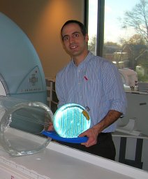 Norman B Konyer合作论文数23
 Dinesh Kumbhare合作论文数Institute of Health Policy, Management and Evaluation, University of Toronto;Division of Physical Medicine and Rehabilitation, Department of Medicine, University of Toronto;KITE21
 Carol Dematteo合作论文数Sch Rehabil Sci, McMaster Univ17
 Eric Smith合作论文数Department of Clinical Neurosciences, University of Calgary13
 Sandra Black合作论文数Institute of Medical Science, Temerty Faculty of Medicine, University of Toronto;Sunnybrook Health Science Centre13Lange Thomas合作论文数12
 Jean-Claude Tardif合作论文数Département de Médecine, Faculté de Médecine, Université de Montréal;Département de Biochimie et Médecine Moléculaire, Faculté de Médecine, Université de Montréal12Greg Wells合作论文数Floor Toronto, ON, Canada M5V 2G511
 Richard Frayne合作论文数Univ Calgary, Calgary, AB, Canada10
 Colm Boylan合作论文数10
 John F Connolly合作论文数Royal College of Surgeons10
 Zukotynski Katherine A合作论文数Dana-Farber Cancer Institute, Harvard Medical School9Elzibak Alyaa H合作论文数9Sharma Bhanu合作论文数Toronto Rehabilitation Institute9
 David Robert Chettle合作论文数Medical Physics and Applied Radiation Science, McMaster University Hamilton9
 Fiona E. Mcneill合作论文数Department of Physics and Astronomy, McMaster University9
 Monica Maly合作论文数School of Rehabilitation Science, McMaster University9Ethan Danielli合作论文数8
 Sabrina Adamo合作论文数Sunnybrook Research Institute, University of Toronto8
 Vesna Sossi合作论文数Department of Physics & Astronomy, The University of British Columbia;PET-MRI Imaging Centre, Faculty of Medicine, The University of British Columbia8
 Maged Goubran合作论文数Department of Medical Biophysics, Temerty Faculty of Medicine, University of Toronto8
 Alexander Thiel合作论文数Department of Neurology & Neurosurgery, Faculty of Medicine and Health Sciences, McGill University;Lady Davis Institute for Medical Research, Jewish General Hospital8
 Jean Paul Soucy合作论文数Montreal Neurological Institute-Hospital, McGill University8
 Frank Prato合作论文数Department of Medical Biophysics, Western University;St. Joseph's Hospital;Lawson Health Research Institute;University of Western Ontario8
 Robert Laforce合作论文数Groupe de Recherche en Réadaptation physique, IRDPQ8
 Michael Borrie合作论文数8
 Robin Hsiung合作论文数Division of Neurology, Department of Medicine, Faculty of Medicine, The University of British Columbia7
 Russel Schachar合作论文数Department of Psychiatry, Temerty Faculty of Medicine, University of Toronto;Department of Psychiatry, The Hospital for Sick Children7
 Salim Yusuf合作论文数Medical School, McMaster University;Population Health Research Institute, Hamilton Health Sciences7Phillip H. Kuo合作论文数Department of Molecular Physiology and Biological Physics, University of Virginia7
 Christian Bocti合作论文数University of Montreal7Noam Soreni合作论文数7
 Demetrios Sahlas合作论文数Faculty of Health Sciences, McMaster University;Hamilton Health Sciences7
 Geoffrey B Hall合作论文数Department of Psychiatry and Behavioural Neurosciences, McMaster University7
 Olga Dona合作论文数bDepartment of Radiation Oncology, University of Rochester Medical Center6
 Anthony A Gatti合作论文数Dept Radiol, Stanford Univ6Chertkow Howard合作论文数6Christopher G. Scott合作论文数6
 James L Gräfe合作论文数6
 Katherine M Morrison合作论文数Department of Pediatrics, McMaster University and Hamilton Health Sciences6
 Thomas E. Doyle合作论文数McMaster University6
 Markad Venkatraya Kamath合作论文数Faculty of Health Sciences, McMaster University6C. Boylan合作论文数5Tm Bray合作论文数5
 Frank J Ong合作论文数Department of Pediatrics, McMaster University5
 Julie Ottoy合作论文数Sunnybrook Research Institute, University of Toronto5
 Alexander Weber合作论文数Graduate Program in Neuroscience, Faculty of Medicine, The University of British Columbia5Poirier Paul合作论文数5
 Koon Teo合作论文数Division of Cardiology, Faculty of Health Sciences, McMaster University5




