Sociality is a defining feature of the human experience: We rely on others to ensure survival and cooperate in complex social networks to thrive. Are there brain mechanisms that help ensure we quickly learn about our social world to optimally navigate it? We tested whether portions of the brain’s default network engage “by default” to quickly prioritize social learning during the memory consolidation process. To test this possibility, participants underwent functional MRI (fMRI) while viewing scenes from the documentary film, Samsara . This film shows footage of real people and places from around the world. We normed the footage to select scenes that differed along the dimension of sociality, while matched on valence, arousal, interestingness, and familiarity. During fMRI, participants watched the “social” and “nonsocial” scenes, completed a rest scan, and a surprise recognition memory test. Participants showed superior social (vs. nonsocial) memory performance, and the social memory advantage was associated with neural pattern reinstatement during rest in the dorsomedial prefrontal cortex (DMPFC), a key node of the default network. Moreover, it was during early rest that DMPFC social pattern reinstatement was greatest and predicted subsequent social memory performance most strongly, consistent with the “prioritization” account. Results simultaneously update 1) theories of memory consolidation, which have not addressed how social information may be prioritized in the learning process, and 2) understanding of default network function, which remains to be fully characterized. More broadly, the results underscore the inherent human drive to understand our vastly social world.
From stage magicians to the Vulcan mind meld, mind reading has been the stuff of magic and science fiction. Recent developments in neuroimaging might be bringing it one step closer to reality, however, as increasingly sophisticated analysis techniques move toward the decoding of mental states from functional imaging data in humans. Two companies even offer fMRI-based lie detectors1. Although legal applications are still premature, these pattern classification techniques represent a new way of looking at neuroimaging data2 and may extend the power of functional imaging substantially. Conventional neuroimaging analysis correlates external regressors such as task condition with activity in specific brain areas. Pattern classification inverts this methodology and instead predicts the external stimulus based on neuroimaging data. Unlike conventional analyses, these pattern-based analyses take into account the full spatial pattern of brain activity rather than concentrating on specific regions. Thus, even if activity at a particular voxel does not distinguish different cognitive states, the pattern of activity distributed over many regions can do so, increasing sensitivity. This multivariate approach generates pattern vectors corresponding to specific cognitive states, and a classifier is trained to discriminate between these states. This classifier can then be used to predict the cognitive state on the basis of brain activity alone. Such approaches have been used to predict what percept is dominant in a binocular rivalry protocol3 or what orientation subjects are viewing4, even when they are not consciously aware of the stimulus. These techniques make it easier to evaluate responses to naturalistic stimuli, as pattern classification algorithms are designed to analyze activity over the whole brain without attempting to localize function. In a recent competition at the University of Pittsburgh (http://www.ebc.pitt.edu/competition.html), participants were given fMRI data and subjective ratings from observers as they viewed two short film clips. Competitors then had to produce an algorithm that could predict what the subjects were seeing based on a third fMRI data set. The winning entries achieved correlations as high as 0.86 for basic features such as the presence of music. Uri Hasson from New York University, one of the researchers who judged the competition, says, "I am much more optimistic as to the power of fMRI to read and predict human experience. Many of the participants managed to predict the twelve features chosen for the competition (such as language, music, emotion), as well as the specific observers who coded the movie. Moreover, a few groups managed to predict the identity of the actor being seen or the location the subject is watching." The power of this approach extends beyond just predicting cognitive states from brain activity. Pattern classification techniques can provide clues about how this information is processed as well. For example, this technique shows that object categories with shared image-based attributes have shared neural representation, even when multiple views of objects are included or when line drawings without much detail are used5. This kind of information would be difficult to uncover using a conventional analysis technique. James Haxby of Princeton University says, "I find myself working with a whole new community of people: computer scientists, electrical engineers, scientists who come from an applied physics and mathematics background. The fact that we are finally looking for patterns of activity across the whole population rather than areas with different function means that we are taking a more information-based approach. This is a fundamental shift." Pattern classification can also overcome another criticism of functional imaging: that it lacks the temporal resolution of other whole-brain imaging techniques such as EEG. Pattern classification has been used to identify distributed patterns of activity associated with different categories of objects6. During later free recall, the patterns of activity associated with the specific object category reappear several seconds before the verbal recall of the object. The greater sensitivity of pattern classification techniques compared to conventional imaging analysis results in a temporal resolution approaching that of EEG. As yet, however, these techniques cannot generate the sort of brain maps that we all know from conventional neuroimaging studies, showing spots of localized activity associated with particular functions. Efforts are underway to develop such maps based on pattern classification techniques, but conventional image analysis is likely to remain the preferred method for generating activity maps to understand where in the brain a process is occurring. Localization via conventional brain imaging complements the information about how a process is occurring that can be provided by pattern classification techniques, so both approaches will continue to be useful. Researchers are attempting to use pattern classification techniques to predict brain states in real-world applications, such as lie detection, but this endeavor seems much less promising. One problem is that activity is more likely to be variable but, more importantly, an fMRI lie detector would rely crucially on the compliance of its subjects. To train a classifier to categorize lying and truth telling, a suspect would essentially be asked to calibrate the instrument for his own conviction, potentially violating the fifth amendment to the United States Constitution, which protects people from being forced to incriminate themselves. Even though the accuracy of the technology is likely to improve, it is unclear if such an fMRI lie detector can surpass conventional polygraph and EEG lie detectors, with which it is likely to share drawbacks such as noise introduced by arousal or emotional responses. Although the applicability of pattern classification techniques to lie detection is uncertain, their influence on basic research is likely to be important. Neuroimaging's obsession with localization has often led to accusations that it is little more than phrenology. By using population responses across the whole brain to ask how rather than where information is processed, neuroimaging may be starting to come of age.
On a wintery afternoon over 60 years ago, I was browsing the Baker Library stacks at Dartmouth College and stumbled across a small book with an arresting name: "What Is Life?" [Schrödinger, E. . Cambridge, United Kingdom: Cambridge University Press, 1944]. This small volume contained numerous concepts, which would transform the future of the biological sciences, giving rise to new fields, dogmas, approaches, and debates. Here, I present the core concepts of Schrödinger's book, the influence they have had on biology, and the influence they may continue to have on the cognitive neurosciences.
In Chapter 12, Michael Gazzaniga tells us: “We are . . . animals with brains that carry out every . . . action automatically and outside our ability to describe how it works . . .. a soup of dispositions controlled by genetic mechanisms, some weakly and some strongly expressed.” He also tells us: “We humans have something called the interpreter, located in our left brain, that weaves a story about why we feel and act the way we do.” Gazzaniga explores the concepts of free will and moral responsibility in light of such facts, arguing that we all remain personally responsible for our actions because responsibility arises out of each person’s interaction with the social layer she is embedded in. “Responsibility is not to be found in the brain,” he concludes, rather it is “a needed consequence of more than one individual interacting with another.”
How we make decisions that have direct consequences for ourselves and others forms the moral foundation of our society. Whereas economic theory contends that humans aim at maximizing their own gains, recent seminal psychological work suggests that our behavior is instead hyperaltruistic: We are more willing to sacrifice gains to spare others from harm than to spare ourselves from harm. To investigate how such egoistic and hyperaltruistic tendencies influence moral decision making, we investigated trade-off decisions combining monetary rewards and painful electric shocks, administered to the participants themselves or an anonymous other. Whereas we replicated the notion of hyperaltruism (i.e., the willingness to forego reward to spare others from harm), we observed strongly egoistic tendencies in participants' unwillingness to harm themselves for others' benefit. The moral principle guiding intersubject trade-off decision making observed in our study is best described as egoistically biased altruism, with important implications for our understanding of economic and social interactions in our society.
The search for memory is one of the oldest quests in written human history. For at least two millennia, we have tried to understand how we learn and remember. We have gradually converged on the brain and looked inside it to find the basis of knowledge, the trace of memory. The search for memory has been conducted on multiple levels, from the organ to the cell to the synapse, and has been distributed across disciplines with less chronological or intellectual overlap than one might hope. Frequently, the study of the mind and its memories has been severely restricted by technological or philosophical limitations. However, in the last few years, certain technologies have emerged, offering new routes of inquiry into the basis of memory. The 2016 Kavli Futures Symposium was devoted to the past and future of memory studies. At the workshop, participants evaluated the logic and data underlying the existing and emerging theories of memory. In this paper, written in the spirit of the workshop, we briefly review the history of the hunt for memory, summarizing some of the key debates at each level of spatial resolution. We then discuss the exciting new opportunities to unravel the mystery of memory.
The split-brain literature offers a unique perspective on theories of consciousness. Since both the left and right hemispheres of split-brain patients remain conscious following split-brain surgery, any theory that attempts to explain consciousness in neurotypical individuals must also be able to explain the dual consciousness of split brain patients. This commentary examines illusionism the theory that phenomenal properties are illusory through the lens of the split-brain literature. Based on evidence that both hemispheres of split-brain patients are capable of introspection and both hemispheres can experience and maintain illusions, it is theoretically possible that phenomenal properties are illusions created by distorted introspection, in accordance with illusionism. However, in order to appropriately evaluate whether illusionism is a valid explanation of consciousness in split-brain patients, it is imperative that neural mechanisms are proposed that explain how introspection gives rise to illusory phenomenal properties.
The corpus callosum anatomically and functionally connects the two cerebral hemispheres. Despite its important role in interhemispheric communication however, severing the corpus callosum produces few—if any—noticeable cognitive or behavioral abnormalities. Incredibly, split-brain patients do not report any drastic changes in their conscious experience even though nearly all interhemispheric communication ceases after surgery. Extensive research has shown that both hemispheres remain conscious following disconnection and the conscious experience of each hemisphere is private and independent of the other. Additionally, the conscious experiences of the hemispheres appear to be qualitatively different, such that the consciousness of the left hemisphere is more enriched than the right. In this chapter, we offer explanations as to why split-brain patients feel unified despite possessing dual conscious experiences and discuss how the divided consciousness of split-brain patients can inform current theories of consciousness.
Dr. Michael Gazzaniga is the Director of the Sage Center for the study of Mind at the University of California, Santa Barbara. In 1964 he received a Ph.D from the California Institute of Technology, where he worked under the guidance of Roger Sperry, with primary responsibility for initiating human split-brain research. In his subsequent work he has made important advances in our understanding of functional lateralization in the brain and how the cerebral hemispheres communicate with one another. He has published many books accessible to a lay audience, such as The Social Brain, Mind Matters, Nature's Mind, The Ethical Brain, Human and Who's in Charge? Free Will and the science of the brain. Dr. Gazzaniga's teaching and mentoring career has included beginning and developing Centers for Cognitive Neuroscience at Cornell University Medical Center, University of California-Davis, and Dartmouth College He founded the Cognitive Neuroscience Institute and the Journal of Cognitive Neuroscience, of which he is the Editor-in-Chief Emeritus. Dr. Gazzaniga is also prominent as an advisor to various institutes involved in brain research, and was a member of the President's Council on Bioethics from 2001-2009. He is a member of the American Academy of Arts and Science, the Institute of Medicine and the National Academy of Sciences. His new book is Tales from Both Sides of the Brain.
OBJECTIVEPsychopathy is a personality disorder with symptoms that include lack of empathy or remorse, antisocial behavior, and excessive self-focus. Previous neuroimaging studies have linked psychopathy to dysfunction in the default mode network (DMN), a brain network that deactivates during externally focused tasks and is more engaged during self-referential processing. Specifically, the DMN has been found to remain relatively active in individuals with psychopathic tendencies during externally focused tasks, suggesting a failure to properly deactivate. However, the exact extent and nature of task-induced DMN dysfunction is poorly understood, including (a) the degree to which specific DMN subregions are affected in criminal psychopaths, and (b) how activity in these subregions relates to affective/interpersonal and antisocial/lifestyle traits of psychopathy.METHODWe performed a group independent component analysis to assess DMN activation during a Go/NoGo task in a group of 22 high-psychopathy and 22 low-psychopathy prisoners. The identified group-level DMN was parcellated into 6 subregions, and group differences in task-induced activity were examined.RESULTSIn general, DMN subregions failed to deactivate beneath baseline in the high-psychopathy group. A group comparison with the low-psychopathy group localized this attenuated task-induced deactivation to the posteromedial cortical (mPC) region of the DMN. Moreover, multiple regression analyses revealed that activity in the mPC was associated with affective/interpersonal traits of psychopathy.CONCLUSIONThese findings suggest that attenuated deactivation of the mPC subregion of the DMN is intrinsic to psychopathy, and is a pattern that may be more associated with affective psychopathic traits, including lack of concern for others.
From bugs to humans, the rate at which we have accumulated information about nervous systems in the last 100 y has been astonishing. Nonetheless, if one always adopted the intellectual style of first learning all there is to know about a topic before studying its new dimensions, future progress would be slow. Unlike many other disciplines, neuroscience does not currently enjoy the luxury of an agreed on set of next questions to be answered. It has been the Wild West, untamed and reinless, and, in many ways, continues to be.
'Departmerit c?f Psycldogy, Utiiversity of Califortiia, Berkeley, rid 'Center for Neirroscietice, Utiiversity of Cnl$ortiia, Dmis Abstract-The tieirral ~iieclia~ii~~ii~ of limb coorditiofioti were itir-estigated by testing callosototriy patierits atid riorriial cotitrol srrhjccts oti hitiiatirral tiiovemetits. Nortiid siibjects prodirced deviations iti the trrrjeitories wiieti spatial detricitidsfr,r the two hatids were different, despite tetiipornl syticlirotiy iti the otiset of hitriatiuril trioveltietits. Calhsototriy pritietits did tid prodirce spntid deviutiotis, ciltlioiigli their liarids tiimed with tiorriiul tenipord syriclirotiy. Noritid sirhjects hirt riot ccillosototiiy pci- tietits cxliibited Irirge iticrenses iti phitiitig cirid execiition time for ttiovettietits with differetit spntinl rleriioridsj?)r the two limds relative to triovetrierits with idetitictil spotid denicinds f.r the two hnwds. This tieiirril dissocintioti indicciies that spciticil ititer- feretice in tnovemerits resirlts frotit ccillosal cotiticetioris, dierecis tetriporcil sytichrony in triovetiietit otiset does riot rely oti the corpirs callosirtii. A remarkable faculty of humans is the ability to produce coherent actions by coordinating the limbs to achieve a com- mon goal. such as in grasping ajug, tying one's shoelaces, or playing a musical instrument. Although the subcortical and cor- tical processes that underlie single-limb movements have been studied extensively, the neural mechanisms that govern coor- dination between the limbs have received relatively little atten- tion. Each cerebral hemisphere exerts primary control of the contralateral limbs, but the spatial and temporal properties of bimanual movements reveal strong interactions between the limbs. It is difficult, for example, to produce different temporal I synchrony is so powerful that the patients are virtually unable to turn handheld knobs using two different temporal patterns when visual feedback of the limbs is removed. Without such feedback, they resort to moving with identical temporal pat- terns (Preilowski, 1972). The strong focus on temporal coupling and the limited use of spatially demanding tasks in these inves- tigations have left open the question of whether callosotomy patients demonstrate spatial coupling. To investigate the neural processes of spatial and temporal coupling, we applied to callosotomy patients a standard para- digm for examining bimanual movements. When normal sub- jects attempt to produce movements requiring different spatial forms (i.e., lines combined with circles) or directional proper- ties (i.e., horizontal movements combined with vertical move- ments) for the two hands, spatial interference in the trajectory paths of both hands is exhibited. These tasks prove to be spa- tially demanding even when subjects move at a preferred rate (Franz et al., 1991). In the present study, a callosotomy patient and normal control subjects were tested on repeated cycles of trajectories that required movements in either the same orien- tation or orthogonal orientations for the two hands. We also tested complex spatial tasks in a discrete paradigm using con- trol subjects and two callosotomy patients to elucidate whether the observed spatial effects would generalize to a more complex spatial pattern.
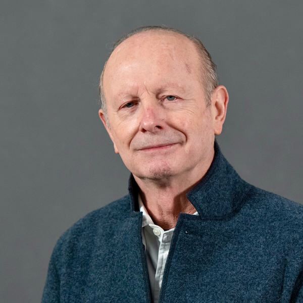 Joseph E. Ledoux合作论文数Department of Child and Adolescent Psychiatry, Grossman School of Medicine, New York University;Department of Psychiatry, Grossman School of Medicine, New York University;Center for Neural Science, New York University25
 Richard Koppenaal合作论文数23
 Barbara Freitas合作论文数23
 Michael Miller合作论文数Department of Psychological and Brain Sciences, University of California23Robert Fendrich合作论文数21
 Scott Grafton合作论文数Department of Psychological & Brain Sciences, College of Letters & Science, University of California, Santa Barbara20Donald H. Wilson合作论文数19
 Margaret Funnell合作论文数18
 Bruce T. Volpe合作论文数Department of Neurology & Neuroscience, Weill Medical College, Cornell University;Center for Biomedical Science, Feinstein Institute for Medical Research;Institute of Molecular Medicine, Feinstein Institutes for Medical Research16C. Mark Wessinger合作论文数15Mark Jude Tramo合作论文数14Kathleen Baynes合作论文数13
 George (Ron) Mangun合作论文数Center for Mind and Brain, University of California, Davis;School of Medicine, University of California, Davis13
 Jack Van Horn合作论文数Department of Bioengineering, School of Engineering, University of California, Los Angeles11John J. Sidtis合作论文数11
 Steven Hillyard合作论文数University of California, San Diego10
 Todd Handy合作论文数Department of Psychology, Faculty of Arts, The University of British Columbia10
 Matt Roser合作论文数Department of Psychological and Brain Sciences, Dartmouth College10William C. Loftus合作论文数10
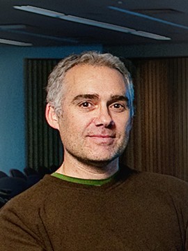 Alan Kingstone合作论文数Brain, Attention and Reality Lab, Department of Psychology, Faculty of Arts, The University of British Columbia9J D Holtzman合作论文数8JJ Hutsler合作论文数8
 Paul Corballis合作论文数School of Psychology, Faculty of Science, The University of Auckland8Cs Smylie合作论文数7A Francis合作论文数7
 David John Turk合作论文数Department of Psychological and Brain Sciences;Center for Cognitive Neuroscience;Dartmouth College;Center for Cognitive Neuroscience, Dartmouth College7TC Handy合作论文数6
 Steve Luck合作论文数Center for Neuroscience, University of California, Davis6Green R L合作论文数6
 Todd Heatherton合作论文数Department of Psychological and Brain Sciences, Dartmouth College6Gail L. Risse合作论文数6
 Kevin Niall Dunbar合作论文数Department of Human Development and Quantitative Methodology, College of Education, University of Maryland;Department of Psychology, University of Toronto;Laboratory for Thinking, Reasoning, Creativity & Educational Neuroscience, College of Education, University of Maryland6Mary K. Colvin合作论文数6
 Elizabeth A. Phelps合作论文数Department of Psychology, Harvard University6
 Jonathan A Fugelsang合作论文数Univ Waterloo6SH Johnson合作论文数5NEA Kroll合作论文数5
 C. Neil Macrae合作论文数School of Psychology, University of Aberdeen5
 Charles Spence合作论文数Department of Experimental Psychology, Medical Sciences Division, University of Oxford5
 Souheil Inati合作论文数Dartmouth College5Sally P. Springer合作论文数5
 Karl W. Doron合作论文数Harold Goodglass Boston University Aphasia Research Center, Boston University School of Medicine and the Veterans Affairs Boston Healthcare System5
 Stephen M Kosslyn合作论文数Department of Psychology, Harvard University;Active Learning Sciences, Inc;Minerva Schools, Keck Graduate Institute5
 George Wolford合作论文数Department of Psychology, Dartmouth College5William M. Kelley合作论文数5
 Jim Eliassen合作论文数5James C. Eliassen合作论文数4
 Walter Sinnott-Armstrong合作论文数Department of Philosophy, Trinity College of Arts & Sciences, Duke University4




