PurposeAdult Growth Hormone Deficiency (AGHD) is a complex and under-recognized condition, mainly managed in outpatient settings and characterized by fragmented and heterogeneous clinical data. Population registries provide high-quality evidence but require long timeframes and substantial resources. The aim of this study was to design and validate an artificial intelligence (AI)-driven methodology for the construction of a disease-specific AGHD Data Mart from routine clinical data within a single high-volume center, to support real-world evidence generation and future advanced analytics.MethodsA standardized Data Science framework, based on automated extraction from hospital data warehouses and electronic medical records, integrating structured data and unstructured clinical narratives through natural language processing, was implemented. AGHD patients were identified using a combination of ICD-9 codes and text-mining applied to outpatient reports. A multidisciplinary workflow ensured clinical validation of extracted data. The Data Mart described patient identification from first hospital access (T0) and included diagnostic modality, etiology, biochemical data, comorbidities, and growth hormone replacement therapy.ResultsAmong 210 identified patients, 188 were validated as AGHD after expert review. Diagnoses were based on dynamic testing (28.2%), panhypopituitarism with low IGF-1 (54.3%), or AI-assisted identification (17.6%). Etiology was retrieved in 87.8% of cases, with post-surgical causes being the most frequent. 37.8% of patients were receiving rhGH therapy. Specific trends for IGF-1 values for single patients were described.ConclusionThis study represents the first AI-driven AGHD Data Mart and demonstrates the feasibility of constructing the Data Mart from routine clinical data. This approach offers a complementary framework for structured RWD extraction and may support future longitudinal analyses and AI-based clinical support in AGHD.
Human follicular development depends on coordinated communication between granulosa cells (GCs) and oocytes through endocrine cues, direct contacts, and extracellular vesicles (EVs). Exosomes are key EV mediators of intrafollicular signaling, but their cargo and functions in gonadotropin-stimulated GCs remain poorly defined. The human granulosa-like tumor cell line KGN was used to investigate exosome secretion and protein composition following human chorionic gonadotropin (hCG) stimulation. Exosomes were isolated by ultracentrifugation, characterized via nanoparticle tracking analysis (NTA), Scanning Electron Microscopy (SEM) and Western blotting, and analyzed using high-resolution mass spectrometry. Comparative proteomics integrating exosomal profiles with the whole secretome were performed, followed by bioinformatic analyses of protein networks, gene ontology, and pathway enrichment. hCG reshaped exosomal cargo, identifying 59 proteins enriched in exosomes, including Integrin α3 (ITGα3), Galectin-3-binding protein (LGALS3BP), tetraspanins (CD63, CD151), and proteasome subunits. Functional enrichment indicated roles in extracellular matrix remodeling, integrin signaling, proteostasis, and steroidogenesis. Comparison with the secretome revealed distinct protein distributions, supporting selective exosomal packaging. Western blot confirmed increased ITGα3 and LGALS3BP levels in exosomes upon hCG treatment. In conclusion, hCG modulates exosome cargo composition in granulosa cells, uncovering a novel mechanism of extracellular regulation.
BACKGROUND:Male accessory gland inflammation (MAGI) comprises a heterogeneous group of inflammatory conditions that may differentially affect the seminal microenvironment and sperm function. However, whether transrectal ultrasound (TRUS)-defined MAGI phenotypes correspond to distinct biological profiles remains insufficiently characterized. OBJECTIVE:To evaluate whether TRUS-defined phenotypes of MAGI can be discriminated using seminal inflammatory and oxidative stress biomarkers and sperm proteomic profiles, and to assess their potential clinical relevance for disease stratification. MATERIALS AND METHODS:In this case-control study, 20 men with MAGI and 10 healthy controls were enrolled. Patients were classified by TRUS as hypertrophic-congestive (acute) or fibro-sclerotic (chronic) phenotypes. Semen analysis, seminal plasma antioxidant capacity, and sperm lipid peroxidation were assessed. Seminal inflammatory biomarkers were quantified and evaluated by receiver operating characteristic analysis. Proteomic profiling of purified spermatozoa was performed with multivariate and differential expression analyses, followed by functional enrichment and immunofluorescence validation. RESULTS:Both MAGI phenotypes showed reduced sperm concentration and motility compared with controls, whereas abnormal morphology was more pronounced in the fibro-sclerotic phenotype. Antioxidant capacity was reduced, and lipid peroxidation increased in both groups, with greater oxidative imbalance in the hypertrophic-congestive phenotype. SuPAR discriminated MAGI patients from controls with high specificity, whereas sST2 accurately differentiated acute and chronic phenotypes. Proteomic analysis identified distinct, nonoverlapping molecular signatures across groups. Pathway analysis showed downregulation of proteins involved in sperm function and fertilization, with additional impairment of motility-related processes in the chronic phenotype. Immunofluorescence confirmed reduced expression of a key flagellar structural protein, most markedly in fibro-sclerotic MAGI. DISCUSSION AND CONCLUSION:TRUS-defined MAGI phenotypes are associated with distinct biomarker and proteomic profiles, consistent with different inflammatory states and patterns of sperm molecular injury. These findings support a biomarker-assisted approach to phenotype stratification and provide mechanistic insight into inflammation-driven male reproductive dysfunction.
Male accessory gland inflammation (MAGI) can impair male fertility through inflammation-driven oxidative stress and direct sperm damage; nutraceutical approaches may be useful when antibiotics are not indicated. Here, we evaluated a 3-month treatment with a Graminex™-based dietary supplement (Deprox-HP) in twenty MAGI patients integrating conventional semen analysis and oxidative stress assessment with sperm proteomics before and after therapy. After treatment, total and progressive sperm motility increased significantly, whereas sperm concentration and sperm morphology showed a non-significant upward trend. Sperm lipid peroxidation decreased markedly, while the antioxidant capacity showed a non-significant increase. Analysis of the sperm proteome demonstrated a clear PRE-POST clustering, consistent with treatment-associated remodeling. POST samples showed upregulation of proteins linked to sperm motility, redox homeostasis, mitochondrial metabolism and membrane remodeling. Two pregnancies occurred during the treatment period; in both cases, lipid peroxidation decreased along with an increase of morphologically typical spermatozoa, and sperm proteomics showed a concordant post-treatment shift enriched in flagellar and mitochondrial respiratory/redox compartments. Moreover, we found a selective enrichment POST treatment in these two patients of TEX50, a crucial protein involved in acrosome/head-stability during epididymal transit. Overall, Deprox-HP was associated with reduced oxidative membrane damage and a coordinated sperm proteomic shift consistent with improved motility.
Bone metabolism is typically impaired in patients with acromegaly due to increased bone turnover, increased bone resorption, and impaired bone neoformation. The pathogenetic mechanisms underlying skeletal fragility in patients with acromegaly remain not fully clarified. We aim to compare the bone proteome of patients with acromegaly to that of a control group of patients with non-secreting pituitary tumors (NSPTs). A Liquid Chromatography-Mass Spectrometry was conducted on ethmoid samples (after processing and digestion of the sample) of five patients with acromegaly and five patients with NSPTs, to identify and assay the proteome. Biological functions were investigated for proteins that were found quantitatively up- and down-regulated in the bone of acromegalic patients, with a ratio of variation based on Fold-Change (FC) >|1.50| and statistical significance (p-value < 0.05). 312 proteins belonging to each group were identified. Six proteins with positive FC (up-regulated) and 12 proteins with negative FC (down-regulated) significantly differ in patients with acromegaly than in patients with NSPTs. Among up- and down-regulated proteins, profilin-1, isoform 5 of the periostin, apolipoprotein E, and caveolin-1 were known to be involved in bone metabolism. In our cohort, a positive correlation was detected between the profilin-1, the isoform 5 of the periostin, GH, and IGF-I levels; and a negative correlation was detected between caveolin-1 and serum GH and IGF-I levels, and with apolipoprotein E and serum IGF-I levels. Our results proved that the bone of patients with acromegaly is characterized by a specific proteomic profile, which is closely correlated to GH and IGF-I hypersecretion.
Background/Objectives: Testicular germ cell tumors, particularly seminoma, represent the leading cause of cancer in men aged 15-40 years. The decision about adjuvant therapy relies on histological features with uncertain prognostic value. The Pituitary-Tumor-Transforming Gene 1 (PTTG1), which encodes the securin protein, is crucial in sister chromatid separation. Our previous in vitro studies demonstrated that PTTG1 nuclear expression promotes invasiveness, dedifferentiation, and neolymphangiogenesis in testicular seminoma. Methods: We conducted a hypothesis-generating retrospective observational study on 51 patients (aged 23-68) with testicular seminoma, with (43%) or without (57%) lymph node involvement, evaluating potential correlations between PTTG1 and currently known prognostic factors. Clinical and pathological data were collected, including lymph node involvement, recurrence, necrosis, rete testis invasion, vascular invasion, and adipose tissue invasion. An immunohistochemical scoring system assessing intranuclear PTTG1 expression was developed. Results: The PTTG1 score was related to lymph node metastasis and adipose tissue invasion. ROC curve analysis showed that the PTTG1 immunohistochemical score showed good discriminatory ability in identifying lymph node involvement (AUC = 0.939); the optimal cut-off was 4.0 (sensitivity 90.5%; specificity 57.1%), while the ROC curve for adipose tissue invasion was inadequate. Lymph node metastasis also correlated with necrosis; however, logistic regression confirmed that PTTG1 score was independently associated with nodal involvement (p = 0.002), regardless of tumor size and necrosis. Conclusions: Our findings suggest a correlation between PTTG1 expression and lymphadenopathy at diagnosis, independent of tumor size and T stage. It may reflect biological features associated with lymphatic dissemination and requires further investigation in larger prospective studies.
Microfluidic platforms have emerged as critical technologies for exploring sperm chemotaxis, providing precise gradient control, and facilitating in-depth behavioral assessment. We designed a novel, user-friendly microfluidic device that is optimized for human sperm morphology and motility. The device was validated using two well-established sperm chemoattractants, progesterone and bourgeonal, demonstrating its reliability and reproducibility. Given the key role of olfactory receptors (ORs) in mediating sperm chemotaxis, the newly developed device was employed to identify additional receptors that may contribute to sperm behavior. Using the Atlas database, we identified OR2H1 as a candidate receptor. It is enriched in testis-derived cells, particularly in early and late spermatids, and it is broadly expressed across human spermatozoa. We demonstrated that OR2H1’s ligand, methional, a sulfur-containing aldehyde naturally found in vaginal fluid and biosynthesized by Lactococcus lactis, significantly enhances sperm migration and progressive motility. Methional stimulation also triggered increased intracellular calcium levels, indicating receptor activation. Computer-assisted sperm analysis revealed that methional treatment improved sperm linearity, straightness, and wobble without affecting the average velocity, suggesting enhanced directional movement. These findings provide evidence that methional promotes sperm chemotaxis via OR2H1 and highlight the potential role of the vaginal microbiome in influencing human fertility.
Background: Varicocele is a common condition involving the dilation of veins in the scrotum, often linked to male infertility and testicular dysfunction. This study aimed to elucidate the molecular effects of successful varicocele treatment on sperm proteomes following percutaneous sclero-embolization. Methods: High-resolution tandem mass spectrometry was performed for proteomic profiling of pooled sperm lysates from five patients exhibiting improved semen parameters before and after (3 and 6 months) varicocele sclero-embolization. Data were validated by Western blot analysis. Results: Seven proteins were found exclusively in varicocele patients before surgery—such as stathmin, IFT20, selenide, and ADAM21—linked to inflammation and oxidative stress. After sclero-embolization, 55 new proteins emerged, including antioxidant enzymes like selenoprotein P and GPX3. Thioredoxin (TXN) and peroxiredoxin (PRDX3) were upregulated, indicating restoration of key antioxidant pathways. Additionally, the downregulation of some histones and the autophagy-related protein ATG9A suggests a shift toward an improved chromatin organization and a healthier cellular environment post-treatment. Conclusions: Varicocele treatment that improves sperm quality and fertility parameters leads to significant proteome modulation. These changes include reduced oxidative stress and broadly restored sperm maturation. Despite the limited patient cohort analyzed, these preliminary findings provide valuable insights into how varicocele treatment might enhance male fertility and suggest potential biomarkers for improved male infertility treatment strategies.
INTRODUCTION:Recurrent pregnancy loss (RPL), defined as two or more consecutive pregnancy losses before 24 weeks of gestation, affects up to 1%-2% of couples. Aim of this retrospective cohort study was to report the main causes and pregnancy outcomes of a cohort of women with RPL and the efficacy of a personalized work-up and treatment in terms of live birth rate. MATERIAL AND METHODS:Women with primary (pRPL) and secondary (sRPL) RPL underwent a complete work-up and personalized therapeutic management. Data related to clinical findings and subsequent pregnancy outcomes were collected. A retrospective comparison between clinical findings and pregnancy outcomes of pRPL vs sRPL was performed by Mann-Whitney U or Chi-square test. RESULTS:Main findings after diagnostic work-up in pRPL (n = 157) vs sRPL (n = 138) couples were hormonal and metabolic factors (75% vs. 90%, p < 0.01), autoimmunity (52% vs. 59%, p = 0.2), acquired uterine/endometrial factors (43% vs. 34%, p = 0.2), vaginal and/or cervical infections (19% vs. 49%; p < 0.0001), congenital Mullerian anomalies (15% vs. 9%; p = 0.1), inherited thrombophilias (13% vs. 21%; p = 0.1), female karyotype abnormalities (2% vs. 2%; p = 0.9), sperm infections (27% vs. 22%; p = 0.1), abnormal semen analysis (17% vs. 14%; p = 0.1), male karyotype abnormalities (2% vs. 0%; p = 0.1). Higher pregnancy and fetal loss rate was observed in pRPL compared with sRPL (85% vs. 56%, p < 0.0001and 9% vs. 0%, p < 0.01, respectively). Higher live birth rate was found in pRLP vs sRPL women (76% vs. 56%, p < 0.001). Increased live birth rate was observed among pRPL women aged <40 years (OR 2.76; CI 1.36-5.64, p < 0.01) and/or with an AMH >1 ng/mL (OR 3.96; CI 1.34-12.52, p < 0.05). Among sRPL women, the age < 40 years was significantly associated to higher live birth rate (OR 3.23; 1.55-6.94, p < 0.01). CONCLUSIONS:RPL is a heterogeneous multifactorial syndrome. A customized management can lead to a good pregnancy outcome in more than a half of cases. Age <40 and AMH >1 ng/mL are the major positive predictors of live birth rate in RPL women.
In the management of medullary thyroid carcinoma (MTC), current prognostic tools have limited discriminatory power, and no widely accepted tissue markers are available aside from proliferative activity and tumour necrosis, recently validated in a grading system. Herein, we assess the prognostic value of selected immunohistochemical biomarkers in MTC: proliferation index Ki-67, insulinoma-associated protein 1 (INSM1), and somatostatin receptor subtype 2A (SSTR2A). We retrospectively analysed 43 patients diagnosed at our centre between October 2003 and July 2024 with histologically confirmed MTC (mean follow-up, 52.5 months). Expression of Ki-67 (> 3
Pituitary tumor-transforming gene 1 (PTTG1), discovered in 1997 by Pei and Melmed, takes part in cellular replication, cell cycle control, DNA repair mechanisms, organogenesis, metabolism regulation, cellular transformation, and senescence. Its biological actions include protein–protein interactions, modulation of gene transcription, and other than intracellular and autocrine mechanisms, even paracrine activities. For the reasons mentioned above, PTTG1 stands out as a multifaceted regulator of cancer biology; it is involved in genomic and chromosomal instability, local invasiveness, neo-lymphangiogenesis, and metastatic spreading. In solid neoplasms, endocrine neoplasms, although deemed rare, have experienced a significant increase in diagnostic incidence in recent years. Endocrine cancers are still a major challenge in healthcare and research since several questions remain unanswered, even though researchers have made considerable efforts to uncover their causes. Twenty-seven years have passed since PTTG1’s discovery, and several works have been published. However, only the tip of the iceberg has been unveiled. Herein, we review current knowledge of PTTG1’s action in endocrine cancers, such as pituitary, thyroid, testicular, adrenal, pancreatic, and ovarian.
BackgroundIn infertile couples whose male partner has alterations in semen parameters frequently, a comprehensive andrological approach is lacking and approximately 30-50% are classified as idiopathic infertility. These couples are often directly addressed to assisted reproduction techniques (ARTs). However, several clinical conditions may benefit from medical treatment. By acting on etiology and/or risk factors, this aims at improving seminal parameters and restoring natural fertility.ObjectivesTo verify the impact of a comprehensive andrological assessment on the management of infertility (in particular, in couples with isolated male factor infertility) using as the primary outcome the natural pregnancy rate.Materials and MethodsA multicenter retrospective study was conducted between 2015 and 2022 in 1014 couples with primary infertility seeking natural conception (including 266 couples with previous ART failure). Each couple underwent a multidisciplinary evaluation. This involved: a gynecologist and an andrologist both with expertise in infertility, a psychologist when requested, and a fertility awareness practitioner according to a unique diagnostic and therapeutic multidisciplinary protocol.ResultsAn isolated male factor was found in 23% of couples. In 45%, it was associated with female factors also. The comprehensive diagnostic approach reduced the proportion of idiopathic infertility to 8% of the couples. Targeted treatment, based on diagnostic categories, was associated with spontaneous pregnancy in 40.9% of the couples. In the 233 cases without female factors, normal semen parameters were observed only in 13% of patients. Male genital tract inflammation was observed in 48.8% of the patients, genital tract infection in 43.1%, and hypospermatogenesis in 16.7%. Patients with infections were treated with antibiotics and probiotics. If further inflammation was documented, this was followed by low-dose corticosteroids and antioxidants. Follicle stimulating hormone (FSH) treatment was used in patients with hypospermatogenesis, and varicocele repair surgery was performed in four patients.Discussion and ConclusionsOur data underline the efficacy of a comprehensive approach to the diagnostic process of male factor infertility, both in reducing the percentage of idiopathic infertility and in restoring natural fertility based on a targeted treatment.
Granulosa cell (GC) differentiation, stimulated by FSH and LH, drives oocyte maturation and follicle development. FSH promotes GC proliferation, and LH triggers ovulation. In clinical practice, hCG is used to mimic LH. Despite various controlled ovarian stimulation (COS) protocols employing exogenous gonadotropins and GnRH analogs to prevent premature ovulation, their effectiveness and safety remain debated. To identify markers predicting a positive treatment response, the secretome of gonadotropin-stimulated GC using the human granulosa-like tumor cell line (KGN) via proteomics was analyzed. Additionally, a novel 2D-FFT quantitative method was employed to assess cytoskeleton fiber aggregation and polymerization, which are critical processes for GC differentiation. Furthermore, the activation of key kinases, focal adhesion kinase (FAK), and Rho-associated coiled-coil-containing protein kinase 1 (ROCK-1), which are implicated in cytoskeleton dynamics and hormone signaling, was evaluated. The proteomic analysis revealed significant modulation of proteins involved in extracellular matrix organization, steroidogenesis, and cytoskeleton remodeling. Notably, the combined FSH/hCG treatment led to a dynamic upregulation of the semaphorin pathway, specifically semaphorin 7A. Finally, a significant reorganization of the cytoskeleton network and signaling was detected. These findings enhance our understanding of folliculogenesis and suggest potential novel molecular markers for predicting patient responses to gonadotropin stimulation.
Hyperandrogenism is a condition in which the levels of androgen hormones in the blood are significantly increased and could be of an adrenal or ovarian origin. The adrenal androgens, normally secreted by the zona reticularis, are steroid hormones with weak androgen activity. The causes of hyperandrogenism are diverse and could be endogenous and exogenous. Androgen excess affecting different tissues and organs results in clinical features such as acne, hirsutism, virilization, and reproductive dysfunction such as oligomenorrhoea/amenorrhoea. Although androgen excess is rarely associated with adrenal tumours, it is important as it could be predictive of malignancy. A careful evaluation of the androgen pattern, also in patients with clear signs of hyperandrogenism, could be useful. Laboratory evaluation should focus on measuring total testosterone levels, followed by the estimation of other androgens such as dehydroepiandrosterone and androstenedione, and using visualisation procedures in the further management. The treatment of adrenal hyperandrogenism is eminently surgical, in consideration of the frequent malignant origin. The aim of this review is to elaborate and summarize the prevalence and clinical management of hyperandrogenism of an adrenal origin by describing the physiological mechanisms of adrenal androgen steroidogenesis, the clinical manifestations of hyperandrogenism with a special reference to hyperandrogenism in adrenal adenomas and carcinomas, and the diagnostic methods that will lead us to establishing the correct diagnosis and different treatment options to manage this condition according to the clinical presentation of the patient.
Several genetic investigations were conducted to identify germline and somatic mutations in somatotropinomas, a subtype of pituitary tumors. To our knowledge, we report the first acromegaly patient carrying a RET pathogenic variant: c.2410G>A (rs79658334), p.Val804Met. Alongside the fact that the patient’s father and daughter carried the same variant, we investigated the clinical significance of this variant in the context of somatotropinomas and other endocrine tumors, reviewing the RET mutations’ oncogenic mechanisms. The aim was to search for new targets to precisely manage and treat acromegaly. Our case describes a new phenotype associated with the RET pathogenic variant, represented by aggressive acromegaly, and suggests consideration for RET mutation screening if NGS for well-established PitNET-associated gene mutations renders negative.
BackgroundIt is reported that treatment with anaplastic lymphoma kinase (ALK) tyrosine kinase inhibitors (TKIs) induces hypogonadism both in male patients with ALK-positive cancer and in murine models.MethodsIn this study, three groups, including an experimental group of male patients with ALK-positive, advanced nonsmall cell lung cancer (ANSCLC) who were receiving alectinib (cohort A), a control group of female patients with ALK-positive ANSCLC who were receiving alectinib (cohort B), and a control group of male patients with ALK-negative ANSCLC (cohort C), prospectively underwent a full hormone assessment for androgen deficiency at 8 weeks after the start of treatment and in case of reported suspected symptoms. Patients with major sexual dysfunctions were referred to an endocrinologist.ResultsNinety-five patients were consecutively enrolled onto the study. Among sixty-eight male patients, both median total testosterone levels (2.93 vs. 4.92 ng/ml; p = .0001) and free testosterone levels (0.11 vs. 0.17 pg/ml; p = .0002) were significantly lower in ALK-positive ANSCLC patients in cohort A compared with ALK-negative patients in cohort C; conversely, median FSH (10.32 vs. 17.52 mUI/ml; p = .0059) and LH levels (4.72 vs. 7.49 mUI/ml; p = .0131) were significantly higher in cohort C compared to cohort A. Median inhibin B levels were higher in ALK-positive male patients (74.3 vs. 44.24 pg/ml; p = .0038), but all patients had inhibin B values within the normal range. The percentage of male patients who had positive scores on the Androgen Deficiency in Aging Males (ADAM) questionnaire was 62% in cohort A and 26.8% in cohort C, including eight patients who reported at least one major symptom and were referred to Andrology Unit. No significant differences in the endocrine assessment were reported between cohorts A and B.ConclusionsSymptoms of androgen deficiency should be tracked in male patients with ALK-positive ANSCLC who are receiving alectinib, and testosterone replacement should be considered, as appropriate. Male patients with ALK-positive advanced NSCLC treated with first-line alectinib had lower median total testosterone (p = .0001) and free testosterone (p = .0002) levels compared with male patients who had ALK-negative disease and were receiving other anticancer treatments. Symptoms of hypogonadism were more frequent in ALK-positive patients compared with ALK-negative patients (62% vs 26.8% rates of positive score at ADAM questionnaire, respectively), suggesting the need for hormone assessment and referral to an andrologist, as appropriate.
Seminoma is the most common testicular cancer. Pituitary tumor-transforming gene 1 (PTTG1) is a securin showing oncogenic activity in several tumors. We previously demonstrated that nuclear PTTG1 promotes seminoma tumor invasion through its transcriptional activity on matrix metalloproteinase 2 (MMP-2) and E-cadherin (CDH1). We wondered if specific interactors could affect its subcellular distribution. To this aim, we investigated the PTTG1 interactome in seminoma cell lines showing different PTTG1 nuclear levels correlated with invasive properties. A proteomic approach upon PTTG1 immunoprecipitation uncovered new specific securin interactors. Western blot, confocal microscopy, cytoplasmic/nuclear fractionation, sphere-forming assay, and Atlas database interrogation were performed to validate the proteomic results and to investigate the interplay between PTTG1 and newly uncovered partners. We observed that spectrin beta-chain (SPTBN1) and PTTG1 were cofactors, with SPTBN1 anchoring the securin in the cytoplasm. SPTBN1 downregulation determined PTTG1 nuclear translocation, promoting its invasive capability. Moreover, a PTTG1 deletion mutant lacking SPTBN1 binding was strongly localized in the nucleus. The Atlas database revealed that seminomas that contained higher nuclear PTTG1 levels showed significantly lower SPTBN1 levels in comparison to non-seminomas. In human seminoma specimens, we found a strong PTTG1/SPTBN1 colocalization that decreases in areas with nuclear PTTG1 distribution. Overall, these results suggest that SPTBN1, along with PTTG1, is a potential prognostic factor useful in the clinical management of seminoma.
BACKGROUND:The presurgical evaluation of cervical lymph nodes (CLNs) in the management of thyroid malignant lesions is crucial for the extent of surgery or detection of metastases. In these last decades, fine-needle aspiration cytology (FNAC) has been shown to have a central role in the detection of nodal thyroid metastases. It is adopted for the possibility of confirming suspected metastases either in the presurgical phase or in the follow-up of patients after thyroidectomy. However, FNAC from CLNs can be challenging, especially in cystic lesions. In this regard, the combination of FNAC with thyroglobulin measurement in the eluate from FNAC (Tg-FNAC) seems to increase the sensitivity of FNAC in the detection of CLN metastases. The role of FNAC and Tg-FNAC was investigated in this series.METHODS:One hundred fifty-three prospective cytological samples of CLNs were studied along with surgical follow-up in the period between 2020 and 2022. Immunocytochemistry (ICC) was performed on liquid-based cytology-stored material.RESULTS:One hundred fifty-nine enlarged CLNs included 19 central lymph nodes and 140 CLNs. Forty-two thyroidal CLN metastases and 117 reactive lymph nodes were found. Thirty-one CLN dissections were performed in patients with a previous diagnosis of thyroid carcinoma (mostly papillary thyroid carcinoma [PTC]), whereas 128 CLNs with a concomitant suspicious and/or malignant thyroid nodule were found. There was one false-positive case among all the malignant histologically confirmed cases, and two of 117 reactive CLNs (1.7%) had a diagnosis of metastatic PTC. Markedly high Tg-FNAC was found in all metastatic CLNs, including 11 cystic metastatic CLNs detected by Tg-FNAC with a negative FNAC. ICC (including Tg, CK-19, and LCA) recognized nine cases with low Tg-FNAC and scant suspicious thyrocytes. Tg-FNAC plus FNAC diagnosed 94.2% of malignancies.CONCLUSIONS:FNAC represents a valid method for the evaluation of CLNs, especially combined with ICC. Tg-FNAC is an additional method with a useful role in FNAC.
Although precision medicine took its first steps from genomic medicine, it has gone far beyond genomics, considering the full complexity of cellular physiology. Therefore, the present time can be considered as the "post-genomic era". In detail, proteomics captures the overall protein profile of an analyzed sample, whilst metabolomics has the purpose of studying the molecular aspects of a known medical condition through the measurement of metabolites with low molecular weight in biological specimens. In this review, the role of post-genomic platforms, namely proteomics and metabolomics, is evaluated with a specific interest in their application for the identification of novel biomarkers in male hypogonadism and in the identification of new perspectives of knowledge on the pathophysiological function of testosterone. Post-genomic platforms, including MS-based proteomics and metabolomics based on ultra-high-performance liquid chromatography-HRMS, have been applied to find solutions to clinical questions related to the diagnosis and treatment of male hypogonadism. In detail, seminal proteomics helped us in identifying novel non-invasive markers of androgen activity to be translated into clinical practice, sperm proteomics revealed the role of testosterone in spermatogenesis, while serum metabolomics helped identify the different metabolic pathways associated with testosterone deficiency and replacement treatment, both in patients with insulin sensitivity and patients with insulin resistance.
BACKGROUND:Sperm DNA fragmentation was hypothesized to have a role in the pathogenesis of recurrent pregnancy loss. Unfortunately, the quality of already published evidence is low.OBJECTIVES:To investigate the association between sperm DNA fragmentation and idiopathic recurrent pregnancy loss by limiting, as much as possible, the interference of confounding factors.MATERIALS AND METHODS:This was a retrospective multicenter case-control study conducted in two Italian University Hospitals (i.e., Policlinico Gemelli, Rome and Humanitas S. Pio X, Milan) from July 2020 to March 2022. Cases were men belonging to couples affected by first trimester idiopathic recurrent pregnancy loss, defined as the previous loss of two or more pregnancies. Two control groups were selected: (i) men belonging to couples with proven fertility (i.e., at least two previous full-term pregnancies) (control group A); (ii) men belonging to couples with proven infertility (i.e., the failure to achieve a pregnancy after 12 months or more of regular unprotected sexual intercourse) (control group B). The sperm DNA fragmentation index was measured by the terminal deoxynucleotidyl transferase dUTP nick end labeling assay.RESULTS:We included 74 cases, 37 men with proven fertility (control group A) and 100 men belonging to infertile couples (control group B). The median sperm DNA fragmentation index was significantly lower in control group A (17%, interquartile range: 14.3%-20.6%) compared to both case group (24.5%, interquartile range: 17%-32%; p < 0.0001) and control group B (24%, interquartile range: 18.9%-30%; p = 0.001). The rate of subjects with sperm DNA fragmentation index greater than 30% was significantly higher in both case groups (28%, 95% confidence interval [18%-40%]) and control group B (26%, 95% confidence interval [18%, 36%]) compared to control group A (0%, 95% confidence interval [0%-10%]) (p < 0.001). Multivariate regression models yielded a significant association between sperm DNA fragmentation index and recurrent pregnancy loss (adjusted odds ratio 1.13, 95% confidence interval [1.04-1.23], p = 0.006), but failed to show an association between sperm DNA fragmentation index and infertility (adjusted odds ratio 1.13, 95% CI [1-1.29], p = 0.05).CONCLUSIONS:Men within couples affected by recurrent pregnancy loss or infertility had a significantly higher rate of sperm DNA fragmentation compared to fertile controls. However, after adjusting for covariates, sperm DNA fragmentation index was associated only with recurrent pregnancy loss.
 Laura De Marinis合作论文数Department of Endocrinology, Università Cattolica del Sacro Cuore76
 Alfredo Pontecorvi合作论文数61
 Giuseppe Grande合作论文数45Antonella Giampietro合作论文数41A. Bianchi合作论文数34A Mancini合作论文数31Antonio Bianchi合作论文数Dipartimento Medicina Chirurgia Traslazionale, Univ Cattolica Sacro Cuore23Vincenzoni Federica合作论文数22
 Francesca Mancini合作论文数20Sabrina Chiloiro合作论文数19Riccardo Marana合作论文数19G. Grande合作论文数17Francesco Pierconti合作论文数12Andrea Urbani合作论文数11
 Massimo Castagnola合作论文数Laboratorio di Proteomica e Metabonomica, Fondazione Santa Lucia Istituto di Ricovero e Cura a Carattere Scientifico;Istituto di Chimica del Riconoscimento Molecolare, Consiglio Nazionale delle Ricerche11E. Giacchi合作论文数10Linda Tartaglione合作论文数10
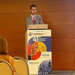 Edoardo Vergani合作论文数Università Cattolica del Sacro Cuore10Emanuela Teveroni合作论文数10Elisabetta Meucci合作论文数10Marilda Mormando合作论文数9
 Guido Rindi合作论文数Dipartimento di Scienze della vita e Sanità Pubblica, Faculty of Medicine and Surgery, Università Cattolica del Sacro Cuore9A. Fusco合作论文数8
 Gian Paolo Littarru合作论文数8Giovanni Luca合作论文数8Silvia Baroni合作论文数Istituto di Chimica e Chimica Clinica, Policlinico A. Gemelli8
 Maurizio Martini合作论文数7Anna Laura Astorri合作论文数7Ge Martorana合作论文数7G Conte合作论文数7Di Nicuolo Fiorella合作论文数7Gaetano Gulino合作论文数7
 Andrea Garolla合作论文数7Giovanni Schinzari合作论文数Endoscopy Unit, Department of Internal Medicine7Riccardo Calafiore合作论文数7
 Michele Perrelli合作论文数7
 Mordente Alvaro合作论文数Institute of Biochemistry, University of Ancona Medical School7A L Astorri合作论文数6R Marana合作论文数6Giuseppina Pompa合作论文数6D Valle合作论文数6
 Ettore Domenico Capoluongo合作论文数Dipartimento di Medicina Molecolare e Biotecnologie Mediche,Faculty of Medicine, Università degli Studi di Napoli Federico II;Federico II University and Federico II Teaching Hospital6
 Irene Messana合作论文数Istituto di Chimica, Universitá Cattolica del Sacro Cuore6
 Giuseppe Maulucci合作论文数Universitá Cattolica S. Cuore6C Vezzosi合作论文数5G Pompa合作论文数5R M Tacchino合作论文数5




