Objective This study aims to evaluate the relationships between the anterior ethmoidal artery (AEA) notch landmark, Keros classification, and the presence and volume of supraorbital ethmoidal cells (SOECs) to determine their anatomical correlations and surgical implications. Materials and Methods A retrospective analysis of 102 paranasal sinus CT scans (204 sides) was performed. The AEA notch landmark was classified into three degrees based on its vertical distance from the skull base. The presence and volume of SOECs were recorded, and Keros classification was assessed. Correlations among these parameters were analyzed using Spearman's rank correlation coefficient, Chi-square, and Fisher's exact tests. Results SOECs were identified in 39.22% of cases, with a mean volume of 0.3535 +/- 0.109 cm(3) on the right side and 0.3399 +/- 0.054 cm(3) on the left. A significant correlation was observed between Keros classification and the AEA notch landmark (Spearman's rho = 0.637, p < 0.05). Additionally, SOEC volume showed a weak but significant correlation with the AEA notch landmark (Spearman's rho = 0.161, p < 0.05). However, there was no significant correlation between SOEC volume and Keros classification (p > 0.05). Conclusion AEA positioning is significantly influenced by both its distance from the skull base and SOEC volume, though SOEC volume does not affect Keros classification. These insights contribute to a better understanding of endoscopic sinus surgery anatomy and may help minimize surgical risks.
Inflammatory and infectious conditions of the larynx and hypopharynx have diverse etiologies, some of them rather uncommon, making clinical diagnosis challenging. Imaging plays a crucial role in assessing the characteristics, extent, and patterns of involvement of the disease, thereby contributing to the establishment of an accurate diagnosis. In acute infections with severe airway involvement, rapid and accurate treatment is imperative before further investigation. This review covers a wide range of pathologies, demonstrating their CT and/or MR findings. Knowledge of these differential possibilities will aid radiologists in accurately interpreting imaging studies.Learning Objective: To recognize clinical aspects and imaging findings of inflammatory and infectious diseases of the larynx and hypopharynx, providing support to differentiate them from neoplastic processes.
It is with great pleasure that we introduce to our readership Dr Camilo Jaimes and Dr Jungwhan del Rosario in Bogota, Colombia. Upon graduation, Dr Jaimes completed Diagnostic Radiology residency and Neuroradiology fellowship at Massachusetts General Hospital (MGH)-Harvard Medical School, followed by a fellowship in Pediatric Neuroradiology at Boston Children's Hospital where he started his brilliant career in 2019. Dr Camilo Jaimes served as Director of Faculty Affairs, Associate Fellowship Program Director, and Associate Chief of MRI. In his current role at MGH, Dr Jaimes is the Chief of Pediatric Neuroradiology, Co-Director of the Pediatric Imaging
 F. Allan Midyett合作论文数40
 Mohannad Ibrahim合作论文数Division of Neuroradiology, Department of Radiology, Medical School, University of Michigan18
 Gaurang Shah合作论文数Department of Radiology, University of Michigan16
 Ashok Srinivasan合作论文数Department of Radiology, Medical School, University of Michigan14
 Hemant Parmar合作论文数Division of Neuroradiology, Department of Radiology, Medical School, University of Michigan14
 Dheeraj Gandhi合作论文数Neurology (DG),, Johns Hopkins Hospital13
 Thomas Chenevert合作论文数Department of Radiology, Medical School, University of Michigan;Michigan Institute for Data & AI in Society, University of Michigan;Rogel Cancer Center, University of Michigan12
 Sachin K. Gujar合作论文数11Mauricio Castillo合作论文数10
 Avraham Eisbruch合作论文数Department of Radiation Oncology, University of Michigan10Bradford Moffat合作论文数Department of Radiology, Melbourne Medical School, Faculty of Medicine, Dentistry and Health Sciences, The University of Melbourne8Abdel Razek Ahmed Abdel Khalek合作论文数8
 Alnawaz Rehemtulla合作论文数Alnawaz Rehemtulla Laboratory, Department of Radiation Oncology, Medical School, University of Michigan;Department of Radiology, Medical School, University of Michigan7
 Charles Meyer合作论文数Medical School, University of Michigan;College of Engineering, University of Michigan7
 Brian Ross合作论文数Department of Biological Chemistry, Medical School, University of Michigan6
 Theodore S Lawrence合作论文数Department of Radiation Oncology, Medical School, University of Michigan6
 Theodoros N. Teknos合作论文数Case Comprehensive Cancer Center, Case Western University;Department of Otolaryngology, School of Medicine, Case Western University;University Hospitals Seidman Cancer Center6
 Douglas Chepeha合作论文数Institute of Medical Science, Temerty Faculty of Medicine, University of Toronto;Mount Sinai Hospita5
 Rajan Jain合作论文数Department of Neurosurgery, Grossman School of Medicine, New York University5
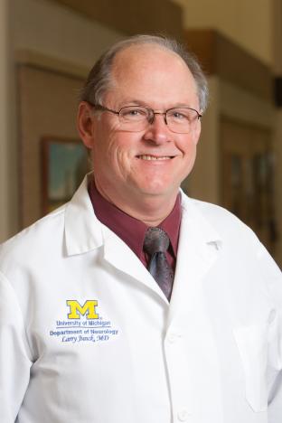 Larry Junck合作论文数Department of Neurology, Medical School, University of Michigan5
 Timothy D. Johnson合作论文数Department of Biostatistics, School of Public Health, University of Michigan-Ann Arbor5
 Gregory T. Wolf合作论文数Department of Otolaryngology-Head and Neck Surgery, University of Michigan Medical Center5Jenny T. Bencardino合作论文数4
 Douglas Quint合作论文数4
 Ellen Hoeffner合作论文数Department of Radiology, Medical School, University of Michigan;University of Michigan Frankel Cardiovascular Center4
 Lubomir Hadjiyski合作论文数Department of Radiology, Medical School, University of Michigan, Ann Arbor;Rogel Cancer Center, University of Michigan, Ann Arbor4
 Patricia Lou Robertson合作论文数Department of Pediatrics, Medical School, University of Michigan;Department of Neurology, Medical School, University of Michigan;Pediatric Neurology Clinic, C. S. Mott Children's Hospital;Pediatric Neurosurgery Clinic, C. S. Mott Children's Hospital4
 Jay S. Cooper合作论文数Maimonides Medical Center4
 Carol R. Bradford合作论文数Department of Otolaryngology - Head and Neck Surgery, Michigan Medicine, University of Michigan4Stephen S. Gebarski合作论文数4
 John A. Ridge合作论文数Temple University Lewis Katz School of Medicine;Fox Chase Cancer Center4
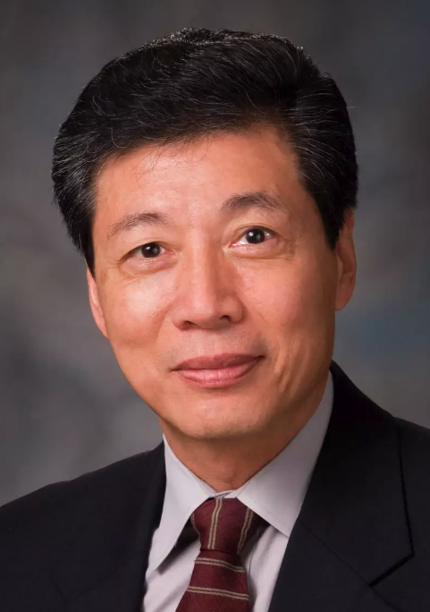 Kian Ang合作论文数Department of Radiation Oncology, The University of Texas M. D. Anderson Cancer Center4
 Heang-Ping Chan合作论文数Computer-Aided Diagnosis-Artificial Intelligence Research Laboratory, Department of Radiology, Medical School, University of Michigan4
 Berkman Sahiner合作论文数US Food and Drug Administration;Advanced Research Projects Agency for Health4
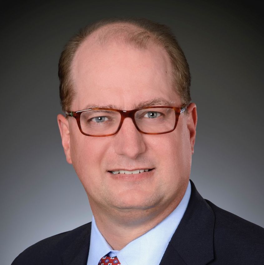 Daniel Allan Hamstra合作论文数Baylor College of Medicine4FA Pameijer合作论文数3I Case合作论文数3
 Qian Dong合作论文数Department of Radiology, Medical School, University of Michigan3
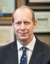 Mark E. P. Prince合作论文数Department of Otolaryngology-Head and Neck Surgery, Medical School, University of Michigan;University of Michigan Medical Center3
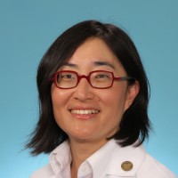 Christina Tsien合作论文数Washington University in St. Louis3
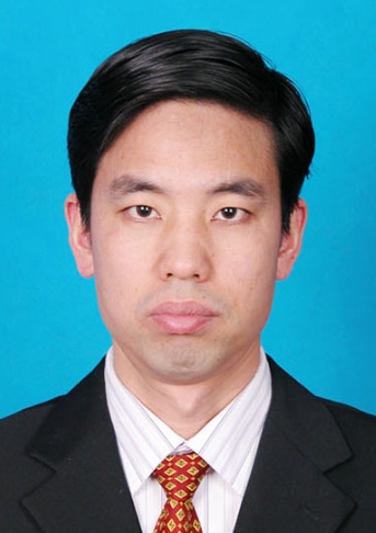 Junfang Xian (鲜军舫)合作论文数Beijing Tongren Hospital, Capital Medical University;Graduate School of Capital Medical University3
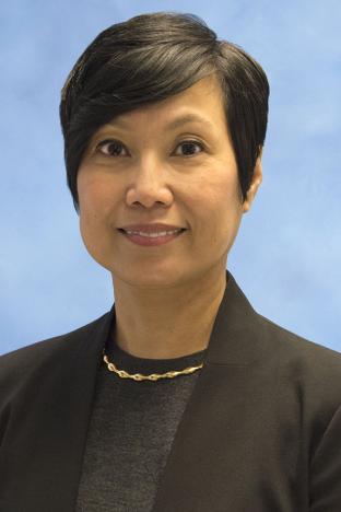 Ruth Carlos合作论文数Department of Radiology, Division of Abdominal Radiology, University of Michigan3Joshi Varsha M合作论文数Department of Radiology, Apollo Hospital3
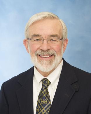 Thomas E. Carey合作论文数Department of Otolaryngology-Head and Neck Surgery, Medical School, University of Michigan;Kresge Hearing Research Institute, Medical School, University of Michigan;Department of Pharmacology, Medical School, University of Michigan3
 William M. Mendenhall合作论文数Department of Radiation Oncology, College of Medicine, University of Florida3
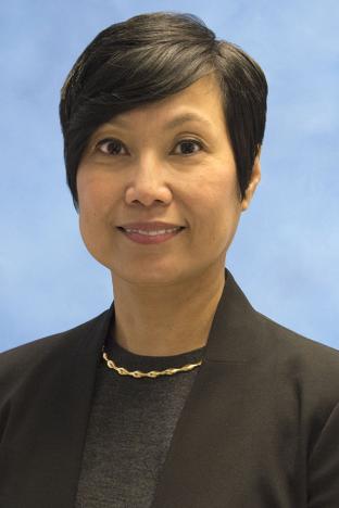 Ruth C. Carlos合作论文数Department of Radiology and the Program for Imaging Comparative Effectiveness and Health Services Research, University of Michigan3
 Anthony A. Mancuso合作论文数Department of radiology, College of Medicine, University of Florida3Robert Hermans合作论文数3Vincent F.H Chong合作论文数3




