Purpose: We investigated the results of endoscopic fenestration for deeply located intracranial cysts (DLICs), risk factors for reoperation, and symptom improvement. Materials and Methods: We included 51 patients with DLICs who underwent endoscopic fenestration between November 2006 and October 2022. The median age was 5 +/- 20 years (6 days-67 years), and 36 (70.6%) patients were aged <20 years. The male-tofemale ratio was 1.3:1. The ventriculoscope was used to fenestrate the cysts, which had diameters under 4.5 mm. The volume of DLICs was measured separately on serial magnetic resonance imaging, and the patients were followed up for 32 +/- 40 months. Results: The mean preoperative volume of DLICs was 63.5 +/- 87.4 cm(3), which decreased to 23.7 +/- 56.2 cm3 postoperatively, with a 45.4%+/- 32.1% decrease rate in 32 months. All DLICs were approached appropriately, avoiding the eloquent areas. Overall, 39 (76.5%) patients showed symptom improvement after a single operation, which was preserved without recurrence, whereas 12 (23.5%) underwent a second operation [shunting (17.6%) or repeating the endoscopic fenestration (5.9%)] owing to symptom aggravation and recurrent cysts. Patients aged <12 months showed 7.4 times more re-operation rate (p=0.046) and 7.4 times less symptom improvement (p=0.038) compared to those with older age. Females showed 6.5 times more re-operation rate (p=0.037) and 7.1 times less symptom improvement (p=0.027) than males. No patients experienced complications such as cerebrospinal fluid leakage, postoperative hemorrhage, or infection. Conclusion: Endoscopic surgery is feasible for the treatment of DLICs. Female sex and age <12 months are risk factors for re-operation and less symptom improvement.
This paper addresses the gaps in understanding green regional path development and the role of firm-level agency in driving regional green transitions. By analysing a large patent dataset covering 30 years, we provide a systematic account of green regional path development in 70 regions across four Nordic countries. We identify six types of green path development - extension, stagnation, extinction, diversification, renewal and creation - and explore how different types of organisations - incumbents and new entrants - contribute to these paths. We show that regions often have multiple green paths, and the dominant types are mostly driven by incumbents.
Purpose: To develop an artificial intelligence (AI) model for the diagnosis of breast cancer on digital breast tomosynthesis (DBT) images and to investigate whether it could improve diagnostic accuracy and reduce radiologist reading time. Materials and Methods: A deep learning AI algorithm was developed and validated for DBT with retrospectively collected examinations (January 2010 to December 2021) from 14 institutions in the United States and South Korea. A multicenter reader study was performed to compare the performance of 15 radiologists (seven breast specialists, eight general radiologists) in interpreting DBT examinations in 258 women (mean age, 56 years +/- 13.41 [SD]), including 65 cancer cases, with and without the use of AI. Area under the receiver operating characteristic curve (AUC), sensitivity, specificity, and reading time were evaluated. Results: The AUC for stand-alone AI performance was 0.93 (95% CI: 0.92, 0.94). With AI, radiologists' AUC improved from 0.90 (95% CI: 0.86, 0.93) to 0.92 (95% CI: 0.88, 0.96) (P = .003) in the reader study. AI showed higher specificity (89.64% [95% CI: 85.34%, 93.94%]) than radiologists (77.34% [95% CI: 75.82%, 78.87%]) (P < .001). When reading with AI, radiologists' sensitivity increased from 85.44% (95% CI: 83.22%, 87.65%) to 87.69% (95% CI: 85.63%, 89.75%) (P = .04), with no evidence of a difference in specificity. Reading time decreased from 54.41 seconds (95% CI: 52.56, 56.27) without AI to 48.52 seconds (95% CI: 46.79, 50.25) with AI (P < .001). Interreader agreement measured by Fleiss kappa increased from 0.59 to 0.62. Conclusion: The AI model showed better diagnostic accuracy than radiologists in breast cancer detection, as well as reduced reading times. The concurrent use of AI in DBT interpretation could improve both accuracy and efficiency.
Abstract Background Artificial intelligence (AI) algorithms for the independent assessment of screening mammograms have not been well established in a large screening cohort of Asian women. We compared the performance of screening digital mammography considering breast density, between radiologists and AI standalone detection among Korean women. Methods We retrospectively included 89,855 Korean women who underwent their initial screening digital mammography from 2009 to 2020. Breast cancer within 12 months of the screening mammography was the reference standard, according to the National Cancer Registry. Lunit software was used to determine the probability of malignancy scores, with a cutoff of 10% for breast cancer detection. The AI’s performance was compared with that of the final Breast Imaging Reporting and Data System category, as recorded by breast radiologists. Breast density was classified into four categories (A–D) based on the radiologist and AI-based assessments. The performance metrics (cancer detection rate [CDR], sensitivity, specificity, positive predictive value [PPV], recall rate, and area under the receiver operating characteristic curve [AUC]) were compared across breast density categories. Results Mean participant age was 43.5 ± 8.7 years; 143 breast cancer cases were identified within 12 months. The CDRs (1.1/1000 examination) and sensitivity values showed no significant differences between radiologist and AI-based results (69.9% [95% confidence interval [CI], 61.7–77.3] vs. 67.1% [95% CI, 58.8–74.8]). However, the AI algorithm showed better specificity (93.0% [95% CI, 92.9–93.2] vs. 77.6% [95% CI, 61.7–77.9]), PPV (1.5% [95% CI, 1.2–1.9] vs. 0.5% [95% CI, 0.4–0.6]), recall rate (7.1% [95% CI, 6.9–7.2] vs. 22.5% [95% CI, 22.2–22.7]), and AUC values (0.8 [95% CI, 0.76–0.84] vs. 0.74 [95% CI, 0.7–0.78]) (all P < 0.05). Radiologist and AI-based results showed the best performance in the non-dense category; the CDR and sensitivity were higher for radiologists in the heterogeneously dense category (P = 0.059). However, the specificity, PPV, and recall rate consistently favored AI-based results across all categories, including the extremely dense category. Conclusions AI-based software showed slightly lower sensitivity, although the difference was not statistically significant. However, it outperformed radiologists in recall rate, specificity, PPV, and AUC, with disparities most prominent in extremely dense breast tissue.
Abstract Background Remimazolam, a newer benzodiazepine that targets the GABAA receptor, is thought to allow more stable blood pressure management during anesthesia induction. In contrast, propofol is associated with vasodilatory effects and an increased risk of hypotension, particularly in patients with comorbidities. This study aimed to identify medications that can maintain stable vital signs throughout the induction phase. Methods We conducted a single-center, two-group, randomized controlled trial to investigate and compare the incidence of hypotension between remimazolam- and propofol-based total intravenous anesthesia (TIVA). We selected patients aged between 19 and 75 years scheduled for neurosurgery under general anesthesia, who were classified as American Society of Anesthesiologists Physical Status I–III and had a history of hypertension. Results We included 94 patients in the final analysis. The incidence of hypotension was higher in the propofol group (91.3%) than in the remimazolam group (85.4%; P = 0.057). There was no significant difference in the incidence of hypotension among the various antihypertensive medications despite the majority of patients being on multiple medications. In comparison with the propofol group, the remimazolam group demonstrated a higher heart rate immediately after intubation. Conclusions Our study indicated that the hypotension incidence of remimazolam-based TIVA was comparable to that of propofol-based TIVA throughout the induction phase of EEG-guided anesthesia. Both remimazolam and propofol may be equally suitable for general anesthesia in patients undergoing neurosurgery. Trial registration Clinicaltrials.gov (NCT05164146).
The purposes of this study were to develop an artificial intelligence (AI) model for future breast cancer risk prediction based on mammographic images, investigate the feasibility of the AI model, and compare the AI model, clinical statistical risk models, and Mirai, a state of-the art deep learning algorithm based on screening mammograms for 1–5-year breast cancer risk prediction. We trained and developed a deep learning model using a total of 36,995 serial mammographic examinations from 21,438 women (cancer-enriched mammograms, 17.5%). To determine the feasibility of the AI prediction model, mammograms and detailed clinical information were collected. C-indices and area under the receiver operating characteristic curves (AUCs) for 1–5-year outcomes were obtained. We compared the AUCs of our AI prediction model, Mirai, and clinical statistical risk models, including the Tyrer–Cuzick (TC) model and Gail model, using DeLong’s test. A total of 16,894 mammograms were independently collected for external validation, of which 4002 were followed by a cancer diagnosis within 5 years. Our AI prediction model obtained a C-index of 0.76, with AUCs of 0.90, 0.84, 0.81, 0.78, and 0.81, to predict the 1–5-year risks. Our AI prediction model showed significantly higher AUCs than those of the TC model (AUC: 0.57; p < 0.001) and Gail model (AUC: 0.52; p < 0.001), and achieved similar performance to Mirai. The deep learning AI model using mammograms and AI-powered imaging biomarkers has substantial potential to advance accurate breast cancer risk prediction.
To examine the discrepancy in breast density assessments by radiologists, LIBRA software, and AI algorithm and their association with breast cancer risk. Among 74,610 Korean women aged ≥ 34 years, who underwent screening mammography, density estimates obtained from both LIBRA and the AI algorithm were compared to radiologists using BI-RADS density categories (A–D, designating C and D as dense breasts). The breast cancer risks were compared according to concordant or discordant dense breasts identified by radiologists, LIBRA, and AI. Cox-proportional hazards models were used to determine adjusted hazard ratios (aHRs) [95
PURPOSE:The Korean Society of Pediatric Neuro-Oncology (KSPNO) conducted treatment strategies for children with medulloblastoma (MB) by using alkylating agents for maintenance chemotherapy or tandem high-dose chemotherapy (HDC) with autologous stem cell rescue (ASCR) according to the risk stratification. The purpose of the study was to assess treatment outcomes and complications based on risk-adapted treatment and HDC.MATERIALS AND METHODS:Fifty-nine patients diagnosed with MB were enrolled in this study. Patients in the standard-risk (SR) group received radiotherapy (RT) after surgery and chemotherapy using the KSPNO M051 regimen. Patients in the high-risk (HR) group received two and four chemotherapy cycles according to the KSPNO S081 protocol before and after reduced RT for age following surgery and two cycles of tandem HDC with ASCR consolidation treatment.RESULTS:In the SR group, 24 patients showed 5-year event-free survival (EFS) and overall survival (OS) estimates of 86.7% (95% confidence interval [CI], 73.6 to 100) and 95.8% (95% CI, 88.2 to 100), respectively. In the HR group, more infectious complications and mortality occurred during the second HDC than during the first. In the HR group, the 5-year EFS and OS estimates were 65.5% (95% CI, 51.4 to 83.4) and 72.3% (95% CI, 58.4 to 89.6), respectively.CONCLUSION:High intensity of alkylating agents for SR resulted in similar outcomes but with a high incidence of hematologic toxicity. Tandem HDC with ASCR for HR induced favorable EFS and OS estimates compared to those reported previously. However, infectious complications and treatment-related mortalities suggest that a reduced chemotherapy dose is necessary, especially for the second HDC.
Breast cancer is a significant cause of cancer-related mortality in women worldwide. Early and precise diagnosis is crucial, and clinical outcomes can be markedly enhanced. The rise of artificial intelligence (AI) has ushered in a new era, notably in image analysis, paving the way for major advancements in breast cancer diagnosis and individualized treatment regimens. In the diagnostic workflow for patients with breast cancer, the role of AI encompasses screening, diagnosis, staging, biomarker evaluation, prognostication, and therapeutic response prediction. Although its potential is immense, its complete integration into clinical practice is challenging. Particularly, these challenges include the imperatives for extensive clinical validation, model generalizability, navigating the "black-box" conundrum, and pragmatic considerations of embedding AI into everyday clinical environments. In this review, we comprehensively explored the diverse applications of AI in breast cancer care, underlining its transformative promise and existing impediments. In radiology, we specifically address AI in mammography, tomosynthesis, risk prediction models, and supplementary imaging methods, including magnetic resonance imaging and ultrasound. In pathology, our focus is on AI applications for pathologic diagnosis, evaluation of biomarkers, and predictions related to genetic alterations, treatment response, and prognosis in the context of breast cancer diagnosis and treatment. Our discussion underscores the transformative potential of AI in breast cancer management and emphasizes the importance of focused research to realize the full spectrum of benefits of AI in patient care.
연구 목적: 본 연구는 내면화된 수치심과 대인관계문제의 관계에서 부정적 평가에 대한 두려움과 완벽주의적 자기제시의 매개효과를 검증하고자 하였다.연구 방법: 만 18세 이상 만 38세 이하의 성인 326명을 대상으로 온라인 자기보고식 설문조사를 실시해 자료를 수집하였고, SPSS Process macro 3.3 model 6을 이용해 분석하였다.연구 내용: 내면화된 수치심과 대인관계문제의 관계에서 부정적 평가에 대한 두려움의 매개효과는 유의하지 않았으나 완벽주의적 자기제시의 매개효과는 유의한 것으로 나타났다. 부정적 평가에 대한 두려움과 완벽주의적 자기제시는 순차적 매개효과를 지니는 것으로 나타났다.결론 및 제언: 본 연구는 내면화된 수치심이 부정적 평가에 대한 두려움과 완벽주의적 자기제시를 매개로 대인관계문제에 영향을 미친다는 것을 확인하였다. 이러한 결과를 통해 자기 수용과 자각을 바탕으로 대인관계문제를 개선시키는 개입을 제안해 볼 수 있겠다.
Recently, deep learning models have shown the potential to predict breast cancer risk and enable targeted screening strategies, but current models do not consider the change in the breast over time. In this paper, we present a new method, PRIME+, for breast cancer risk prediction that leverages prior mammograms using a transformer decoder, outperforming a state-of-the-art risk prediction method that only uses mammograms from a single time point. We validate our approach on a dataset with 16,113 exams and further demonstrate that it effectively captures patterns of changes from prior mammograms, such as changes in breast density, resulting in improved short-term and long-term breast cancer risk prediction. Experimental results show that our model achieves a statistically significant improvement in performance over the state-of-the-art based model, with a C-index increase from 0.68 to 0.73 (p < 0.05) on held-out test sets.
Purpose: This study aimed to analyze the effect of foramen magnum decompression with C1 laminectomy (C1L) for Chiari malfor-mation type 1 (CM-1) in terms of improving clinical symptoms, expanding posterior fossa volume, and decreasing syrinx volume.Materials and Methods: Between January 2007 and June 2019, 107 patients with CM-1 were included. The median patient age was 13 +/- 13 years (range: 9 months-60 years), female-to-male ratio was 1:1, and average length of tonsil herniation was 13 +/- 5 mm (range: 5-24 mm). Surgical techniques were divided into four groups based on duraplasty or C1L usage. Among the study subjects, 38 pa-tients underwent duraplasty and had their syrinx volumes measured separately on serial magnetic resonance imaging. A three-di-mensional visualization software was used to evaluate the syrinx-volume decrease rate.Results: Bony decompression exhibited a mere 20% volume expansion of the lower-half posterior fossa. C1L offered a 3% addition-al volume expansion, which rose to 5% when duraplasty was added (p=0.029). There were no significant differences in complica-tion rate when C1L was combined with duraplasty (p=0.526). Syrinx volumes were analyzed in 38 patients who had undergone du-raplasty. Among them, 28 patients who had undergone duraplasty without C1L demonstrated a 5.9% monthly decrease in syrinx volume, which was 7.5% in the remaining 10 patients with C1L (p=0.040).Conclusion: C1L was effective in increasing posterior fossa volume expansion, both with and without duraplasty. A more rapid decrease in syrinx volume occurred when C1L was combined with duraplasty.
Post-hemorrhagic hydrocephalus (PHH) in preterm infant is common, life-threatening and the main cause of bad developmental outcomes. Ventriculoperitoneal (VP) shunt is used as the ultimate treatment for PHH. Low birth weight and low gestational age are the combination of worse prognostic factors while the single most important prognostic factor of VP shunting is age. Aggressive and early intervention have better effect in intraventricular hemorrhage and intracranial pressures control. It reduces infection rate and brain damage resulted in delayed shunt insertion. It is extremely important to let PHH infants get older and gain weight to have internal organs to be matured before undergoing VP shunt. As premature infants undergo shunt after further growth, shunt-related complications would be reduced. So temporary surgical intervention is critical for PHH infants to have them enough time until permanently shunted.
Purpose:To develop an efficient deep neural network model that incorporates context from neighboring image sections to detect breast cancer on digital breast tomosynthesis (DBT) images. Materials and Methods:The authors adopted a transformer architecture that analyzes neighboring sections of the DBT stack. The proposed method was compared with two baselines: an architecture based on three-dimensional (3D) convolutions and a two-dimensional model that analyzes each section individually. The models were trained with 5174 four-view DBT studies, validated with 1000 four-view DBT studies, and tested on 655 four-view DBT studies, which were retrospectively collected from nine institutions in the United States through an external entity. Methods were compared using area under the receiver operating characteristic curve (AUC), sensitivity at a fixed specificity, and specificity at a fixed sensitivity. Results:On the test set of 655 DBT studies, both 3D models showed higher classification performance than did the per-section baseline model. The proposed transformer-based model showed a significant increase in AUC (0.88 vs 0.91, P = .002), sensitivity (81.0% vs 87.7%, P = .006), and specificity (80.5% vs 86.4%, P < .001) at clinically relevant operating points when compared with the single-DBT-section baseline. The transformer-based model used only 25% of the number of floating-point operations per second used by the 3D convolution model while demonstrating similar classification performance. Conclusion:A transformer-based deep neural network using data from neighboring sections improved breast cancer classification performance compared with a per-section baseline model and was more efficient than a model using 3D convolutions.Keywords: Breast, Tomosynthesis, Diagnosis, Supervised Learning, Convolutional Neural Network (CNN), Digital Breast Tomosynthesis, Breast Cancer, Deep Neural Networks, Transformers Supplemental material is available for this article. © RSNA, 2023.
Radiologists consider fine-grained characteristics of mammograms as well as patient-specific information before making the final diagnosis. Recent literature suggests that a similar strategy works for Computer Aided Diagnosis (CAD) models; multi-task learning with radiological and patient features as auxiliary classification tasks improves the model performance in breast cancer detection. Unfortunately, the additional labels that these learning paradigms require, such as patient age, breast density, and lesion type, are often unavailable due to privacy restrictions and annotation costs. In this paper, we introduce a contrastive learning framework comprising a Lesion Contrastive Loss (LCL) and a Normal Contrastive Loss (NCL), which jointly encourage models to learn subtle variations beyond class labels in a self-supervised manner. The proposed loss functions effectively utilize the multi-view property of mammograms to sample contrastive image pairs. Unlike previous multi-task learning approaches, our method improves cancer detection performance without additional annotations. Experimental results further demonstrate that the proposed losses produce discriminative intra-class features and reduce false positive rates in challenging cases.
Hemimegalencephaly (HME) is a rare disease characterized by partial or complete hypertrophy of one cerebral hemisphere.It is associated with intractable seizures, developmental delay, hemiparesis, and other neurological symptoms.Tuberous sclerosis (TSC) is a neurocutaneous syndrome that affects various organs, including the heart, brain, skin, kidney, and eyes, and is caused by mutations in TSC1 and TSC2 genes.Herein, we report the case of a patient with TSC and HME who required early neurosurgical treatment during the neonatal period.A full-term girl was suspected to have TSC prenatally because of left ventriculomegaly and cardiac masses on fetal ultrasonography.HME was confirmed by postnatal neuroimaging studies.Multiple rhabdomyomas and renal cysts were compatible with TSC.Serially performed electroencephalography (EEG) showed intractable electrical seizures in the left hemisphere with secondary generalization, despite rare clinical convulsions.As anti-epileptic drugs did not improve electrical seizures, corpus callosotomy was performed at 39 days of age.Postoperatively, the frequency of secondary generalization of seizures was significantly reduced on EEG.A novel frameshift mutation in c. 1743_1744insCAAGG (p.Thr582GlnfsTer49) in the TSC1 gene was confirmed using targeted next-generation sequencing.
As the strong demand for higher resolution and new functionality is rapidly increasing in the mobile CMOS Image Sensor (CIS) market, we have seen the emergence of: submicron pixels, >200M pixels, fast readout, global shutter, high dynamic range, and phase-detection autofocus (PDAF) [1 – 3]. Among these, PDAF is an essential feature of cutting-edge CIS for accurate autofocus at extremely low-light situations, and dual-pixel technology has been widely used for AF of the entire image area [4]. To implement high pixel resolution in a limited optical size, the pixel size has continued to shrink, and the pixel structure has evolved to maintain high image quality. However, for a dual pixel, integrating two photodiodes (PDs) in one pixel by backside deep trench isolation (BDTI) has technical limitations and causes degradation of image AF performance as well as image quality.
BACKGROUND: Moyamoya disease, a rare chronic cerebrovascular disease with a fragile vascular network at the base of the brain, can cause ischemic or hemorrhagic strokes or seizures. Precise blood pressure control and adequate analgesia are important for patients with moyamoya disease to prevent neurological events such as ischemia and hemorrhage. This study aimed to compare the intraoperative mean arterial pressure of pregnant women with moyamoya disease according to the mode of anesthesia (general anesthesia versus spinal anesthesia) used during cesarean delivery. METHODS: We retrospectively reviewed the medical records of 87 cesarean deliveries in 74 patients who had been diagnosed with moyamoya disease before cesarean delivery. The primary outcome, intraoperative maximum mean arterial pressure during anesthesia, was compared according to the type of anesthesia administered (general versus spinal anesthesia). Other perioperative hemodynamic data (lowest mean arterial pressure, incidence of hypotension, vasopressor use, and antihypertensive agent use), maternal neurologic symptoms, neonatal outcomes (Apgar scores <7, ventilatory support, and intensive care unit admission), maternal and neonatal length of stay, postoperative pain scores, and rescue analgesic use were assessed as secondary outcomes. RESULTS: While the lowest blood pressure during anesthesia and incidence of hypotension did not differ between the 2 groups, the maximum mean arterial pressure during anesthesia was lower in the spinal anesthesia group than that in the general anesthesia group (104.8 ± 2.5 vs 122.0 ± 4.6; P = .002). Study data did not support the claim that maternal neurologic symptoms differ according to the type of anesthesia used (5.6% vs 9.3%; P = .628); all patients recovered without any sequelae. The postoperative pain scores were lower, and fewer rescue analgesics were used in the spinal anesthesia group than in the general anesthesia group. Other maternal and neonatal outcomes were not different between the 2 groups. CONCLUSIONS: Compared with general anesthesia, spinal anesthesia mitigated the maximum arterial blood pressure during cesarean delivery and improved postoperative pain in patients with moyamoya disease.
To investigate the feasibility and clinical effectiveness of performing multiple burr hole surgery in pediatric moyamoya patients as a response to failed modified encephaloduroarteriosynangiosis (mEDAS). From January 2014 to May 2018, multiple burr hole surgery (MBS) was conducted on 16 hemispheres in 12 patients as a secondary treatment following mEDAS. The male-to-female ratio was 1:2 and the average age at the time of mEDAS was 6 years old. The average patient age was 9 ± 3 years olds (range 7–17) at the time of MBS which occurred an average of 46 months after mEDAS. An average of 10 ± 1 holes (range 8–13) were made. Time-to-peak (TTP) magnetic resonance images (MRI) were taken along 20 axial cuts. Of these cuts, two consecutive cuts on the lateral ventricle were selected to calculate the average value of the region of interest (ROI). The value of the cerebellum was subtracted from the average value of two consecutive cuts. The ROI value was analyzed using a paired t test by SPSS 20 (SPSS Inc., Chicago, IL, USA). All 16 cases presented improvement of clinical symptoms as determined by ROI analysis of the TTP MRI images. The average ROI value was 5.03 ± 6.36 before MBS and − 15.54 ± 9.42 after MBS. The average change in the ROI value was − 20.58 ± 12.59. The ROI value decreased in all cases after MBS. Magnetic resonance angiography (MRA) also showed a positive effect on vascularization. In pediatric moyamoya patients, MBS is recommended as secondary option as a response to failed mEDAS. Its clinical effectiveness was shown by analyzing TTP images and assisted by MRA and digital subtraction angiography.
 Kyu won Shim合作论文数Brain Research Institute, Yonsei University35
 Dong Seok Kim合作论文数31Bae Jin Ha合作论文数23chun suk nam合作论文数22
 Bo Kyung Seo合作论文数Department of Radiology, Korea University;Department of Radiology, Ansan Hospital14Ok Hee Woo合作论文数14s h lee合作论文数11
 Dong-Seok Kim合作论文数10Kum Suk Kang合作论文数10Hee Jin Kim合作论文数EM&QP Lab, Kyung Hee University10
 Sung Eun Song合作论文数Korea University, Korea University College of Medicine10
 Christian R. Østergaard合作论文数Dept . of Business Studies;Aalborg University;Dept . of Business Studies, Aalborg University10Jang Soon Kim合作论文数9Chan Oh Yoon合作论文数9bu gi jung合作论文数8Kim Ju-Seong合作论文数8Kim Sun Ho合作论文数8
 Jin Wook Kim合作论文数8Jong Myung Ha合作论文数8Su Jae Lee合作论文数7eo jin lee合作论文数7Jaehyung Cha合作论文数7Yong Oock Kim合作论文数7Kim Se Hoon合作论文数Department of Pathology, Yonsei University College of Medicine7Kim Eui Hyun合作论文数7
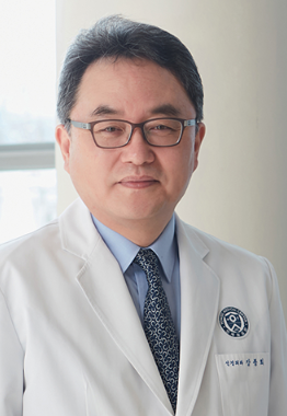 Jong Hee Chang合作论文数Department of Neurosurgery, Severance Hospital, Yonsei University Health System;Brain Tumor Center, Severance Hospital, Yonsei University Health System7Yong-Kil Hong合作论文数7
 Yong-Min Huh合作论文数7donggeun lee合作论文数6Jin-Kyoung Shim合作论文数6Jae Gol Choe合作论文数6Kyu-Won Shim合作论文数5Hyeonsoo Lee合作论文数5Hye-Jin Shin合作论文数5Yun In Sik合作论文数5
 Ji-Hyun Lee合作论文数5
 Thijs Kooi合作论文数Department of Information and Computer Science, Keio University5Pyung Seong Kwon合作论文数5
 Byung-Ok Choi合作论文数SKKU-SOM Newsletter School of Medicine, Sungkyunkwan University;Samsung Medical Center, Sungkyunkwan University;Samsung Advanced Institute for Health Science & Technology, Sungkyunkwan University;Institute for Medical Device Management & Research Center, Sungkyunkwan University5Ah Young Park合作论文数4
 Ki-Hwan Kim合作论文数4sung chan park合作论文数4Oh Sun Young合作论文数4Joong Uhn Choi合作论文数4Yun Ho Lee合作论文数4Hoon Chul Kang合作论文数4
 Ki Wha Chung合作论文数4
 Sang-Hyeon Lee合作论文数4Sung Bum Cho合作论文数4




