Abstract Total knee arthroplasty has evolved from a purely mechanical procedure towards a more personalized restoration of each patient's native knee function. This narrative review explores the physiological and pathological patterns of knee laxity, the spectrum of alignment phenotypes and their interplay with ligament balancing strategies. It summarizes current evidence on physiological laxity envelopes, osteoarthritic alterations and alignment classifications such as Coronal Plane Alignment of the Knee, functional and laxity‐based phenotypes. The review further examines how alignment philosophies affect soft‐tissue tension and postoperative stability. Recent advances in robotic and sensor‐assisted techniques have enabled objective intraoperative assessment of ligament balance, allowing reproducible quantification of medial–lateral gaps and compartmental pressures. These technologies reveal that individualized ligament behaviour often diverges from bony alignment, underscoring the need to define reproducible physiological balance targets. Functional outcomes appear optimized when medial stability and controlled lateral laxity are restored, supporting the concept of reproducing physiological asymmetry rather than enforcing symmetric gaps. However, existing studies remain heterogeneous and often underpowered, with conflicting results regarding the clinical impact of intraoperative balance on outcomes. Future research should rely on larger, prospective, high‐level evidence studies to better define optimal alignment–balancing strategies and their translation into functional benefit. Level of Evidence Level V.
INTRODUCTION:Instability is a leading cause of revision total hip arthroplasty (rTHA). This study aimed to (1) determine rates of recurrent dislocation and re-revision following rTHA for instability and (2) identified patient- and surgical risk factors associated with failure. METHODS:Patients who underwent revision THA at our institution between 2016 and 2024 for a primary indication of instability were included. A total of 242 patients who met inclusion criteria, including 10 (4.1%) using a direct anterior approach, whereas the remaining 232 were performed using a postero-lateral approach (95.9%). Outcomes of interest included recurrent dislocation and re-revision for instability. Multivariable logistic regression was performed to identify independent predictors of failure. RESULTS:There were 36 patients (14.9%) who experienced at least one dislocation after rTHA and 27 patients (11.2%) who required re-revision after a mean of 1.3 dislocations, at a median of 126 days from revision surgery. On adjusted multivariable analyses, prior spinal fusion was independently associated with an increased risk of redislocation (OR [odds ratio] 3.51, 95% CI [confidence interval] 1.40 to 8.58; P < 0.01), while use of a preoperative hip-spine workflow was associated with reduced risk of redislocation (OR 0.43, 95% CI 0.18 to 0.97; P < 0.05) and re-revision (OR 0.38, 95% CI 0.13 to 0.96; P < 0.05). Smaller femoral head size (≤32 mm) increased the risk of re-revision (OR 5.20, 95% CI 1.26-16.19; P = 0.04), whereas technology-assisted surgery and dual-mobility articulations were not associated with improved outcomes. CONCLUSION:In this large cohort, rTHA performed for instability was associated with substantial rates of recurrent dislocation and re-revision at a mean follow-up of 4.1 years. These findings emphasize the multifactorial nature of instability post-revision and support the integration of patient-specific spino-pelvic assessment into surgical decision-making to optimize stability after rTHA.
Arthrofibrosis following total knee arthroplasty (TKA) is a challenging complication that contributes to failure to achieve functional range of motion and results in physical impairment, pain, and revision surgery. Despite increasing recognition of arthrofibrosis as a biological disease process rather than a purely mechanical complication, there is no single document that integrates current evidence on diagnosis, surgical management, and preventive strategies. In June 2025, the Stavros Niarchos Foundation Complex Joint Reconstruction Center at Hospital for Special Surgery convened an international symposium to address arthrofibrosis following TKA. Invited experts in orthopedic surgery, basic science, biomechanics, and outcomes research were organized into 3 panels focused on (1) basic science, diagnosis, and risk stratification; (2) surgical management and postoperative rehabilitation; and (3) prevention strategies and adjunctive therapies. Each panel reviewed the literature, addressed predefined clinical questions, and developed consensus through structured discussion. This summary describes the recommendations from each panel, emphasizing early, multispecialty evaluation, stage-specific intervention, and procedure-specific rehabilitation strategies. Gaps in the evidence and priorities for future research are highlighted. These recommendations aim to standardize care, guide clinical decision-making, and shift the focus from late salvage toward early identification and prevention of arthrofibrosis following TKA. Level of Evidence: Level V: expert opinion.
Arthrofibrosis and stiffness are challenging complications after total knee arthroplasty (TKA), often treated with Manipulation Under Anesthesia (MUA). Robotic-assisted TKA (RA-TKA) aims to enhance surgical precision, though its impact on stiffness remains unclear. This study examined the association between surgical technologies and MUA rates using propensity score matching (PSM) and assessed whether surgeon experience affects MUA risk. We conducted a retrospective case-control study of 25,611 primary unilateral TKAs (2016–2024), stratified by manual, computer-assisted (CA), or RA techniques. MUA cases within 90 days were matched 1:2 to controls using PSM. Conditional logistic regression assessed the association between surgical technologies and MUA risk. A learning curve analysis of each surgeon’s first 100 RA-TKAs assessed surgical proficiency progression. Odds ratios were compared before and after the learning phase to evaluate its impact on MUA rates. Both CA-TKA (OR = 1.11, P = 0.33) and RA-TKA (OR = 1.24, P = 0.12) revealed a higher trend in MUA risk compared to manual. Learning curve analysis of 10 surgeons demonstrated a distinct learning breakpoint at case 11.8 (95
Aims:This Delphi consensus study aimed to achieve international expert agreement on clinical and radiological criteria for diagnosing aseptic component loosening in total hip arthroplasty (THA). The consensus involved revision hip surgeons from diverse healthcare settings worldwide, aiming to standardize diagnostic criteria and enhance clinical decision-making and future research comparability. Methods:A three-round Delphi study was conducted in accordance with Conducting and Reporting of Delphi Studies guidelines. A panel of 125 orthopaedic surgeons from 32 countries was approached based on geographical representation and experience in revision THA. In Round 1, experts listed clinical and imaging criteria; in Rounds 2 and 3, derived statements were rated on a Likert scale. Consensus was a priori defined as ≥ 70% agreement. A total of 43 experts completed Round 1, and 37 completed all three Delphi rounds. Results:Consensus was achieved on 25 clinical and radiological statements. Investigations for periprosthetic joint infection was unanimously considered the first step. Limping and pain were general indicators of loosening. Pain during weightbearing, hip rotation, start-up, or localized to the thigh was linked to femoral loosening. Pain at start-up, in the groin, or during activities was associated with acetabular loosening. Radiographs were the primary imaging modality, supported by serial radiographs and CT in uncertain cases. Key radiological indicators included progressive radiolucency, osteolysis, implant migration, broken screws, and cortical thickening. Conclusion:The consensus statements from this study establish key clinical and radiological parameters for diagnosing aseptic loosening in THA, recommending plain radiographs as the primary imaging modality and CT as secondary. The results lay the groundwork for more standardized diagnostic protocols and improved consistency in future clinical practice and research.
For many years, providing patients with a total knee arthroplasty (TKA) prosthesis which is stable with a neutrally aligned lower limb using a mechanical alignment philosophy has represented the primary technical goal of TKA. However, it has previously been reported that only 50% of patients have neutral mechanical axis. Furthermore, despite continuous engineering developments with respect to implants used in TKA, contemporary philosophies, computer-assisted navigation and robotic-assisted TKA, registry data suggest that 1 in 5 patients undergoing TKA report overall dissatisfaction with their procedure in the medium to long term. Therefore, the purpose of this review was to first describe and compare outcomes of current alignment philosophies in TKA. Additionally, the authors sought to review the outcomes of computer-assisted navigation and robotic-assisted TKA procedures, while also evaluating the role of sensors and tensioners with respect to TKA alignment. Finally, the authors aimed to review potential future developments within this ever-developing space in the literature.
Aims: The ability of a surgeon to provide accurate visual estimates of intraoperative gaps during total knee arthroplasty (TKA) is not well understood. This study evaluated: 1) the accuracy of gap estimation in extension and in flexion; 2) the accuracy of gap estimation in the medial and lateral compartments, also in extension and flexion; 3) the differences in accuracy among surgeons; and 4) the frequency of clinically significant errors in gap estimation, defined as greater than 1 mm. Methods: A posterior stabilized TKA was performed on seven cadaveric knees. Five fellowship-trained arthroplasty surgeons and one orthopaedic resident manually stressed each knee, and visually assessed the medial and lateral gaps in full extension and 90° of flexion. Gaps were objectively measured via a motion capture system. Gap estimation error was calculated as the difference between the surgeons’ visual assessment and the measured gaps. Results: Across all surgeons and knees, the mean gap estimation error was -0.4 mm (SD 0.7), with the majority (72%) of gaps being underestimated. Errors were greater in extension (-0.7 mm (SD 0.8)) than in flexion (-0.2 mm (SD 1.0)) (p < 0.001). Lateral gap error was less in flexion (-0.1 mm (SD 1.0)) than extension (-0.7 mm (SD 0.8)). Gap estimation error pooled for all assessments differed between surgeons, ranging from a mean error of -0.8 mm (SD 0.8) to 0.2 mm (SD 1.2) (p < 0.001). Clinically significant gap estimation errors (> 1 mm) occurred in 33% of assessments in extension and 26% in flexion (p = 0.315, not statistically different). The frequency of such errors varied by surgeon ranging from 18% to 42% (p = 0.370). Conclusion: Surgeons tend to underestimate intraoperative gaps during TKA, particularly in extension. Clinically meaningful gap estimation errors (> 1 mm) occurred in up to 33% (26/78) of exams, supporting the need to enhance gap assessment accuracy. Cite this article: Bone Jt Open 2026;7(3):417–424.
Periprosthetic femoral fracture (PFF) is a common early complication after primary total hip arthroplasty. Collared stems reduce but do not eliminate the incidence of PFF, and exhibit variable biomechanical effectiveness, emphasizing our limited understanding of the local bone-implant interaction mechanics. Our goal was to elucidate the relationship between the local strains at the bone-implant interface and the experimental fracture patterns and loads for collared and collarless stems. Six pairs of women cadaveric femurs implanted with a collarless or collared stem were loaded to failure under simulated stumbling to determine the location of PFF and the load to fracture, which we related to the collar-calcar separation. Corresponding specimen-specific FE models were developed to determine the strain at the bone-implant interface and to predict the fracture onset load and location of fracture, which we related to the experimental location of PFF and the load-to-fracture. Load to fracture was greater for collared stems and was inversely correlated with the collar-calcar separation at the time of implantation (r = -0.80, p = 0.055). Fractures occurred in areas of high strain. The experimental fracture location coincided with the first yielding cortical element in five cases. The load at which the first cortical element yielded (i.e., onset of fracture was moderately correlated with the experimental fracture load (R2 = 0.43, RMSE = 1231 N). Our results emphasize the importance of initial calcar contact to realize the benefits of the collar. Localized load transfer was a precursor of macroscopic fracture, which initiated in areas of high tensile strain.
Aims:While aspirin has been widely adopted as an effective method for deep vein thrombosis (DVT) prevention following primary total knee arthroplasty (TKA), concerns remain with regard to its efficacy in the setting of revision TKA (rTKA). However, the risk for thromboembolic events must be weighed against the increased potential for bleeding with more potent anticoagulants. This study aimed to compare venous thromboembolism (VTE) and transfusion rates between patients receiving aspirin with non-aspirin anticoagulation following aseptic rTKA. Methods:We identified patients undergoing aseptic rTKA from 1 January 2016 to 30 April 2023 in a national database. Those with infection, fracture, prior VTE, coagulopathy, or preoperative anticoagulant use were excluded. Patients were grouped by postoperative prophylaxis: aspirin alone or non-aspirin anticoagulants. Patients were allocated to one of two categorical treatment groups. Crossover between groups was permitted where clinically indicated. Propensity score matching (1:1) was performed on demographic details, insurance, comorbidities, postoperative home health use, hospital length of stay ≤ one day, surgery year, and Current Procedural Terminology (CPT) code, yielding 4,585 patients per group. Outcomes included 30- and 90-day DVT, pulmonary embolism (PE), overall VTE, and transfusion. Results:In the matched cohort, aspirin use was associated with lower 90-day DVT (0.8% (n = 38) vs 2.1% (n = 96)), PE (0.6% (n = 26) vs 2.2% (n = 102)), and VTE (1.1% (n = 51) vs 3.3% (n = 152); all p < 0.001). Transfusion was also lower in the aspirin group at 30 days (0.6% (n = 28) vs 1.2% (n = 56); p = 0.003) and 90 days (0.7% (n = 33) vs 1.4% (n = 65); p = 0.002). DVT ultrasound use within 30 days was significantly higher in the non-aspirin group (7.1%; n = 326) compared with aspirin (5.1%; n = 235); p < 0.001), which may partly account for the higher VTE detection. Regression controlling for CPT code confirmed reduced odds of VTE with aspirin. Conclusion:Aspirin is a safe, effective, and noninferior option for VTE prophylaxis following aseptic rTKA in appropriately selected patients.
Aseptic loosening is one of the main causes of failure for both primary and revision total knee arthroplasty (rTKA). Bone loss and bone quality can vary significantly across anatomical regions, potentially challenging long-term fixation. In recent years, zonal fixation in rTKA has become increasingly popular; this approach divides areas of fixation based on anatomy into 3 zones: epiphysis, metaphysis, and diaphysis. This system emphasizes the importance of preoperative planning and encourages surgeons to carefully select the fixation method for each respective zone. To plan proper fixation, adequate imaging is required to document bone defects and areas of sclerosis. While anteroposterior and lateral radiographs remain the gold standard for defect classification, computed tomography imaging has facilitated 3-D defect evaluation and bone density assessment. For many years cemented and hybrid fixation have been the main modes of fixation in rTKA, but recently these modes of fixation have been augmented by the use of uncemented cones and sleeves. Through bone ingrowth this type of fixation provides lasting fixation in the metaphysis and seems to reduce the need for longer stem fixation in the diaphysis. This narrative review provides an overview of advanced imaging techniques, defect grading, and principles of implant fixation using the concept of zonal fixation.
BACKGROUND:The proportion of revision surgeries among all arthroplasties performed annually suggests a higher revision burden for total knee arthroplasty (TKA) than for total hip arthroplasty (THA). This study analyzed temporal trends in the knee/hip (K/H) Revision Burden Ratio (RBR) over the past decade, evaluated hip and knee RBRs separately, compared trends across four national arthroplasty registries, and assessed changes in revision causes for THA and TKA over time. METHODS:A retrospective, registry-based observational study was conducted using publicly available annual reports from four national arthroplasty registries in the United Kingdom, Australia, the United States, and Sweden. Annual numbers of primary and revision THA and TKA procedures were extracted. Hip and knee RBRs and K/H RBRs were calculated annually. Temporal trends were assessed using linear regression, and revision causes common to all registries were analyzed for the 2018 to 2023 period. RESULTS:Across registries, primary arthroplasty volumes increased substantially, with knee procedures exceeding hip procedures. Globally, the K/H RBR increased significantly (β = 3.63 percentage points/year, R2 = 0.876, P < 0.001), driven by a decrease in hip RBR (β = -0.165, P = 0.006) and an increase in knee RBR (β = 0.207, P < 0.001). Similar trends were observed in the United Kingdom, Australia, and Sweden, whereas no significant temporal change was identified in the United States. The proportion of revisions due to aseptic loosening declined significantly, whereas infection and periprosthetic fractures increased over time, particularly after TKA. CONCLUSIONS:Revision burden is progressively shifting from hip to knee arthroplasty, driven by declining aseptic loosening and a concomitant rise in infection-related revisions, particularly after TKA, where infection is becoming the predominant failure mechanism. These findings have major implications for clinical practice and highlight the need for infection-focused prevention and treatment strategies. LEVEL OF EVIDENCE:Level III (retrospective comparative observational study).
BACKGROUND:Evaluation and management of bone loss in revision total knee arthroplasty (rTKA) poses a demanding challenge. The rising number of knee revisions and rerevisions has led to increasing bone loss severity, and previous bone loss classification systems fail to provide adequate assessment of the diaphysis. The Scuderi classification was recently introduced to better assess diaphyseal bone loss and underwent internal validation. The purpose of this study was to perform an external validation using preoperative radiographs of failed stemmed rTKA and describe the interobserver and intraobserver reliability. METHODS:From our institutional database, 128 preoperative radiographs of failed rTKAs with stemmed prostheses awaiting rerevision were identified. There were 120 stemmed femoral prostheses (60 cemented, 60 hybrid fixation) and 120 stemmed tibial prostheses (60 cemented, 60 hybrid fixation) that were subject to analysis. There were two readers who had similar experience levels who scored the cases according to the Scuderi classification, and each reader performed two independent reads at least two weeks apart from the first read. The levels of interobserver and intraobserver reliability were determined by the intraclass correlation coefficients. RESULTS:Strong intraclass correlation coefficient for both femur (total: 0.96) and tibia (total: 0.89) has been demonstrated among the graders. Intraobserver reliability of 0.86 (grader 1) and 0.79 (grader two) for femur and 0.8 (grader 1) and 0.71 (grader 2) for tibia demonstrated satisfactory agreement. Absolute score congruency between the readers was 86.7% for the femur and 89.2% for the tibia, showing excellent grading reproducibility for this new classification. CONCLUSIONS:This study provided an external validation of this bone loss classification in revision prosthesis with strong to near-perfect interobserver and intraobserver reliability. Based on our reported results, we advocate the use of this intuitive grading scheme, which can facilitate bone loss evaluation and may provide a future foundation to assist with implant selection in these complex cases.
BACKGROUND:Dislocation remains one of the most challenging complications after total hip arthroplasty (THA). Previous studies suggested that three or more dislocations were a threshold for revision; however, these data predate contemporary implants and modern surgical approaches. We used a contemporary cohort of primary THA patients to evaluate the relationship between dislocation recurrence, timing of instability, and subsequent revision for instability. METHODS:There were 40,315 patients who underwent primary THA at our institution from 2016 to 2024. Among these patients, those who sustained a dislocation event were included. Keyword and diagnosis code search were performed to identify patients, and charts were reviewed to confirm dislocation. A total of 285 patients (0.7%) were included at a mean 5.7-year follow-up, of whom 242 (84.9%) ultimately underwent revision for instability. Instability was categorized by recurrence (one, two, or ≥ three dislocations) and timing ([early ≤ 90] versus late [greater than 90 days)]). Multivariable logistic regressions were performed to identify factors independently associated with risk of revision. RESULTS:Revision risk increased sharply with recurrence, rising from 69.1% after one dislocation to 93.9% after two and 98.5% after three or more. Patients who had two dislocations had significantly greater odds of revision than those who had one (odds ratio 7.70, 95% confidence interval 3.28 to 20.49, P < 0.001), while risk did not significantly increase beyond the second dislocation. Late dislocations were more likely to result in revision than early events (97.0 versus 74.9%, P < 0.001) and remained independently associated with revision (odds ratio 7.76, 95% confidence interval 2.46 to 35.10, P = 0.002). The association between recurrence, timing, and revision risk was consistent across both approaches. CONCLUSIONS:In contemporary THA practice, revision frequently occurred after a second dislocation, and late dislocations were more likely to require revision than early events. Future multicenter studies are needed to further refine instability management strategies.
Introduction STAR total hip arthroplasty (THA) approach preserves piriformis whilst providing sufficient visualization of the acetabulum, potentially comparable to technology-assisted systems. This multicenter study compared acetabular cup positioning within the Lewinnek safe zone between manual and technology-enabled STAR cohorts. We hypothesized comparable accuracy between methods, suggesting that the preservation of the piriformis does not obstruct the optimal access to the acetabulum in the manual STAR THA. Methods A total of 772 primary STAR THAs from two institutions were analyzed. Patients were matched 1:1 for age, sex, BMI, ASA class, and operative side, resulting in 386 manual and 386 technology-enabled cases. Technology-enabled THA included both computer-assisted and robotic systems. Cup inclination and anteversion were measured on postoperative supine anteroposterior pelvic radiographs using the Widmer method. A subset of 20 radiographs independently reviewed by an external observer, demonstrated excellent inter-observer reliability. Cup positioning was evaluated based on compliance with the Lewinnek safe zone criteria (inclination 30°–50°, anteversion 5°–25°). Groups were compared using two-sample proportion tests with significance set at p < 0.05. Results Groups were well matched across baseline variables (standardized mean differences < 0.10). Mean cup inclination was 42.8° ± 4.9° in the manual group and 44.3° ± 3.7° in the technology-enabled group. Mean anteversion was 20.1° ± 7.7° and 23.6° ± 5.4°, respectively. 65 % of manual and 68.4% of technology-enabled THAs were within the Lewinnek safe zone (absolute difference 3.4%, p = 0.36, 95% CI −10.3% to 3.5%). No significant differences in cup orientation or distribution were observed. Conclusion THA performed via the manual STAR approach, did not significantly differ by the technology-enabled systems with regards of the acetabular component positioning within the Lewinnek safe zone. These findings indicate that the STAR approach itself provides reliable and reproducible visualization, enabling precise cup placement, while preserving the piriformis.
BACKGROUND:The coronal plane alignment of the knee (CPAK) classification proposes nine knee phenotypes based on constitutional limb alignment and joint line obliquity. However, there is considerable confusion and variability in defining types, and its impact on surgical decision-making is unclear. A simple modified CPAK system is proposed to improve accuracy and facilitate decision-making. Herein, we applied a deep learning model to automate knee phenotyping and analyzed modified CPAK distributions in a large cohort. METHODS:We included 972 patients (1,944 healthy and arthritic knees) who had full-limb radiographs from a large publicly available dataset. A previously validated deep learning model was used to measure the lateral distal femoral angle and the medial proximal tibial angle. Knee phenotypes were determined using a two-level classification framework designed to capture both overall limb alignment, as measured by arithmetic hip-knee-ankle, and femoral and tibial joint-line contribution, as measured by lateral distal femoral angle and medial proximal tibial angle. RESULTS:There were seven phenotypic patterns identified, with five accounting for 99.2% of knees. The most frequent phenotypes were varus knees with valgus femur/varus tibia (26.9%), followed by neutral knees (25.6%), and valgus knees with valgus femur/varus tibia (24.2%). Less common were valgus knees with valgus femur/valgus tibia (12.2%) and varus femur/varus tibia in varus knees (10.3%). Varus phenotypes were predominantly tibial-driven (99.2%), while valgus phenotypes were femoral-driven (98.9%). Among neutral knees, 95% demonstrated a valgus femur with varus tibia configuration, while perfectly neutral combinations accounted for fewer than 2% of the total cohort. CONCLUSIONS:The Modifed CPAK offers a simplified and precise framework for knee phenotyping. Knee alignment demostrates a consistent pattern, with varus primarly driven by the tibia and valgus by the femur. This system provides a surgically relevant assessment of the knee, supporting alignment-based surgical planning.
BACKGROUND:Cementless fixation is increasingly popular for total knee arthroplasty (TKA). Prior research suggested that volumetric bone mineral density (vBMD) measured in preoperative computerized tomography (CT) scans could be useful to identify suitable candidates for cementless TKA with sufficient bone strength to avoid aseptic loosening. However, the clinically relevant thresholds of vBMD for cementless knees have not been defined. As a step toward defining such thresholds, we sought to relate the preoperative vBMD to the migration of tibial baseplates after TKA as a marker of aseptic loosening. METHODS:We prospectively enrolled 15 patients undergoing unilateral primary TKA with cementless tibial baseplates and cruciate-retaining inserts. Patients received a preoperative CT scan as standard of care, including a BMD reference phantom, and postoperative CT scans the day of surgery, at 6 weeks postoperatively, and at 6 months postoperatively. We calculated the implant motion relative to the day of surgery scan and related it to the vBMD in the four mm immediately under the baseplate. RESULTS:The maximum total point motion (MTPM) ranged from 0.1 to 0.9 mm at 6 weeks and between 0.2 and 1.1 mm at 6 months. The largest motion occurred vertically and, on average, was consistent with the posterior tilt of the baseplate. The preoperative vBMD was highest under the posterior-medial quadrant (mean: 229.8 mg/cm3 range, 113 to 329.2), and the medial-to-lateral vBMD ratio was proportional to the preoperative arithmetic hip-knee-ankle angle. However, neither the vBMD under the implant nor the limb alignment was related to any metric of implant motion at any time point. CONCLUSIONS:This is, to our knowledge, the first study to characterize the postoperative implant motion relative to a pre-weight-bearing CT scan and relate such motion to the vBMD directly under the tibial baseplate. Our results suggest that BMD alone cannot predict early migration of tibial baseplates.
Purpose:Offset measurement is critical in total hip arthroplasty (THA) for guiding restoration of native anatomy. However, measurements are time-consuming and measurer-dependent, creating obstacles for large cohort analyses. We aim to create an objective and reliable offset measurement algorithm using deep learning. Materials and Methods:Five hundred radiographs from the Osteoarthritis Initiative (OAI) were segmented with identification of the teardrop, femoral head, implant head, and femoral diaphysis. A U-Net convolutional neural network was trained to identify these landmarks and optimized using the multi-class Dice coefficient metric. Femoral axis and femoral/implant head center of rotation were calculated with the model predictions, and measurements of offset were compared against two trained readers on an independent testing cohort. Results:The optimized model had a Dice coefficient of 0.96 and a foreground mask accuracy of 96.2%. The model measured femoral, acetabular, and global offset on both limbs at a rate of 1.67 sec/image. On an independent cohort (n=90), the intraclass correlation coefficient between readers and the algorithm was 0.86 (95% confidence interval [CI] 0.80-0.91) for femoral offset, 0.87 (95% CI 0.78-0.91) for acetabular offset, and 0.94 (95% CI 0.91-0.96) for global offset. When applied to the entire OAI cohort (n=4,188), all relevant anatomical features (femoral axis, implant/femoral center of rotation, inter-teardrop line) were correctly calculated in 83.0% of images. Conclusion:We report the development of an accurate and rapid offset measurement model using deep learning that can be applied before and after THA. Future work will involve external model validation.
BACKGROUND:Massive femoral bone loss with collateral insufficiency in revision total knee arthroplasty is often managed with distal femoral replacement (DFR), but this procedure sacrifices bone stock and carries notable risks. We described outcomes of a bone-preserving alternative (the "DFR downgrade") that pairs bicondylar, metadiaphyseal-engaging femoral cones with a hinge construct. METHODS:We performed a retrospective review of consecutive patients who underwent revision total knee arthroplasty using a "DFR downgrade" technique for severe femoral bone loss between February 2016 and July 2024. There were 20 patients (mean age, 69 years (range, 58 to 86); 65% men) included, with a mean follow-up of 14.5 months (range, 0.9 to 43.7). Indications were aseptic loosening in 75%, infection in 10%, ligamentous instability in 10%, and arthrofibrosis in 5%. Demographics, Anderson Orthopaedic Research Institute classification, operative details, postoperative complications, and clinical and radiographic outcomes were collected. The Kaplan-Meier method was used to estimate reoperation-free and rerevision-free survivorship. Femoral bone loss was classified as Anderson Orthopaedic Research Institute (IIA in 10%, IIB in 30%, and III in 60% of cases. RESULTS:Survivorship free from revision for aseptic loosening was 100% at one and two years. Survivorship free from any rerevision was 85.1% at one and two years. Survivorship free from any reoperation was also 85.1% at one and two years. There were three knees that required reoperation: one for a periprosthetic femoral fracture revised to a DFR; one for infection treated with two-stage revision and reimplantation of a DFR downgrade construct; and one for patellar button exchange after an implant recall. CONCLUSIONS:In patients who have severe femoral bone loss and condylar compromise, a "DFR downgrade" consisting of a bicondylar cone-hinge construct achieved favorable survivorship. While further study is warranted, this appears to be a pragmatic, bone-preserving alternative to DFR.
Purpose:To quantitatively assess relative arterial contributions to the patellar tendon (PT) across predefined anatomic regions with 7-Tesla quantitative magnetic resonance imaging (7T-qMRI), algorithm-based histological analysis and high-resolution computed tomography (micro-CT) in a cadaveric model. Methods:Seven fresh-frozen human cadaveric knee pairs (mean age 41.9 ± 15.5 years) underwent limited vascular dissection and arterial cannulation. Pre- and post-contrast 7T-qMRI, with a volumetric interpolated breath-hold examination (VIBE) three-dimensional T1-weighted gradient echo pulse sequence, quantified tendonous vascularity by measuring contrast enhancement. Subsequent quantitative algorithm-based histologic analysis with hematoxylin and eosin (H&E) staining followed, and two additional specimens underwent high-resolution (98 μm) micro-CT for qualitative vascular assessment. Results:In the transverse analysis, 7T-qMRI demonstrated the highest mean relative arterial contributions in the medial region (42.4%) compared with the middle region (30.2%; p = 0.035) and higher, though not significant, than the lateral region (32.0%). The central PT demonstrated greater relative arterial contributions (37.5%) than the proximal (26.5%) or distal (29.3%) thirds (p > 0.05) in the longitudinal analysis. At the patellar enthesis, the middle third exhibited higher contributions (35.3%) than medial (28.8%) or lateral (29.6%), without significance, while the tibial tuberosity showed greater contributions along the lateral region (37.2%; p > 0.05). Histology confirmed significantly greater medial arterial contribution, with 8.3% higher supply than lateral (p = 0.018). Micro-CT revealed a robust vascular network along the medial PT with smaller branches laterally. Distal to the inferior patellar pole, a peripatellar circular network, extending medially into the posterior PT layers, was qualitatively identified. Conclusion:7T-qMRI and histological analyses demonstrated significantly greater arterial supply along the medial border of the PT, while micro-CT revealed a medial and peripatellar circular vascular network extending from the medial margin and the inferior patellar pole into the posterior tendon layers. These findings identify the medial margin as the main vascular source for the PT, with implications for surgical preservation and reducing PT devascularization risk. Level of Evidence:N/A.
 David J Mayman合作论文数Hospital for Special Surgery77
 Brian P. Chalmers合作论文数Department of Orthopaedic Surgery, Adult Reconstruction and Joint Replacement, Hospital for Special Surgery, New York, New York64
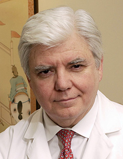 Thomas Peter Sculco合作论文数Hospital for Special Surgery;Weill Cornell Medical College, Cornell University62
 Alex Gu合作论文数61
 Susan M. Goodman合作论文数Department of Rheumatology, Hospital for Special Surgery;Weill Cornell Medical College56Malahias Michael-Alexander合作论文数2nd Orthopaedic Department, Athens University54
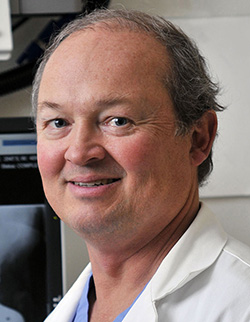 Mark P. Figgie合作论文数Orthopedic, Hospital for Special Surgery49
 Tim Wright合作论文数Department of Biomechanics, Hospital for Special Surgery48
 Cynthia A Kahlenberg合作论文数Feinberg School of Medicine, Northwestern University48Vigdorchik Jonathan合作论文数46Jerabek Seth A合作论文数41
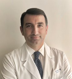 De Martino Ivan合作论文数Department of Orthopaedic Surgery, Hospital for Special Surgery41
 Geoffrey H. Westrich合作论文数Medical Center, Cornell University40
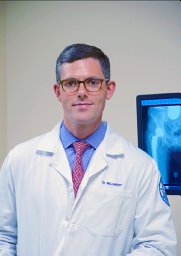 McLawhorn Alexander S合作论文数Department of Orthopedic Surgery, Hospital for Special Surgery34Allina A Nocon合作论文数33
 Michael B Cross合作论文数31
 Elizabeth Gausden合作论文数Division of Pediatric Orthopaedic Surgery, Hospital for Special Surgery, New York, New York, USA.30Blevins Jason L合作论文数28
 Friedrich Boettner合作论文数Hospital for Special Surgery, 535 East 70th Street, New York, NY 10021, USA28
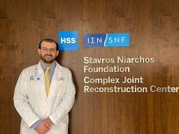 Ioannis Gkiatas合作论文数School of Medicine, University of Ioannina25
 Bella Mehta合作论文数24
 Linda A Russell合作论文数24
 Alejandro Della Valle合作论文数24
 Mathias P. Bostrom合作论文数Hospital for Special Surgery;Weill Cornell Medical College;NewYork-Presbyterian Hospital23Imhauser Carl W合作论文数Department of Mechanical Engineering and Mechanics, Drexel University20
 Michael P Ast合作论文数20
 Jiabin Liu合作论文数19
 Eytan M Debbi合作论文数Department of Orthopedics, Assaf Harofeh Medical Center19
 Seong Jun Jang合作论文数18
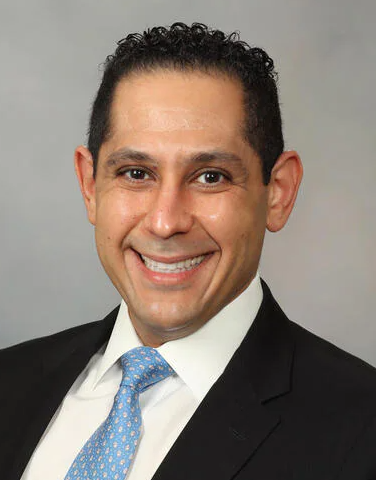 Matthew P. Abdel合作论文数Department of Orthopedic Surgery, Mayo Clinic18
 David L Helfet合作论文数18Richardson Shawn合作论文数17Joseph D Lipman合作论文数16
 Dimitrios A Flevas合作论文数Dept Arthroscopy & Orthopaed Surg, Metropolitan Hosp16
 Craig E. Klinger合作论文数16
 William Xiang合作论文数15Safa C Fassihi合作论文数15Chapman Wei合作论文数15Lazaro Lionel E合作论文数15Carli Alberto合作论文数15
 Ajay Premkumar合作论文数Department of Orthopaedic Surgery, Emory University15
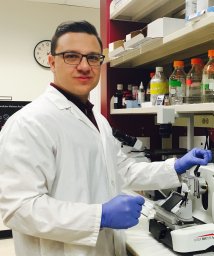 Troy D Bornes合作论文数Li Ka Shing Centre for Health Research Innovation, University of Alberta14Dean G. Lorich合作论文数14Sonia K. Chandi合作论文数13Tania Pannellini合作论文数Hosp Special Surg, Res Div, 535 E 70th St, New York, NY 10021 USA13
 Fernando J Quevedo González合作论文数Hosp Special Surg, 535E 71st St, New York, NY 10021 USA13
 Laura Donlin合作论文数HSS Research Institute;Hospital for Special Surgery, Weill Cornell Medicine13
 Shady Elmasry合作论文数Biomechanics Research Laboratory, University of Miami13
 T. David Tarity合作论文数13




