Sex differences in obesity are well recognized; however, the identification of sex-specific obesity genes and the mechanisms through which they affect obesity development remain elusive. Here, we identify a germ-cell-specific gene C2orf74, whose expression is responsive to high-fat diet (HFD) and promotes HFD-induced obesity in male mice by restraining lipolysis and limiting the browning of white adipocytes through suppression of androgen receptor signaling, but not in females. Additionally, C2orf74's expression increases with aging, contributing to aging-related obesity and metabolic comorbidities in chow-fed male mice. We demonstrate that C2orf74 is an ER-residing transmembrane protein that anchors and stabilizes dolichol phosphate mannose synthase 1 (Dpm1) on the ER membrane, facilitating Dpm1-mediated glycosylation and secretion of Bpifa3 from germ cells. As a paracrine regulator, Bpifa3 transcriptionally suppresses the expression of testosterone biosynthesis enzymes in Leydig cells. Therapeutically, we demonstrate that antisense oligonucleotide (ASO) targeting C2orf74 protects male mice from HFD-induced obesity. Thus, our study defines a role for germ-Leydig cell crosstalk, mediated by C2orf74, in the white adipocytes browning in both age-associated and diet-induced obesity.
OBJECTIVE:To test the hypothesis that women assigned to a natural ovulation regimen before frozen embryo transfer compared with a programmed regimen would have an increased chance of a healthy live birth and a reduced risk of pre-eclampsia or eclampsia. DESIGN:Multicentre, randomised, parallel group, assessor blinded clinical trial. SETTING:24 academic fertility centres in China. PARTICIPANTS:4376 ovulatory women (aged 20-40 years) planning to undergo a frozen single blastocyst transfer. INTERVENTIONS:Eligible participants were randomised (1:1) to receive a natural ovulation regimen or a programmed regimen of hormone replacement for endometrial preparation. Endometrial preparation and frozen embryo transfer timing were determined in the natural ovulation regimen group by monitoring natural follicle development and measuring serum levels of luteinising hormone, oestradiol, and progesterone. In the programmed regimen group, endometrial preparation was achieved by sequential administration of oestrogen and progesterone. MAIN OUTCOMES AND MEASURES:Primary outcomes were a healthy live birth and pre-eclampsia or eclampsia after a frozen embryo transfer. Secondary outcomes were cycle cancellation, biochemical pregnancy, clinical pregnancy, ongoing pregnancy, pregnancy loss, ectopic pregnancy, live birth, birth weight, and maternal, fetal, and neonatal complications. RESULTS:In the intention-to-treat analyses, 910 (41.6%) of 2185 patients in the natural ovulation regimen group and 890 (40.6%) of 2191 in the programmed regimen group achieved a healthy live birth (relative ratio 1.03 (95% confidence interval (CI) 0.96 to 1.10); P=0.49). The risk of pre-eclampsia was lower in the natural ovulation regimen group among patients who achieved clinical pregnancy than in the programmed regimen group (2.9% (38 of 1302) v 4.6% (61 of 1326); 0.63 (0.43 to 0.94); P=0.02). The incidences of early pregnancy loss (12.1% (158 of 1302) v 15.2% (201 of 1326); 0.80 (0.66 to 0.97)), placental accreta spectrum (1.8% (24 of 1302) v 3.6% (48 of 1326); 0.51 (0.31 to 0.83)), caesarean section (69.5% (776 of 1117) v 75.6% (831 of 1100); 0.92 (0.87 to 0.97)), and postpartum haemorrhage (2.0% (22 of 1117) v 6.1% (67 of 1100); 0.32 (0.20 to 0.52)) were lower in the natural ovulation regimen group. No differences between groups were observed for birth weight or neonatal complications. The rate of cycle cancellation was higher in the natural ovulation regimen (16.2% (354 of 2185) v 11.5% (251 of 2191), P<0.001). The prespecified per protocol and subgroup analyses yielded results consistent with the intention-to-treat analyses. CONCLUSIONS:In ovulatory women, a natural ovulation regimen for endometrial preparation was as effective as programmed regimen in terms of achieving a healthy live birth after frozen embryo transfer, but with a lower risk of maternal complications during pregnancy. TRIAL REGISTRATION:Chinese Clinical Trial Registry ChiCTR2200057990.
Abstract Multiple morphological abnormalities of the sperm flagella (MMAF) represent a severe form of asthenozoospermia that leads to male infertility, and it is commonly associated with genetic defects affecting flagellar components. Although the gene CFAP65 has been implicated in MMAF, its full mutational spectrum and clinical relevance within highly consanguineous populations remain poorly characterized. To elucidate the genetic basis of this condition, whole-exome sequencing followed by Sanger sequencing was performed in two infertile individuals from a consanguineous Pakistani family. Sperm morphology was assessed using hematoxylin-eosin (H&E) staining; while scanning and transmission electron microscopy (SEM and TEM) were utilized to evaluate ultrastructural defects. A novel homozygous frameshift mutation in CFAP65 (c.582_587delinsCG; p.Q194Hfs*4) was identified. This mutation introduces a premature stop codon within the transmembrane helix domain. Morphological analysis revealed classic MMAF features, including absent, short, bent, and coiled flagella in over 97% of sperm. TEM further demonstrated severe axonemal disorganization, with absent or disrupted microtubule doublets observed in 85% of the cross-sections. Based on clinical history and chest X-ray, the affected individuals reported no chronic respiratory symptoms and had no situs inversus, suggesting an isolated sperm-specific phenotype. In conclusion, this study identifies a novel domain-specific CFAP65 mutation associated with MMAF and male infertility in a consanguineous Pakistani family, thereby expanding the known genetic mutational landscape of MMAF. These findings reinforce the crucial role of CFAP65 in sperm flagellar morphogenesis and underscore its significance in the genetic diagnosis of male infertility.
STUDY QUESTION Do variants in YTH N6-methyladenosine RNA binding protein C2 (YTHDC2) cause male infertility in humans, and what is the underlying pathogenic mechanism? SUMMARY ANSWER Biallelic pathogenic missense variants in YTHDC2 disrupt the mitotic-to-meiotic transition, causing meiotic arrest and non-obstructive azoospermia (NOA) or severe oligozoospermia in humans. WHAT IS KNOWN ALREADY YTHDC2 is a male germ cell-specifically expressed RNA helicase essential for meiotic progression. In mice, loss of Ythdc2 leads to meiotic arrest at the early prophase. However, clinical evidence linking YTHDC2 variants to human male infertility and the underlying mechanisms involved remains to be established. STUDY DESIGN, SIZE, DURATION This study utilized a large cohort comprising 56 consanguineous families and 89 sporadic infertile men diagnosed with NOA or severe oligozoospermia. Through extensive genetic screening, we specifically identified five infertile men from three unrelated families who harbored candidate pathogenic variants in the YTHDC2 gene. The overall study design encompassed genetic screening followed by in vivo functional validation using a knock-in mouse model. PARTICIPANTS/MATERIALS, SETTING, METHODS Whole-exome sequencing (WES) and bioinformatic analyses were performed on the patient cohort to screen for candidate pathogenic variants. Human meiotic defects were characterized via histological analyses and immunofluorescence staining of testicular sections. To validate the pathogenicity of the identified variant, a knock-in mouse model harboring the equivalent variant found in patients was generated by CRISPR/Cas9 technology, and analyzed for spermatogenesis and meiosis using spermatocyte spreading and immunofluorescence staining, quantitative real-time PCR, and western blotting. MAIN RESULTS AND THE ROLE OF CHANCE Two homozygous missense variants in YTHDC2 were identified in four NOA patients from two unrelated consanguineous families (MT1: c.3491A>T, p. E1164V; MT2: c.2639G>A, p. R880H), and compound-heterozygous missense variants were identified in a sporadic patient with severe oligozoospermia (MT3: c.1145A>G, p. D382G; MT4: c.292A>G, p. R98G). The MT1 variant (p.E1164V) is not located in any annotated domains, the other three variants reside within known functional domains of YTHDC2 protein. The knock-in mouse model carrying the MT1 variant recapitulated the patient's phenotype, with both exhibiting meiotic prophase arrest during spermatogenesis. Mechanistically, significantly decreased levels of MEIOC and RBM46, two YTHDC2-interacting proteins required for meiotic transcriptome reprogramming, were observed in the patient's testes. Concurrently, mitotic cell cycle regulators such as CCNA2, CCND1, and WEE1 were aberrantly upregulated in the patient's testicular cells expressing meiosis markers, indicating a failure to silence the mitotic program upon meiotic entry. LIMITATIONS, REASONS FOR CAUTION This study is limited by the small sample size of patients with pathogenic YTHDC2 variants. While the MT1 variant was functionally validated in vivo, the specific pathogenic mechanisms of the other variants identified require further investigation. WIDER IMPLICATIONS OF THE FINDINGS Our findings provide direct clinical evidence establishing the pathogenicity of YTHDC2 variants in human NOA. The study reveals a conserved role for YTHDC2 in safeguarding the mitotic-to-meiotic transition by suppressing mitotic gene expression while maintaining the meiotic program. These findings expand the genetic spectrum of male infertility and suggest YTHDC2 screening as a promising approach for the genetic diagnosis of male infertility. STUDY FUNDING/COMPETING INTEREST(S) This work was supported by the National Key Research and Developmental Program of China (2022YFA0806303 to H.Z., and 2024YFC2706801 to H.J.); the National Natural Science Foundation of China (32470898 to H.Z., W2412028 and 32330032 to Q.S., 32470915 to B.S.); the State Key Laboratory of Reproductive Medicine and Offspring Health, Nanjing Medical University (SKLRM-K202405); and the Open Research Project of Fuyang Normal University (FYKFKT24023). The authors declare no competing interests. TRIAL REGISTRATION NUMBER N/A.
STUDY QUESTION What is the role of calmodulin-regulated spectrin-associated protein 1 (CAMSAP1) in human spermatogenesis?SUMMARY ANSWER Biallelic CAMSAP1 variants cause human male infertility with multiple sperm head deformities and MMAF-like phenotypes by disrupting manchette microtubule dynamics and the microtubule-spectrin-actin scaffold essential for sperm head shaping.WHAT IS KNOWN ALREADY Sperm head shaping requires coordinated acrosome formation, chromatin condensation, and manchette-driven nuclear remodeling. Although several genes have been implicated in globozoospermia, macrozoospermia, and multiple morphological abnormalities of the sperm flagella (MMAF), the genetic causes of other forms of abnormal sperm head morphology remain largely unknown. Camsap1-deficient mice exhibit severe spermatogenic defects, including abnormal sperm head shaping and flagellar malformations resulting from disruption of the acrosome-acroplaxome-manchette complex, ultimately leading to male infertility. However, whether CAMSAP1 plays a conserved role in human sperm development has not been established.STUDY DESIGN, SIZE, DURATION The CAMSAP1 variants were identified by whole-exome sequencing in a cohort of 3757 male infertility patients, and all participants were recruited from 2015 to 2025. In vitro functional assays, immunoprecipitation-mass spectrometry, and sperm morphological analyses were performed.PARTICIPANTS/MATERIALS, SETTING, METHODS Three infertile men with severely impaired sperm morphology and motility were recruited. Bioinformatic assessment, cell-based expression assays, immunostaining, and co-immunoprecipitation (Co-IP) coupled with liquid chromatography-tandem mass spectrometry (LC-MS/MS) using human testicular extracts were used to characterize the functional impact of the variants.MAIN RESULTS AND THE ROLE OF CHANCE Homozygous CAMSAP1 variants were identified in two individuals from consanguineous families (Chinese and Pakistani), and compound heterozygous missense variants in a third individual from a non-consanguineous Chinese family through whole-exome sequencing and further confirmed by Sanger sequencing. Sperm from affected individuals displayed MMAF phenotypes and diverse head abnormalities, including pyriform, amorphous, small, and globozoospermic forms. All four CAMSAP1 variants were predicted to be damaging and exhibited markedly reduced protein stability in vitro. Proteomic analysis revealed that CAMSAP1 interacts with proteins involved in actin cytoskeleton organization, acrosomal vesicle trafficking, microtubule bundle assembly, and calmodulin regulation. Mechanistically, CAMSAP1 associates with CALM1 and SPTBN1 to couple manchette microtubules with the perinuclear spectrin-actin network, ensuring proper force transmission for nuclear shaping. Mutations disrupted these interactions, leading to defective manchette architecture, abnormal acrosome assembly, and nuclear deformation.LIMITATIONS, REASONS FOR CAUTION Although CRISPR-Cas9 knock-in cell lines carrying patient-derived CAMSAP1 variants were generated, this in vitro model could not reproduce the highly dynamic processes of manchette assembly and nuclear remodeling that occur uniquely during spermiogenesis, and therefore cannot fully capture the mechanistic sequence leading to sperm head deformation. Additionally, the number of affected individuals remains limited, and larger multi-center, multi-ethnic cohorts will be required to validate the pathogenicity of CAMSAP1 variants and further delineate the associated clinical spectrum. WIDER IMPLICATIONS OF THE FINDINGS This work identifies CAMSAP1 as a previously unrecognized cause of human asthenoteratozoospermia and expands the genetic landscape of sperm head deformities. The findings underscore the essential role of CAMSAP1 in manchette-mediated nuclear remodeling and provide new insights for genetic diagnosis, counseling, and management of male infertility.STUDY FUNDING/COMPETING INTEREST(S) This work was supported by the National Natural Science Foundation of China (grant no. 82301815), the National Key R&D Program of China (grant no. 2022YFC2702603), the Sichuan Province Science and Technology Innovation Talent Project (grant no. 2024JDRC0006), the 2024 Key Open Project of Sichuan Provincial Key Laboratory for Human Disease Gene Research (grant no. 2024kflx002), and the China Postdoctoral Science Foundation (grant nos. 2023M732468 and GZC20231835). The authors declare no competing interests.TRIAL REGISTRATION NUMBER N/A.
BACKGROUND:Oligoasthenoteratozoospermia (OAT), characterized by reduced sperm count, impaired motility, and abnormal morphology, is a major cause of male infertility with substantial genetic heterogeneity. However, the underlying genetic etiology remains unresolved in a large proportion of affected individuals. OBJECTIVES:This study aimed to identify novel genetic causes of human OAT and to investigate the molecular and clinical consequences of the identified variants. MATERIALS AND METHODS:Whole-exome sequencing was performed in infertile men diagnosed with severe OAT. Identified variants were evaluated by population database screening and functional analyses, including quantitative mRNA assessment, label-free proteomics, immunofluorescence staining, and transmission electron microscopy (TEM). Clinical outcomes following assisted reproductive treatment were also documented. RESULTS:We identified CCDC189 as a novel pathogenic gene associated with human OAT. Two unrelated infertile men carried homozygous variants in CCDC189, including a frameshift variant (c.709_711del, p.W237Afs*3) and a missense variant (c.898C>T, p.R300W), both of which were rare in population databases. Clinically, both individuals exhibited severe OAT with profound acrosomal abnormalities and multiple morphological defects of the sperm flagella. TEM analysis revealed acrosomal detachment, cytoplasmic retention, loss of radial spokes, and absence of the central pair complex, indicating severe disruption of axonemal architecture. Consistently, CCDC189 mRNA levels were markedly reduced, and proteomic and immunofluorescence analyses confirmed the loss of CCDC189 protein along with its interacting partner CABCOCO1. Despite severe structural abnormalities, intracytoplasmic sperm injection (ICSI) using ejaculated sperm from one patient resulted in normal fertilization, high-quality embryo development, and an ongoing clinical pregnancy. CONCLUSION:Our findings establish CCDC189 as a pathogenic gene underlying human OAT and expand the spectrum of coiled-coil domain-containing proteins essential for sperm morphogenesis. The favorable ICSI outcome underscores the clinical importance of genetic diagnosis in CCDC189-related infertility and supports ICSI as an effective therapeutic option for affected couples.
As a specialized form of cell division for the generation of haploid gametes, meiosis is characterized by the separation of homologous chromosomes in meiosis I. Two key events take place in meiotic prophase I to ensure the proper segregation of homologous chromosomes, including the formation of the synaptonemal complex (SC) and the initiation and repair of programmed DNA double-strand breaks (DSBs). Both of these events are essential for the generation of crossovers. The SC serves as the structural basis of all the chromosome behaviors in meiotic prophase I. It not only reorganizes the chromosomes into a highly ordered stem-loop structure and holds the homologous chromosomes together when DSBs are being repaired, but also works as a platform for the recruitment of proteins playing essential roles in the formation and repair of DSBs, as well as the checkpoint monitoring the proper meiosis progression. Given the critical role of the SC in meiosis, questions concerning its structure and function are well studied in the field of meiosis research. Here, we not only reviewed the studies concerning the assembly of the SC, the function of the SC during male meiosis, and how mutations related to the SC coding genes affect male fertility, but also summarized the critical questions to be solved in future research.
Infertility has emerged as a global health concern, impacting around 8%-12% of couples during their reproductive years. Due to limitations in obtaining human biological samples, mouse models have been widely used for investigating gene functions. Fertility assessment in mouse models is a critical component in reproductive biology for studying gene function and elucidating mechanisms of reproductive disorders. However, natural mating observation of mice may yield inconsistent results, especially in the absence of standard guidelines, prolonged experimental cycles, and operational complexity. This protocol establishes a comprehensive breeding strategy for evaluating murine fertility through systematic vaginal plug monitoring and litter size quantification within defined timeframes. Key steps include (1) standardized male-female pairing protocols, (2) daily vaginal plug inspection, and (3) longitudinal tracking of pregnancy outcomes. This protocol presents a straightforward and easily implementable protocol for mouse mating cage setup and statistical analysis, enabling reliable fertility assessment under natural breeding conditions. Key features • Standardized natural mating protocol combining vaginal plug monitoring (daily) and litter size tracking. • Time-optimized workflow completes fertility phenotyping in 2 months. • An easy and custom-constructed vaginal plug inspection tool for optimized vaginal plug inspection.
Multiple morphological abnormalities of the sperm flagella (MMAF) is a severe form of male infertility, linked to defective spermiogenesis. Several flagella-associated proteins have been identified as crucial for the proper organization of the sperm flagellar axoneme. We identify a novel homozygous mutation in the CFAP58 gene (c.562C > T, p. R188*) that co-segregates with the multiple morphological abnormalities of the flagella (MMAF) phenotype in two unrelated consanguineous families from Pakistan. To validate the pathogenicity of this mutation, we developed a Cfap58 mutant mouse model to mimic the patient mutation. The Cfap58M/M mice exhibited infertility and recapitulated the MMAF phenotype observed in human patients. Transmission electron microscopy (TEM) analysis revealed the absence of the central pair (CP) of microtubules in the axonemal structure of sperm flagella. Further analysis demonstrated that the CFAP58 mutation disrupts CP assembly during spermiogenesis, leading to disorganization of axonemal proteins in both human and mouse sperm flagella. Our findings underscore the essential and conserved role of CFAP58 in sperm axoneme assembly and suggests that CFAP58 can serve as a genetic screening marker in the diagnosis and genetic counseling of MMAF and male infertility.
PIWI-interacting RNAs (piRNAs) are small regulatory RNAs (21-35 nucleotides) exclusively expressed in germ cells, where they play a critical role in transposable element repression and post-meiotic gene regulation. The poly(A)-specific RNase-like domain-containing 1 (PNLDC1) protein is essential for piRNA maturation, specifically in 3'-end trimming. Disruption of PNLDC1 has been implicated in nonobstructive azoospermia (NOA) and male infertility. Through whole-exome sequencing, we identified a compound heterozygous mutation (MT1 c.449G > A, p.Trp150* and MT2 c.821A > G, p.His274Ala) in a Chinese NOA patient (P9241) and a homozygous nonsense mutation (MT3 c.1288C > T, p.Arg430*) in a Pakistani NOA patient (II:2) born to a consanguineous couple. Mutant PNLDC1 mRNA was detected, but not the corresponding protein, was detected in the testes of P9241. In contrast, truncated PNLDC1 protein was observed in HEK293T cells transfected with a plasmid harboring mutation MT3. To investigate the functional consequences, we generated a Pnldc1KI/KI mouse model mimicking the MT3 using CRISPR/Cas9 genome editing, which exhibited infertility due to spermiogenesis arrest, phenocopying the NOA condition in patient II:2. Notably, Pnldc1KI/KI testes showed significant derepression of the retrotransposon LINE1 and increased spermatid apoptosis. These findings provide strong functional evidence that PNLDC1 mutations disrupt piRNA biogenesis, impair spermatogenesis, and underlie NOA in both humans and mice.
Male infertility, particularly non-obstructive azoospermia (NOA), remains a major clinical challenge. Although the testis is one of the most transcriptionally complex organs, stage-specific changes in total RNA abundance remain poorly characterized. Through total RNA staining, we identified distinct RNA content profiles across spermatogenic stages, supporting its value as a discriminative marker. Based on this, we developed RNA-DNA dual-parameter flow cytometry (RD-Cyto), a novel method that simultaneously measures RNA and DNA content at single-cell resolution for high-precision classification of testicular cells. Compared to conventional Hoechst-based cytometry, RD-Cyto offers superior resolution, enabling clear separation of pachytene (P) and diplotene (D) spermatocytes, first-time identification of metaphase I spermatocytes (MI), and subdivision of metaphase II spermatocytes (MII) into two subsets. To assess its utility in mechanistic research, we applied RD-Cyto to investigate the RNA-binding protein (RBP) DEAD-Box Helicase 17 (DDX17) and found that its deletion causes RNA accumulation in preleptotene spermatocytes and meiotic failure—demonstrating RD-Cyto's sensitivity to stage-specific regulatory disruptions. We further applied RD-Cyto to the clinical diagnosis of azoospermia. Combined with testicular fine-needle aspiration cytology (FNAC), RD-Cyto accurately evaluated spermatogenesis in all enrolled patients, showing 100 % concordance with testicular sperm retrieval outcomes. It successfully identified Sertoli cell-only syndrome (SCOS) in NOA, predicted sperm retrieval in rare NOA cases with residual spermatogenesis, and revealed spermatogenic and morphological abnormalities linked to pathogenic mutations in oligoasthenospermia (OAS). These findings establish RD-Cyto as a robust platform for both mechanistic investigation and diagnostic stratification in male infertility.
During meiosis, at least one crossover is selectively generated per pair of homologous chromosomes through homologous recombination to ensure their faithful segregation. The molecular mechanisms controlling meiotic recombination, particularly in XY chromosomes that share a tiny region of homology (i.e., the pseudoautosomal region, PAR), remain poorly understood. Here, we identify S100PBP as a key modulator of both XY and autosomal recombination in mice. S100pbp- knockout mice exhibit male infertility and spermatogenesis arrest at meiotic metaphase I, resulting from a drastic reduction in XY crossovers. This failure in XY crossover formation is due to a reduction in TEX11/M1AP-bound recombination intermediates at the PAR. By contrast, disruption of S100PBP significantly increases the number of recombination intermediates and crossovers on autosomes. Co-immunoprecipitation mass spectrometry revealed that S100PBP interacts with the nucleoporin TPR. Furthermore, S100PBP is localized specifically to the nuclear pores of meiocytes, likely in a TPR-dependent manner. These findings demonstrate that S100PBP promotes XY crossover formation while limiting excess autosomal crossovers and shed light on the potential role of nuclear pores in regulating meiotic recombination.
Spermatogenesis is a process of self-renewal of spermatogonial stem cells and their proliferation and differentiation to generate mature sperm. This process involves interactions between testicular somatic (mainly Sertoli cells) and spermatogonial cells at their different stages of development. The functionality of Sertoli cells is regulated by hormones and testicular autocrine/paracrine factors. In this study, we investigated the effects of follicle-stimulating hormone (FSH) and testosterone addition on Sertoli cell cultures that undergo hypotonic shock, with a primary focus on Sertoli cell activity. Cells were enzymatically isolated from testicular seminiferous tubules of 7-day-old mice. These cells were cultured in vitro for 3 days. Thereafter, some cultures were treated with hypotonic shock to remove germ cells. After overnight, fresh media without (control; CT) or with FSH, testosterone (Tes), or FSH+T were added to the hypotonic shock-treated or untreated (CT) cultures for 24 h. The morphology of the cultures and the presence of Sertoli cells and germ cells were examined. The expression of growth factors (CSF-1, LIF, SCF, GDNF) or other specific Sertoli cell factors [transferrin, inhibin b, androgen receptor (AR), androgen binding protein (ABP), FSH receptor (FSHR)] was examined by qPCR. Our immunofluorescence staining showed depletion/major reduction in VASA-positive germ cells in Sertoli cell cultures following hypotonic shock (HYP) treatment compared to untreated cultures (WO). Furthermore, the expression of the examined growth factors and other factors was significantly increased in HYP cultures compared to WO (in the CT). However, the addition of hormones significantly decreased the expression levels of the growth factors in HYP cultures compared to WO cultures under the same treatment. In addition, the expression of all other examined Sertoli cell factors significantly changed following HYP treatment compared to WO and following treatment with FSH and or T. However, the expression levels of some factors remained normal following the treatment of Sertoli cell cultures with one or both hormones (transferrin, Fsh-r, Abp, Ar). Thus, our results demonstrate the crucial role of germ cells in the functionality of Sertoli cells and the possible role of FSH and T in maintaining, at least partially, the normal activity of Sertoli cells following germ cell depletion in vitro by hypotonic shock treatment.
A-kinase anchoring proteins (AKAPs) constitute a structurally diverse family of scaffold proteins with significant functional relevance. Various AKAP family members localize to distinct subcellular compartments, such as sperm flagella, via their specific domains. In this study, we investigated the physiological function of AKAP14, a potential functional gene for male fertility due to its high expression in human and mouse testicular tissues. We generated Akap14 knockout mice(Akap14ins/Y) using CRISPR/Cas9 technology to assess the role of AKAP14 in male fertility. Our results demonstrate that Akap14ins/Y mice exhibit normal fertility, with a comparable testes-to-body weight ratio, epididymal sperm count, sperm motility, and sperm morphology to wild-type male mice. Furthermore, examination of meiotic prophase I progression revealed a consistent distribution of each substage in Akap14ins/Y testes compared to wild-type testes, indicating that AKAP14 is dispensable for spermatogenesis in mice. In conclusion, despite its high expression level in the testes, AKAP14 depletion does not impact male fertility in mice, suggesting that further in-depth studies of its role in murine spermatogenesis may be unnecessary and could conserve valuable research resources. IN BRIEF: AKAP14 is highly expressed in the testes, but our study using Akap14 knockout mice reveals that it is dispensable for male fertility and spermatogenesis. Despite its expression in testicular tissues, AKAP14 depletion does not impair sperm function or meiotic progression, suggesting no critical role in murine reproductive health.
Infertility has emerged as a global health concern, impacting around 8%-12% of couples during their reproductive years. Due to limitations in obtaining human biological samples, mouse models have been widely used for investigating gene functions. Fertility assessment in mouse models is a critical component in reproductive biology for studying gene function and elucidating mechanisms of reproductive disorders. However, natural mating observation of mice may yield inconsistent results, especially in the absence of standard guidelines, prolonged experimental cycles, and operational complexity. This protocol establishes a comprehensive breeding strategy for evaluating murine fertility through systematic vaginal plug monitoring and litter size quantification within defined timeframes. Key steps include (1) standardized male-female pairing protocols, (2) daily vaginal plug inspection, and (3) longitudinal tracking of pregnancy outcomes. This protocol presents a straightforward and easily implementable protocol for mouse mating cage setup and statistical analysis, enabling reliable fertility assessment under natural breeding conditions. Key features • Standardized natural mating protocol combining vaginal plug monitoring (daily) and litter size tracking. • Time-optimized workflow completes fertility phenotyping in 2 months. • An easy and custom-constructed vaginal plug inspection tool for optimized vaginal plug inspection.
Ion channel-controlled cell volume regulation is of fundamental significance to the physiological function of sperm. In addition to volume regulation, LRRC8A-dependent volume-regulated anion channel (VRAC) activity is involved in cell cycle progression, insulin signaling, and cisplatin resistance. Nevertheless, the contribution of LRRC8A and its dependent VRAC activity in the germ cell lineage remain unknown. By utilizing a spontaneous Lrrc8a mouse mutation (c.1325delTG, p.F443*) and genetically engineered mouse models, we demonstrate that LRRC8A-dependent VRAC activity is essential for male germ cell development and fertility. Lrrc8a-null male germ cells undergo progressive degeneration independent of the apoptotic pathway during postnatal testicular development. Lrrc8a-deficient mouse sperm exhibit multiple morphological abnormalities of the flagella (MMAF), a feature commonly observed in the sperm of infertile human patients. Importantly, we identified a human patient with a rare LRRC8A hypomorphic mutation (c.1634G>A, p.Arg545His) possibly linked to Sertoli cell-only syndrome (SCOS), a male sterility disorder characterized by the loss of germ cells. Thus, LRRC8A is a critical factor required for germ cell development and volume regulation in the mouse, and it might serve as a novel diagnostic and therapeutic target for SCOS patients.
Asthenoteratozoospermia, a prevalent cause of male infertility, lacks a well-defined etiology. DNAH12 is a special dynein featured by the absence of a microtubule-binding domain, however, its functions in spermatogenesis remain largely unknown. Through comprehensive genetic analyses involving whole-exome sequencing and subsequent Sanger sequencing on infertile patients and fertile controls from six distinct families, we unveiled six biallelic mutations in DNAH12 that co-segregate recessively with male infertility in the studied families. Transmission electron microscopy (TEM) revealed pronounced axonemal abnormalities, including inner dynein arms (IDAs) impairment and central pair (CP) loss in sperm flagella of the patients. Mouse models ( Dnah12 -/- and Dnah12 mut/mut ) were generated and recapitulated the reproductive defects in the patients. Noteworthy, DNAH12 deficiency did not show effects on cilium organization and function. Mechanistically, DNAH12 was confirmed to interact with two other IDA components DNALI1 and DNAH1, while disruption of DNAH12 leads to failed recruitment of DNALI1 and DNAH1 to IDAs and compromised sperm development. Furthermore, DNAH12 also interacts with radial spoke head proteins RSPH1, RSPH9, and DNAJB13 to regulate CP stability. Moreover, the infertility of Dnah12 -/- mice could be overcome by intracytoplasmic sperm injection (ICSI) treatment. Collectively, DNAH12 plays a crucial role in the proper organization of axoneme in sperm flagella, but not cilia, by recruiting DNAH1 and DNALI1 in both humans and mice. These findings expand our comprehension of dynein component assembly in flagella and cilia and provide a valuable marker for genetic counseling and diagnosis of asthenoteratozoospermia in clinical practice.
The syndrome of multiple morphological abnormalities of the sperm flagella (MMAF) is one of the most serious kinds of sperm defects, leading to asthenoteratozoospermia and male infertility. In this study, we use whole-exome sequencing to identify genetic factors that account for male infertility in a patient born from a consanguineous Pakistani couple. A homozygous frameshift mutation (c.1399_1402del; p.Gln468ArgfsTer2) in axonemal dynein light chain domain containing 1 (AXDND1) was identified in the patient. Sanger sequencing data showed that the mutation was cosegregated recessively with male infertility in this family. Papanicolaou staining and scanning electron microscopy analysis of the sperm revealed severely abnormal flagellar morphology in the patient. Immunofluorescence and western blot showed undetectable AXDND1 expression in the sperm of the patient. Transmission electron microscopy analysis showed disorganized sperm axonemal structure in the patient, particularly missing the central pair of microtubules. Immunofluorescence staining showed the absence of sperm-associated antigen 6 (SPAG6) and dynein axonemal light intermediate chain 1 (DNALI1) signals in the sperm flagella of the patient. These findings indicate that AXDND1 is essential for the organization of flagellar axoneme and provide direct evidence that AXDND1 is a MMAF gene in humans, thus expanding the phenotypic spectrum of AXDND1 frameshift mutations.
ABSTRACT:Multiple morphological abnormalities of sperm flagella (MMAF) is a severe form of asthenoteratozoospermia, characterized by morphological abnormalities and reduced motility of sperm, causing male infertility. Although approximately 60% of MMAF cases can be explained genetically, the etiology of the remaining cases is unclear. Here, we identified two novel compound heterozygous variants in the gene, dynein axonemal heavy chain 10 ( DNAH10 ), in three patients from two unrelated Pakistani families using whole-exome sequencing (WES), including one compound heterozygous mutation ( DNAH10 : c.9409C>A [p.P3137T]; c.12946G>C [p.D4316H]) in family 1 and another compound heterozygous mutation ( DNAH10 : c.8849G>A [p.G2950D]; c.11509C>T [p.R3687W]) in family 2. All the identified variants are absent or rare in public genome databases and are predicted to have deleterious effects according to multiple bioinformatic tools. Sanger sequencing revealed that these variants follow an autosomal recessive mode of inheritance. Hematoxylin and eosin (H&E) staining revealed MMAF, including sperm head abnormalities, in the patients. In addition, immunofluorescence staining revealed loss of DNAH10 protein signals along sperm flagella. These findings broaden the spectrum of DNAH10 variants and expand understanding of the genetic basis of male infertility associated with the MMAF phenotype.
Infertility is a global concern, and oligoasthenoteratozoospermia (OAT) is the most severe form of male infertility, characterized by reduced sperm count, decreased motility, and increased abnormal morphology. Multiple morphological abnormalities of the sperm flagella (MMAF) characterize the most severe type of OAT and are usually caused by loss-of-function mutations in the genes essential for vital aspects of sperm biology, including concentration, motility, and morphology. The fibrous sheath interacting protein 2 (FSIP2) plays an essential role in sperm flagellar structure and function by regulating such processes as intraflagellar transport and acrosome formation. The present study, employing whole-exome sequencing (WES), identified two FSIP2 mutations in one patient (patient 1), a homozygous missense (c.262C>A, p.P88T) and a homozygous frameshift mutation (c.10948_10951del, p.N3653Nfs*22), as well as a homozygous FSIP2 frameshift mutation (c.15982_15982del, p.I5328Lfs*33) in another patient (patient 2). The results of bioinformatics analysis indicate that the identified missense mutation (c.262C>A) is rare and predicted to have a deleterious effect on FSIP2. Transmission electron microscopy analysis of sperm revealed several abnormalities, including a disorganized mitochondrial sheath, absence of the central pair and some doublets of microtubules, and significant dysplasia of the fibrous sheath. Reverse transcription-polymerase chain reaction (RT-PCR) indicated significantly reduced FSIP2 messenger RNA (mRNA) levels in sperm lysate of the affected individuals. Immunofluorescence staining revealed a complete absence of FSIP2, A-kinase anchor protein 4 (AKAP4), sperm-associated antigen 6 (SPAG6), intraflagellar transport 20 (IFT20) and actin-like 7A (ACTL7A) proteins in the spermatozoa of patients. Thus, the novel FSIP2 variants identified in patient 1 and patient 2 are recognized as pathogenic mutations responsible for MMAF, providing valuable insights for genetic counseling and reproductive decision-making in affected males.
 Huan Zhang (张欢)合作论文数School of Life Sciences, University of Science and Technology of China64
 Cynthia Jiang (蔣曉華)合作论文数School of Biomedical Sciences, Facullty of Medicine, The Chinese University of Hong Kong63
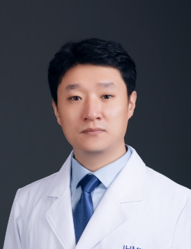 Yuanwei Zhang (张远伟)合作论文数Chinese Academy of Sciences57
 Hui Ma合作论文数43Ranjha Khan合作论文数38Jianteng Zhou合作论文数First Affiliated Hospital of USTC, Hefei National Laboratory for Physical Sciences at Microscale, the CAS Key Laboratory of Innate Immunity and Chronic Disease, School of Basic Medical Sciences, Division of Life Sciences and Medicine, CAS Center for Excellence in Molecular Cell Science, Collaborative Innovation Center of Genetics and Development, University of Science and Technology of China, Hefei 230027, China.32
 Wasim Shah合作论文数31Jiang Hanwei合作论文数30
 Bo Xu (许波)合作论文数School of Medicine, Division of Life Sciences and Medicine, University of Science and Technology of China;The First Affiliated Hospital of University of Science and Technology of China30
 Howard J. Cooke合作论文数MRC Human Genetics Unit, The University of Edinburgh25Sobia Dil合作论文数21
 Ihsan Khan合作论文数University of Science and Technology of China, University of Science and Technology of China21
 Suixing Fan合作论文数School of Basic Medical Sciences, University of Science and Technology of China21Asim Ali合作论文数University of Toronto/The Hospital for Sick Children21
 Furhan Iqbal合作论文数Inst Pure & Appl Biol, Bahauddin Zakariya Univ21
 Tanveer Abbas合作论文数Hefei Natl Lab Phys Sci Micros, Univ Sci & Technol China18Ao Ma合作论文数15
 Baolu Shi合作论文数13Jingwei Ye合作论文数13Beibei Zhang合作论文数From the Hefei National Laboratory for Physical Sciences at Microscale and School of Life Sciences, University of Science and Technology of China, Hefei, Anhui 230027, China.13Ihtisham Bukhari合作论文数13
 Fangpu Han (韩方普)合作论文数State Key Laboratory of Plant Cell and Chromosome Engineering, Institute of Genetics and Developmental Biology, Chinese Academy of Sciences;University of Chinese Academy of Sciences13
 Aurang Zeb合作论文数Nuclear Institute for Food and Agriculture13Daren Zhao合作论文数<institution>The First Affiliated Hospital of USTC, Hefei National Laboratory for Physical Sciences at the Microscale, The CAS Key Laboratory of Innate Immunity and Chronic Diseases, School of Life Sciences, CAS Center for Excellence in Molecular Cell Science, University of Science and Technology of China, Collaborative Innovation Center of Genetics and Development</institution>, Hefei 230027, Anhui, <country>China</country>12Yun Huang合作论文数Department of Pediatric Oncology and Children's Research Centre, University Children's Hospital Zürich12Khalid Khan合作论文数11
 Ansar Hussain合作论文数College of Agriculture and Biology, Zhongkai University of Agriculture and Engineering11
 Manan Khan合作论文数Chinese Academy of Sciences (CAS), University of Science &Technology of China11
 Ghulam Murtaza Dogar合作论文数Department of Pharmacy, COMSATS University Islamabad11Qiaomei Hao合作论文数11
 Li Yang (杨力)合作论文数Institutes of Biomedical Sciences, Fudan University11Asad Khan合作论文数10
 Handong Su (苏汉东)合作论文数College of Plant Science & Technology of Huazhong Agricultural University10Heli Hou合作论文数10
 Basit Shah合作论文数9Fazal Rahim合作论文数9Menglei Yang合作论文数9Chenjia Gong合作论文数9Jianing Gao合作论文数Molecular and Cell Genetics Laboratory, The CAS Key Laboratory of Innate Immunity and Chronic Diseases, Hefei National Laboratory for Physical Sciences at Microscale, School of Life Sciences, CAS Center for Excellence in Molecular Cell Science, University of Science and Technology of China, Hefei, Anhui, 230027, China.9
 Ahsanullah Unar合作论文数USTC, Univ Sci & Technol9Rongjun Ban合作论文数9Liangwen Zhong合作论文数Hefei National Laboratory for Physical Sciences at Microscale and School of Life Sciences, University of Science and Technology of China9
 Muhammad Zubair合作论文数Department of Chemistry, Faculty of Physical Science, Government College University9Jianze Xu合作论文数8
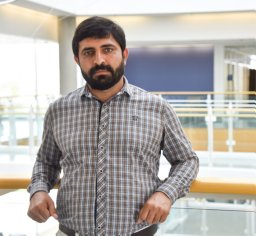 Teka Khan合作论文数8Xianrui Guo合作论文数8Shi Yin合作论文数8
 Long Jiang合作论文数School of Life Sciences, University of Science and Technology of China8




