OBJECTIVE:To measure the relative levels of signal and noise in expert diagnosis of epilepsy. METHODS:Twenty multinational epileptologists independently reviewed 50 vignettes of adult and pediatric patients presenting with suspected seizure(s) on two separate occasions with a ≥30-day washout period. Experts provided a diagnosis of epilepsy or non-epilepsy based on clinical information and, if requested, routine EEG and neuroimaging data. Cases had an established clinical diagnosis of epilepsy or non-epilepsy based on capture of habitual paroxysmal events on video-EEG or long-term clinical follow-up. Experts' judgments were analyzed to decompose variability into different sources: signal (objective differences between cases), level noise (experts' bias toward over/under-diagnosis), pattern noise (experts' idiosyncratic reactions to specific case features), and occasion noise (inconsistency across occasions). RESULTS:The probability of an expert making a different diagnosis for a given case on two different occasions was 16%. The probability of two different experts making a different diagnosis for the same case was 26%. Signal (case "difficulty") accounted for 66-69% of total variation, with 31-34% attributable to noise. Level noise was the largest contributor in the absence of EEG/neuroimaging results (23%), while pattern noise dominated when test results were available (24%). Occasion noise contributed relatively little (1%) but was still sufficient to cause diagnostic reversals in 16-22% between occasions. SIGNIFICANCE:The degree of noise in expert diagnosis of epilepsy is substantial, stemming primarily from physicians' idiosyncratic interpretations of case features and variable dispositions toward over- or under-diagnosis. Strategies to improve reliability are needed, including standardized data collection protocols and structured decision algorithms. For "difficult cases," where expert reliability and accuracy are lowest, our findings support current clinical practice which favors early referral for video-EEG monitoring over reliance on diagnostic anchoring. This diagnostic pathway may become more accessible with advances in EEG technology (e.g., wearable devices) and artificial intelligence.
OBJECTIVE:Quantitative assessment of extent of tissue resection following epilepsy surgery requires accurate delineation of the resection cavity on postoperative magnetic resonance imaging (MRI). Current methods for resection cavity masking are time-consuming and labor-intensive, and existing automated approaches exhibit variable segmentation accuracy, particularly on extratemporal resections. We developed MELD-PostOp, a deep learning tool trained and evaluated on a large, heterogeneous cohort to automatically segment resection cavities. METHODS:The study included 1.5- and 3T postoperative three-dimensional T1-weighted MRI images from the Multicentre Epilepsy Lesion Detection (MELD) project (nsubjects = 969, 27 centers) and from the EPISURG dataset (n = 133). The cohort included children and adults, alongside a range of resection locations, pathologies, and MRI characteristics. Resection cavities were individually segmented in 285 subjects and used to train an nnU-Net prototype model. The prototype model was used to generate an additional 680 resection masks, which were subsequently quality-controlled, edited, and combined with the original 285 to train the final MELD-PostOp model (n = 965). A Stratified Test Cohort (n = 50) and Independent Test Cohort (n = 87) were withheld for model evaluation. Performance was evaluated using Dice similarity coefficient (DSC), 95th percentile Hausdorff distance (HD95), number of predicted clusters, and inference runtime, and compared against established tools (Epic-CHOP, ResectVol, and RESSEG). RESULTS:MELD-PostOp achieved a median DSC of .85 and HD95 of 3.61 on the combined test cohort, outperforming Epic-CHOP (DSC .69, HD95 9.67), ResectVol (DSC .66, HD95 15.05), and RESSEG (DSC .43, HD95 32.67), with significant improvements seen in both temporal and especially extratemporal resections. The model detected 98.5% (135/137) of resection cavities. MELD-PostOp runtime was 17 s per MRI, compared to 612 s (ResectVol), 3205 s (Epic-CHOP), and 4 s (RESSEG). MELD-PostOp performance remained high across clinical and imaging subgroups (median DSC > .8). SIGNIFICANCE:MELD-PostOp is an open-source research tool that provides an accurate, efficient, and generalizable solution for postoperative resection cavity segmentation using only postoperative MRI scans.
Objective Quantitative assessment of extent of tissue resection following epilepsy surgery requires accurate delineation of the resection cavity on postoperative MRI. Current methods for resection cavity masking are time-consuming and labour-intensive, while existing automated approaches exhibit variable segmentation accuracy, particularly on extra-temporal resections. We developed MELD-PostOp, a deep learning tool trained and evaluated on a large, international, heterogeneous cohort to automatically segment resection cavities. Methods The study included 1.5 and 3T postoperative 3D T1-weighted MRI images from the Multicentre Epilepsy Lesion Detection (MELD) project (nsubjects=969, 27 centres) and from the EPISURG dataset (n=133). The cohort included both children and adults, alongside a range of resection locations, pathologies, and MRI characteristics. Resection cavities were individually segmented in 285 subjects and used to train an nnU-Net prototype model. The prototype model was used to generate an additional 680 resection masks, which were subsequently quality-controlled, edited and then combined with the original 285 to train the final MELD-PostOp model (n=965). A Stratified (STC; n=50) and Independent Test Cohort (ITC; n=87) were masked and withheld for model evaluation. Performance was evaluated using Dice Similarity Coefficient (DSC), 95th percentile Hausdorff distance (HD95), number of predicted clusters and inference runtime; and compared against established tools (Epic-CHOP and ResectVol). Results MELD-PostOp achieved a median DSC of 0.85 and HD95 of 3.61 on the combined test cohort, outperforming Epic-CHOP (DSC 0.68, HD95 9.54) and ResectVol (DSC 0.66, HD95 12.07), with significant improvements seen in both temporal and especially extra-temporal resections. The model detected 99% (135/137) of resection cavities. MELD-PostOp runtime was 17s per MRI, compared to 612s (ResectVol) and 3205s (Epic-CHOP). MELD-PostOp performance remained high across clinical and imaging subgroups (median DSC > 0.8). Significance MELD-PostOp provides an accurate, efficient and generalisable solution for postoperative resection cavity segmentation using only postoperative MRI scans. This open-source tool facilitates large-scale quantitative analysis to define what tissue is essential to resect for optimal epilepsy surgical outcomes. ### Competing Interest Statement Alexander G. Weil is a consultant for Monteri. Kate Davis is an advisory board member for NeuroPace, Rapport Therapeutics, Mosaica Therapeutics, UCB. ### Clinical Protocols ### Funding Statement JS, MR and KW are supported by Wellcome Trust (301991/Z/23/Z) and Epilepsy Research Institute UK (P2208). SA is supported by Epilepsy Research Institute UK(P2208). TJO is supported by NHMRC Investigator Grants (APP1176426 & APP2034258). HX is supported by National Institute of Child Health and Human Development (P50HD105328-01). NTC is supported by the National Institute Of Neurological Disorders And Stroke and the National Institutes of Health (Award Number K23NS131522). AGW and AH are supported by Canadian Institute for Health Research , Fonds de recherche du Quebec - Sante, Savoy Foundation, Department of surgery at Universite de Montreal, CHU Sainte-Justine Foundation. AI is supported by grants from the Fogarty International Center, National Institutes of Health, National Institutes of Aging (R01 AG057234, R01 AG075775, R01 AG21051, R01 AG083799, CARDS-NIH, R01 AG057234), Alzheimer's Association (SG-20-725707), Rainwater Charitable Foundation, ANID/FONDECYT Regular (1250091 and 1210176 and 1220995), ANID/PIA/ANILLOS ACT210096, JPI JPND-Care, DISCeRN 2025, FONDEF ID20I10152,ANID/FONDAP 15150012; Wellcome Trust (BRAIN-CLIMA 335293/Z/25/Z), and CliCBrain (Horizon ID: 101236426; DOI 10.3030/101236426, Marie Skłodowska-Curie Actions - MSCA). AC is supported by NIHR & GOSH BRC. MHE is supported by The Sigrid Juselius Foundation. RJP is supported by NIHR & GOSH Children's Charity. CLY is supported by CNPQ (445340/2024-0/; 313263/2025-6). FC is supported by Sao Paulo Research Foundation (FAPESP) grants 2013/07559-3 and 2021/12956-8. The content of this manuscript is solely the responsibility of the authors and does not necessarily represent the official views of any of the funders. The funders had no role in study design, data collection and analysis, decision to publish or preparation of the manuscript. ### Author Declarations I confirm all relevant ethical guidelines have been followed, and any necessary IRB and/or ethics committee approvals have been obtained. Yes The details of the IRB/oversight body that provided approval or exemption for the research described are given below: This study used anonymised MRI data collected as part of the Multi-centre Epilepsy Lesion Detection (MELD) Focal Epilepsies project (https://meldproject.github.io/), which has Health Research Authority ethical approval (IRAS: 301863) I confirm that all necessary patient/participant consent has been obtained and the appropriate institutional forms have been archived, and that any patient/participant/sample identifiers included were not known to anyone (e.g., hospital staff, patients or participants themselves) outside the research group so cannot be used to identify individuals. Yes I understand that all clinical trials and any other prospective interventional studies must be registered with an ICMJE-approved registry, such as ClinicalTrials.gov. I confirm that any such study reported in the manuscript has been registered and the trial registration ID is provided (note: if posting a prospective study registered retrospectively, please provide a statement in the trial ID field explaining why the study was not registered in advance). Yes I have followed all appropriate research reporting guidelines, such as any relevant EQUATOR Network research reporting checklist(s) and other pertinent material, if applicable. Yes Data sharing of the MELD Focal Epilepsies dataset is restricted as consent was not obtained from participants for public sharing of data. EPISURG dataset is available at:https://rdr.ucl.ac.uk/articles/dataset/EPISURG\_a\_dataset\_of\_postoperative\_magnetic\_resonance\_images\_MRI\_for\_quantitative\_analysis\_of\_resection\_neurosurgery\_for\_refractory_epilepsy/9996158?file=26153588 All code for model design, development and statistical analyses alongside the MELD-PostOp model is available at: https://github.com/MELDProject/MELD-PostOp/tree/main
BACKGROUND:People with epilepsy (PWE) may experience seizure clusters, broadly defined as ≥2 seizures that occur in close proximity. In epilepsy monitoring units (EMUs), seizure clusters can spontaneously occur during long-term videoelectroencephalogram monitoring (LTVEM) or as a result of antiseizure medication dose adjustments. In this survey, we examined the experiences and practices of expert clinicians with seizure clusters in EMUs. METHODS:A 55-item survey was sent to members of an Epilepsy Education Council who are epilepsy experts. Items described experiences, treatment practices, and negative outcomes with seizure clusters in EMUs. RESULTS:Of the 15 experts (aged 43-77 y), 14 are physicians and 1 is an advanced practice provider; 14 work at level 4 epilepsy centers. The definition of seizure cluster varied across experts, from 2 seizures in 1 hour to 3 seizures over 24 hours. Twelve experts prescribe immediate-use rescue medication (RM) during EMU stay, usually a benzodiazepine. An intranasal route is preferred by 11 if intravenous access is unavailable. Nine experts have had a presurgical evaluation compromised owing to seizure clusters during LTVEM, and 12 have cared for PWE who required transfer to a higher-level care (eg, intensive care unit) owing to seizure clusters. Thirteen experts indicated they would follow expert consensus recommendations for immediate-use RMs in the EMU if available. CONCLUSIONS:In the EMU, seizure clusters may compromise presurgical evaluations and require higher levels of care. Consensus recommendations are needed to guide patient-specific treatment practices before, during, and after EMU admission.
Introduction Electroconvulsive therapy (ECT) is a highly effective treatment for refractory depression, but it may also cause cognitive side effects. Despite decades of use, the mechanisms by which ECT exerts both its antidepressant and cognitive effects are still poorly understood, with the latter substantially limiting referral and adherence to therapy. ECT induces changes in correlated neural activity—functional connectivity—across various brain networks, which may underlie both its clinical efficacy and associated cognitive side effects. Electroencephalography (EEG) could address these knowledge gaps by identifying biomarkers that predict therapeutic outcomes or cognitive side effects. Such developments could ultimately improve patient selection and adherence. Such markers likely span large-scale functional brain networks or temporal dynamics of brain activity during sleep. We hypothesise that enhancement in slow wave sleep mediates the relationship between antidepressant effects and changes in functional connectivity throughout the course of ECT.Methods and analysis Disruptions of Brain Networks and Sleep by Electroconvulsive Therapy (DNS-ECT) is an ongoing observational study investigating the impact of ECT on large-scale brain functional networks and their relationships to sleep slow waves, an EEG marker linked to synaptic plasticity. The novelty of this study stems from our focus on the assessment of EEG markers during sleep, wakefulness and ECT-induced seizures over the course of therapy. Graph-based network analyses of high-density EEG signals allow characterisation of functional networks locally in specific subnetworks and globally over large-scale functional networks. Longitudinal assessments of EEG alongside clinical and cognitive outcomes provide a unique opportunity to improve our understanding of the circuit mechanisms underlying the development of cognitive impairments and antidepressant effects incurred during ECT.Ethics and dissemination Recruitment for this 5-year study started in March 2023. Dissemination plans include presentations at scientific conferences and peer-reviewed publications. This study has been registered with ClinicalTrials.gov registry under identifier.Trial registration number NCT05905705.
For acute treatment of seizure clusters in patients with epilepsy, intranasal administration of acute seizure therapies has been shown to provide accessibility and ease of use to care partners as well as the potential for self-administration by patients. Diazepam nasal spray (Valtoco®) was approved by the US Food and Drug Administration for acute treatment of intermittent, stereotypic episodes of frequent seizure activity (ie, seizure clusters, acute repetitive seizures) in patients with epilepsy aged ≥6 years. Self-administration consistent with the prescribing information is feasible and was reported by a subgroup of patients (n = 27 of 163) in a long-term phase 3 safety study. Data regarding self-administration among these patients with seizure clusters are examined here to explore the safety profiles and measures of effectiveness, as well as the quality of life of those who self-treated. In addition, this focused look at patients who self-administered diazepam nasal spray may offer some insights into the characteristics of patients who may be appropriate for self-administration.
Abstract Sex differences in drug pharmacokinetics include variations in the expression of the cytochrome P450 enzymes, which are involved in the metabolism of benzodiazepines. It is unclear whether sex influences outcomes associated with intranasally administered drugs. A post hoc analysis of sex differences was conducted to evaluate the effectiveness and safety of diazepam nasal spray, which included examining changes in the number of days between seizure clusters over time (SEIzure interVAL [SEIVAL]). Diazepam nasal spray is approved for acute treatment of seizure clusters in patients with epilepsy aged ≥6 years. Data from a phase 3 safety study were used to determine the proportion of second doses used within 24 h (ie, a proxy for effectiveness) and SEIVAL. Adverse events were recorded. Of 163 treated patients, 89 were female, and 74 were male. Approximately 16% of both sexes self‐administered the study drug. A slightly higher proportion of seizure clusters was treated with a second dose in female (14.7%) than male (9.4%) patients. SEIVAL increased significantly and substantially over a year for all patients. The safety profile was generally similar between the sexes. These results suggest that potential sex differences in benzodiazepine pharmacokinetics do not meaningfully influence outcomes associated with diazepam nasal spray. Plain Language Summary Some drugs may have differences in absorption and metabolism between genders that could translate into differences in safety and effectiveness. This safety study looked at diazepam nasal spray for treating seizure clusters in patients at least 6 years old. It found that safety was about the same for females and males. For both groups, most clusters stopped after only 1 dose of the drug, and the time between treated clusters got longer over a year.
Objective: Central-positive complexes (CPCs) are elicited during electroconvulsive therapy (ECT) as gen-eralized high-amplitude waveforms with maximum positive voltage over the vertex. While these com-plexes have been qualitatively assessed in previous literature, quantitative analyses are lacking. This study aims to characterize CPCs across temporal, spatial, and spectral domains. Methods: High-density 64-electrode electroencephalogram (EEG) recordings during 50 seizures acquired from 11 patients undergoing right unilateral ECT allowed for evaluation of spatiotemporal characteristics of CPCs via source localization and spectral analysis. Results: Peak-amplitude CPC scalp topology was consistent across seizures, showing maximal positive polarity over the midline fronto-central region and maximal negative polarity over the suborbital regions. The sources of these peak potentials were localized to the bilateral medial thalamus and cingulate corti-cal regions. Delta, beta, and gamma oscillations were correlated with the peak amplitude of CPCs during seizures induced during ketamine, whereas delta and gamma oscillations were associated with CPC peaks during etomidate anesthesia (excluding the dose-charge titration). Conclusions: Our findings demonstrate the consistency of CPC presence across participant, stimulus charge, time, and anesthetic agent, with peaks localized to bilateral medial thalamus and cingulate cor-tical regions and associated with delta, beta, and gamma band oscillations (depending on the anesthetic condition).Significance: The consistency and reproducibility of CPCs offers ECT as a new avenue for studying the dynamics of generalized seizure activity and thalamocortical networks.(c) 2022 International Federation of Clinical Neurophysiology. Published by Elsevier B.V. All rights reserved.
With more than 6000 attendees between in-person and virtual offerings, the American Epilepsy Society Meeting 2022 in Nashville, felt as busy as in prepandemic times. An ever-growing number of physicians, scientists, and allied health professionals gathered to learn a variety of topics about epilepsy. The program was carefully tailored to meet the needs of professionals with different interests and career stages. This article summarizes the different symposia presented at the meeting. Basic science lectures addressed the primary elements of seizure generation and pathophysiology of epilepsy in different disease states. Scientists congregated to learn about anti-seizure medications, mechanisms of action, and new tools to treat epilepsy including surgery and neurostimulation. Some symposia were also dedicated to discuss epilepsy comorbidities and practical issues regarding epilepsy care. An increasing number of patient advocates discussing their stories were intertwined within scientific activities. Many smaller group sessions targeted more specific topics to encourage member participation, including Special Interest Groups, Investigator, and Skills Workshops. Special lectures included the renown Hoyer and Lombroso, an ILAE/IBE joint session, a spotlight on the impact of Dobbs v. Jackson on reproductive health in epilepsy, and a joint session with the NAEC on coding and reimbursement policies. The hot topics symposium was focused on traumatic brain injury and post-traumatic epilepsy. A balanced collaboration with the industry allowed presentations of the latest pharmaceutical and engineering advances in satellite symposia.
Focal cortical dysplasia (FCD) type II is a highly epileptogenic developmental malformation and a common cause of surgically treated drug-resistant epilepsy. While clinical observations suggest frequent occurrence in the frontal lobe, mechanisms for such propensity remain unexplored. Here, we hypothesized that cortex-wide spatial associations of FCD distribution with cortical cytoarchitecture, gene expression and organizational axes may offer complementary insights into processes that predispose given cortical regions to harbour FCD. We mapped the cortex-wide MRI distribution of FCDs in 337 patients collected from 13 sites worldwide. We then determined its associations with (i) cytoarchitectural features using histological atlases by Von Economo and Koskinas and BigBrain; (ii) whole-brain gene expression and spatiotemporal dynamics from prenatal to adulthood stages using the Allen Human Brain Atlas and PsychENCODE BrainSpan; and (iii) macroscale developmental axes of cortical organization. FCD lesions were preferentially located in the prefrontal and fronto-limbic cortices typified by low neuron density, large soma and thick grey matter. Transcriptomic associations with FCD distribution uncovered a prenatal component related to neuroglial proliferation and differentiation, likely accounting for the dysplastic makeup, and a postnatal component related to synaptogenesis and circuit organization, possibly contributing to circuit-level hyperexcitability. FCD distribution showed a strong association with the anterior region of the antero-posterior axis derived from heritability analysis of interregional structural covariance of cortical thickness, but not with structural and functional hierarchical axes. Reliability of all results was confirmed through resampling techniques. Multimodal associations with cytoarchitecture, gene expression and axes of cortical organization indicate that prenatal neurogenesis and postnatal synaptogenesis may be key points of developmental vulnerability of the frontal lobe to FCD. Concordant with a causal role of atypical neuroglial proliferation and growth, our results indicate that FCD-vulnerable cortices display properties indicative of earlier termination of neurogenesis and initiation of cell growth. They also suggest a potential contribution of aberrant postnatal synaptogenesis and circuit development to FCD epileptogenicity.
This study aimed to investigate prognostic significance and brain metabolic mechanism of hyponatremia in anti-leucine-rich glioma-inactivated protein 1 (LGI1) encephalitis. After adjusting for confounders, patients with moderate and severe hyponatremia had significantly increased risk of poor functional outcome and sequelae of seizures. In addition, serum sodium was negatively correlated with normalized ratio of the standardized uptake value of medial temporal lobe (MTL), basal ganglia (BG), and hypothalamus on positron emission tomography (PET) and which was further validated using voxel-wise analysis, suggesting an extra-hypothalamus (BG and MTL) localization for hyponatremia.
This exploratory analysis evaluated the safety of second doses of diazepam nasal spray (Valtoco®) within 4 hours of the first dose in a long-term, phase 3, open-label, repeat-dose safety study.
Objective Current diazepam nasal spray labeling requires waiting 4 h before administering a second dose. The objective of the current analyses was to examine safety and pharmacokinetic profiles of second doses of diazepam nasal spray given 0-4 h after the first dose. Methods Two datasets were analyzed. The first, a long-term, repeat-dose safety study of diazepam nasal spray, compared rates of treatment-emergent adverse events (TEAEs), serious TEAEs, and treatment-related TEAEs for patients receiving >= 1 second dose <= 4 h versus all second doses >4 h after the first. The second was a population pharmacokinetic analysis using data from three phase 1 studies to model drug exposure when a second dose of diazepam nasal spray was administered across multiple time points (1 min-4 h) following the first dose. Results In the repeat-dose safety study, a second dose of diazepam nasal spray was administered <= 24 h after the first to treat 485 seizure clusters in 79 patients. Rates of TEAEs were similar between patients receiving >= 1 second dose in <= 4 h (89.5%, n = 38) compared with >4-24 h only (80.5%, n = 41). The most common treatment-related TEAEs were associated with nasal discomfort, which was mild or moderate and transient. There were no reports of respiratory or cardiac depression. The pharmacokinetic simulations of second doses predicted comparable elevations of plasma diazepam concentrations with administrations across a range of intervals after the first dose (1 min-4 h). Significance These data indicate that the safety and pharmacokinetic profiles of a second dose of diazepam nasal spray administered within 4 h of the first dose are consistent with those associated with current labeling. This is potentially important for patients with seizure clusters who have a recurrent seizure within 4 h of first treatment and might benefit from immediate retreatment to reduce the risk of progression to status epilepticus.
This analysis of a long-term safety study examined time to administration and ease of use of diazepam nasal spray (Valtoco®) for patients with epilepsy experiencing seizure clusters.
Objective: Periods of low-amplitude electroencephalographic (EEG) signal (quiescence) are present during both anesthetic-induced burst suppression (BS) and postictal generalized electroencephalographic suppression (PGES). PGES following generalized seizures induced by electroconvulsive therapy (ECT) has been previously linked to antidepressant response. The commonality of quiescence during both BS and PGES motivated trials to recapitulate the antidepressant effects of ECT using high doses of anesthetics. However, there have been no direct electrographic comparisons of these quiescent periods to address whether these are distinct entities. Methods: We compared periods of EEG quiescence recorded from two human studies: BS induced in 29 healthy adult volunteers by isoflurane general anesthesia and PGES in 11 patients undergoing right unilateral ECT for treatment-resistant depression. An automated algorithm allowed detection of EEG quiescence based on a 10-microvolt amplitude threshold. Spatial, spectral, and temporal analyses compared quiescent epochs during BS and PGES. Results: The median (interquartile range) voltage for quiescent periods during PGES was greater than during BS (1.81 (0.22) microvolts vs 1.22 (0.33) microvolts, p < 0.001). Relative power was greater for quiescence during PGES than BS for the 1-4 Hz delta band (p < 0.001), at the expense of power in the theta (4-8 Hz, p < 0.001), beta (13-30 Hz, p = 0.04) and gamma (30-70 Hz, p = 0.006) frequency bands. Topographic analyses revealed that amplitude across the scalp was consistently higher for quiescent periods during PGES than BS, whose voltage was within the noise floor. Conclusions: Quiescent epochs during PGES and BS have distinct patterns of EEG signals across voltage, frequency, and spatial domains. Significance: Quiescent epochs during PGES and BS, important neurophysiological markers for clinical outcomes, are shown to have distinct voltage and frequency characteristics. (C) 2022 International Federation of Clinical Neurophysiology. Published by Elsevier B.V. All rights reserved.
Seizures, transient signs or symptoms caused by abnormal surges of electrical activity in the brain, can result from epilepsy, a neurologic disorder characterized by abnormal electrical brain activity causing recurrent, unprovoked seizures, or from other inciting causes, such as high fever or substance abuse (1). Seizures generally account for approximately 1% of all emergency department (ED) visits (2,3). Persons of any age can experience seizures, and outcomes might range from no complications for those with a single seizure to increased risk for injury, comorbidity, impaired quality of life, and early mortality for those with epilepsy (4). To examine trends in weekly seizure- or epilepsy-related (seizure-related) ED visits† in the United States before and during the COVID-19 pandemic, CDC analyzed data from the National Syndromic Surveillance Program (NSSP).§ Seizure-related ED visits decreased abruptly during the early pandemic period. By the end of 2020, seizure-related ED visits returned almost to prepandemic levels for persons of all ages, except children aged 0-9 years. By mid-2021, however, this age group gradually returned to baseline as well. Reasons for the decrease in seizure-related ED visits in 2020 among all age groups and the slow return to baseline among children aged 0-9 years compared with other age groups are unclear. The decrease might have been associated with fear of exposure to COVID-19 infection in EDs deterring parents or guardians of children from seeking care, adherence to mitigation measures including avoiding public settings such as EDs, or increased access to telehealth services decreasing the need for ED visits (5). These findings reinforce the importance of understanding factors associated with ED avoidance among persons with epilepsy or seizure, the importance that all eligible persons be up to date¶ with COVID-19 vaccination, and the need to encourage persons to seek appropriate care for seizure-related emergencies** to prevent adverse outcomes.
Introduction Electroconvulsive therapy (ECT) is an effective intervention for patients with major depressive disorder (MDD). Despite longstanding use, the underlying mechanisms of ECT are unknown, and there are no objective prognostic biomarkers that are routinely used for ECT response. Two electroencephalographic (EEG) markers, sleep slow waves and sleep spindles, could address these needs. Both sleep microstructure EEG markers are associated with synaptic plasticity, implicated in memory consolidation, and have reduced expression in depressed individuals. We hypothesize that ECT alleviates depression through enhanced expression of sleep slow waves and sleep spindles, thereby facilitating synaptic reconfiguration in pathologic neural circuits. Methods Correlating ECT Response to EEG Markers (CET-REM) is a single-center, prospective, observational investigation. Wireless wearable headbands with dry EEG electrodes will be utilized for at-home unattended sleep studies to allow calculation of quantitative measures of sleep slow waves (EEG SWA, 0.5–4 Hz power) and sleep spindles (density in number/minute). High-density EEG data will be acquired during ECT to quantify seizure markers. Discussion This innovative study focuses on the longitudinal relationships of sleep microstructure and ECT seizure markers over the treatment course. We anticipate that the results from this study will improve our understanding of ECT.
 Enrique Carrazana合作论文数27Adrian L Rabinowicz合作论文数26
 Michael R. Sperling合作论文数Department of Neurology, Sidney Kimmel Medical College, Thomas Jefferson University;Jefferson Comprehensive Epilepsy Center, Thomas Jefferson University Hospital;Clinical Neurophysiology Laboratory, Thomas Jefferson University Hospital25
 James W. Wheless合作论文数Division of Peds Neurology, Department of Pediatrics, College of Medicine, University of Tennessee Health Science Center23
 Tarquinio Daniel C合作论文数19
 Gregory D. Cascino合作论文数Department of Neurology, Mayo Clinic16Ian Miller合作论文数15Dennis J. Dlugos合作论文数Department of Neurology, Perelman School of Medicine, University of Pennsylvania14Jay Desai合作论文数14
 Kore Liow合作论文数14
 Eric Segal合作论文数13
 Luigi Maccotta合作论文数13Nuri B. Farber合作论文数12
 Ben Julian Agustin Palanca合作论文数Sleepy Brain Lab, Department of Anesthesiology, School of Medicine, Washington University11Eisenman Lawrence N合作论文数9
 Terry O’Brien合作论文数Department of Medicine, Faculty of Medicine, Dentistry and Health Sciences, Monash University;Department of Neuroscience, School of Translational Medicine, Monash University9Victor Biton合作论文数Clinical Trials, Inc.9Blanca Vazquez合作论文数Department of Neurology, Department of Medicine, New York University8
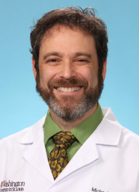 Michael Avidan合作论文数Division of Education, Department of Anesthesiology, School of Medicine, Washington University in St. Louis;Institute of Clinical and Translational Sciences, School of Medicine, Washington University in St. Louis;Roy and Diana Vagelos Division of Biology & Biomedical Sciences, School of Medicine, Washington University in St. Louis8
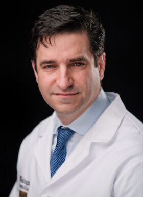 Eric C. Leuthardt合作论文数Department of Neurological Surgery, Washington University School of Medicine in St. Louis;Division of Neurotechnology, Washington University School of Medicine in St. Louis;Barnes-Jewish Hospital;St. Louis Children’s Hospital8
 Mohammadmehdi Kafashan合作论文数7
 Shinung Ching合作论文数Dept Elect & Syst Engn, Washington Univ7
 L Brian Hickman合作论文数David Geffen School of Medicine, UCLA6Steve Chung合作论文数6Ilan Blatt合作论文数5
 Andrea Bernasconi合作论文数Department of Neurology and Neurosurgery, Faculty of Medicine and Health Sciences, McGill University5Misra Sunita N合作论文数5
 Balduin Lawson合作论文数5
 Fernando Cendes合作论文数Department of Neurology, State University of Campinas5B. Keith Day合作论文数4
 Richard D Bucholz合作论文数4Weldon Mauney合作论文数4Emma R Trammel合作论文数4Pavel Klein合作论文数4David Cook合作论文数4Emma R Huels合作论文数4
 Ravnoor S. Gill合作论文数McGill Univ, Montreal Neurol Inst & Hosp, Neuroimaging Epilepsy Lab, Montreal, PQ, Canada4Alyssa K Labonte合作论文数4
 Lei Wang合作论文数Department of Psychiatry and Behavioral Health, College of Medicine, The Ohio State University;Center for Cognitive and Behavioral Brain Imaging, College of Arts and Sciences, The Ohio State University4
 Ingmar Blumcke合作论文数4Bouilleret Viviane合作论文数4Moseley Emily D合作论文数4Clark Annie M合作论文数4
 Bob Anders合作论文数4
 Matthias Koepp合作论文数Department of Clinical and Experimental Epilepsy, Queen Square Institute of Neurology, University College London4Graeme D. Jackson合作论文数4
 Tammie L. S. Benzinger合作论文数Neuroimaging Labs Research Center, Mallinckrodt Institute of Radiology, Washington University School of Medicine in St. Louis4
 Neda Bernasconi合作论文数4
 Eric J. Lenze合作论文数Healthy Mind Lab, Department of Psychiatry, Washington University School of Medicine in St. Louis4




