BACKGROUND: Intracardiac leiomyomatosis is a rare leiomyoma that originates from the uterus. It can be easily misdiagnosed because of its nonspecific manifestations. In this case, we aim to emphasize the complete resection of the ICL with a single-stage laparotomy. CASE PRESENTATION: Here, we report a case of a 36-year-old female who presented with dyspnea and a mobile tumor in the right atrium that was detected in transthoracic echocardiography prior to cosmetic surgery. The results of computed tomography (CT) scan, Cardiovascular magnetic resonance (CMR), and magnetic resonance venography (MRV) revealed a mass in the IVC extending into the right atrium and pelvic veins. One-stage surgery, along with transesophageal echocardiography monitoring, was performed for the patient, and the tumor was resected entirely without the need for open-heart surgery. CONCLUSION: This case report suggests that, in selected cases, one-stage surgery through laparotomy for resecting intravenous leiomyomatosis may be a safer and more efficient approach in comparison with the traditional two-stage operation, due to fewer complications. Based on our case, anticoagulant therapy may also be essential to reduce the risk of embolism during this single-stage laparotomy. Further studies are needed to validate the efficacy and safety of this approach.
Background Reliable preoperative assessment of the aortic annulus is essential for aortic valve replacement and transcatheter aortic valve implantation (TAVI). Transthoracic echocardiography (TTE) and three-dimensional transesophageal echocardiography (3D TEE) are commonly used noninvasive tools for annular sizing; however, their agreement with intraoperative surgical sizing remains uncertain, particularly because surgical sizing reflects a postexcisional functional annular dimension rather than a direct anatomical gold standard. Objective To evaluate and compare the agreement of TTE and 3D TEE with direct surgical sizing of the aortic annulus. Methods This retrospective study included 106 patients who underwent aortic valve replacement between January 2019 and December 2023. The mean patient age was 57.2 +/- 9.8 years (median 57, IQR 46-66), and 68 patients (64.2%) were male. The surgical annulus diameter had a median of 23 mm (IQR 22-25 mm). TTE and 3D TEE yielded lower annular diameters with medians of 22 mm (IQR 20-23 mm) and 22 mm (IQR 21-24 mm), respectively. Compared with surgical sizing, the median differences were -2 mm (IQR -4 to 0 mm, p < 0.001) for TTE and -1 mm (IQR -3 to 1 mm, p = 0.010) for 3D TEE. Bland-Altman analysis demonstrated wide limits of agreement for both modalities (TTE: -6 to +2 mm; 3D TEE: -6 to +4 mm). The correlation between TTE and 3D TEE was strong (r = 0.790, p < 0.001), whereas correlations between each echocardiographic modality and surgical sizing were very weak and statistically nonsignificant (TTE: r = 0.084; 3D TEE: r = 0.038). Conclusion TTE and 3D TEE demonstrated limited agreement with surgical aortic annular sizing, with weak correlations and wide limits of agreement. Although 3D TEE showed smaller mean systematic bias than TTE at the group level, substantial individual-level variability remained. Because surgical sizing is performed after leaflet excision and annular debridement and reflects functional rather than purely anatomical sizing, these findings should be interpreted as agreement with surgical sizing rather than validation against a true anatomical gold standard.
Background: Cardiac tumors are extremely rare conditions encompassing a diverse range of mass lesions, including primary tumors originating from the heart and great vessels, metastatic tumors, and tumor-like lesions that do not fit the conventional definition of a tumor or neoplasm. In contrast to metastatic tumors, primary cardiac tumors and tumorlike lesions are rare, with most primary neoplasms being benign. Methods: A histopathological study was conducted among all patients who presented to Rajaie Cardiovascular Institute between 2020 and 2025. Data were collected from surgical pathology records to identify relevant cases. Results: Among 28 patients diagnosed with benign cardiac tumors and tumor-like conditions (excluding myxomas), 15 were male, and 13 were female. The identified benign tumors included rhabdomyoma, fibroma, papillary fibroelastoma, lipoma, lymphangioma, capillary hemangioma, and cavernous hemangioma. Additionally, two cystic lesions-a pericardial mesothelial cyst and a hydatid cyst-were noted, along with a case of a fungal ball. Conclusions: All cardiac tumors should undergo histologic examination to confirm the diagnosis and rule out malignancy, thereby aiding the development of the most effective treatment strategy.
BACKGROUND:Infective aortitis is a rare complication following coarctoplasty. Unrecognized concomitant congenital defects such as patent ductus arteriosus (PDA) could be devastating. CASE SUMMARY:We describe a young woman presenting with constitutional symptoms, elevated inflammatory markers, and a history of coarctoplasty with stent a decade earlier. Imaging revealed a large previously overlooked PDA and an aortic pseudoaneurysm distal to the stent covered by vegetations. Positron emission tomography/computed tomography demonstrated metabolically active lesions within the pulmonary artery and spleen. Broad-spectrum antibiotics, surgical stent excision, pseudoaneurysm repair with homograft, and PDA ligation achieved favorable outcomes with no residual infection on follow-up. DISCUSSION:Sophisticated diagnostic modalities and a multidisciplinary approach are essential in rare, life-threatening complications. With growing use of endovascular therapies for aortic coarctation, awareness of uncommon complications and their management is crucial. TAKE-HOME MESSAGES:A comprehensive preprocedural assessment to detect coexisting anomalies is essential. Multimodality imaging is key for diagnosis, treatment planning, and follow-up. Combined antibiotic therapy and surgical intervention ensured complete recovery.
ObjectivePectus excavatum and, less commonly, pectus carinatum are congenital chest wall deformities. These may be associated with kyphoscoliosis, pulmonary, and cardiac diseases. However, the incidence of concomitant cardiac disease in patients with pectus deformities is not well-documented. There is no consensus on the optimal age for repair, the most effective technique, or whether a simultaneous or staged approach is preferable. This study presents our experience with combined pectus and cardiac surgery.MethodsThirteen patients (aged 6-32 years) with pectus deformities and concurrent cardiac disease underwent surgery between 2016 and 2024. Eleven had pectus excavatum, one had a mixed deformity, and one had Pouter chest wall deformity. Seven had Marfan syndrome, one had Noonan syndrome, and three had mitral valve regurgitation requiring Bentall and/or valve replacement. The patient with Pouter chest wall deformity had right ventricular outflow tract (RVOT) stenosis, pulmonary valve stenosis, and a patent foramen ovale. Another had a failed Ravitch repair with a right coronary artery to RVOT fistula.ResultsThere was no mortality. All patients were extubated within 72 h, except one requiring reintubation for seven days due to COVID-19 pneumonia. Patients' characteristics are summarized in Table 1. One patient with a previous failed Ravitch repair required bilateral costochondral fixation. Pectus repair outcomes were excellent in 11 patients, while two children developed postoperative pectus carinatum.ConclusionThe choice between simultaneous or staged repair remains debated. Our experience suggests the modified open Nuss procedure is preferable for concomitant pectus and cardiac surgery, except when infeasible.
Left atrial appendage aneurysm (LAAA) is a rare cardiovascular anomaly, with fewer than 200 documented cases. It is often associated with severe complications, such as arrhythmias and thromboembolic events. Recent evidence suggests that viral infections, particularly viral myocarditis, might be an underlying cause of LAAA. We report the case of a 36-year-old woman with a history of asthma who presented with palpitations and atrial tachyarrhythmia two months after a severe upper respiratory infection. Transthoracic echocardiography revealed a large aneurysmal left atrial appendage (LAA) measuring 5.6 × 3.5 cm and a reduced left ventricular ejection fraction of 50%. Cardiac computed tomography confirmed the LAAA and revealed abnormal flow dynamics. Late gadolinium enhancement showed mid-subepicardial hyperenhancement in the posterolateral segments of the left ventricular wall, consistent with a previous myocarditis. The patient underwent a novel, minimally invasive endoscopic thoracoscopic resection of the aneurysm, guided by transesophageal echocardiography. No thrombus was present. The procedure was successfully completed with the aid of cardiopulmonary bypass. This case highlights a potential association between viral myocarditis and LAAA, while also acknowledging the possibility of a congenital and incidentally discovered aneurysm. It underscores the critical role of multimodal imaging in accurate diagnosis and management. The successful minimally invasive surgical resection and subsequent restoration of cardiac function demonstrate the effectiveness of this approach, offering a promising outlook for patients with LAAA. Clinicians should consider viral infections as potential contributors to LAAA development and advocate for early diagnosis and intervention to improve clinical outcomes.
The DeVega annuloplasty procedure involves a double-running suture line along the circumference of the tricuspid annulus, encircling the anterior and posterior leaflets. A rare mechanical complication of this technique is the detachment of the suturing from the annulus, which can be referred to as the "bowstring sign" or "guitar string sign."
Background: Mastocytosis is a systemic disease involving the clonal expansion of mast cells in multiple organs. Given that immune system overreaction and excessive histamine release are among the most prominent events in mastocytosis, the incidence of complications caused by immune reactions is expected to increase across various organs. While systemic manifestations of mastocytosis have been reported frequently, cardiac complications are less often discussed. These Cardiac complications can be early indicators of the disease but such uncommon features may lead to delays in diagnosis. The significance of mast cells and histamine release in the cardiovascular system is acknowledged in prior studies. Case Presentation: This study presents a case of recurrent prosthetic mitral valve malfunction in a 52-year-old patient with a history of cutaneous mastocytosis, who underwent mitral valve replacement three times over ten years. Despite being on appropriate anticoagulation therapy (INR: 2.5-3.5), the patient experienced recurrent prosthetic valve thrombosis. This is, to our knowledge, the first report of prosthetic mitral valve thrombosis in a patient with mastocytosis. Conclusion: Interestingly, cardiac complications may be the first presentation of systemic mastocytosis, diagnosed long after the initial symptoms. The majority of such cases had no visible cutaneous manifestations (table 1). Regarding our case report, we recommend our colleagues to closely monitor and remain vigilant for possible cardiac symptoms of mastocytosis patients with prosthetic cardiac valves.
Background: Spontaneous coronary artery dissection is a rare and important cause of myocardial infarction, especially in young women without other coronary artery disease . This arterial dissection can occur within or between any of the 3 layers. Its predisposing factors include connective tissue diseases (Marfone syndrome, Ehlers-Danlos syndrome), vasculitis (polyarteritis nodosa, systemic lupus erythematosus, and Kawasaki disease), atherosclerosis and fibromuscular dysplasia. Clinical presentations of spontaneous coronary artery dissection are wide spectrum from asymptomatic to acute coronary disease, sustained ventricular arrhythmia and sudden cardiac death. Case presentation: We describe A 33-year-old man with history of Hodgkin's lymphoma five years earlier that became a candidate for Patent foramen ovale closure due to recurrent embolic cerebrovascular accident . Before the intervention, coronary angiography incidentally showed dissection in the left main and all major coronary arteries. Conclusions: Based on our hypothesis, chemoradiotherapy-induced arteriopathies could be consider as a predisposing factor for spontaneous coronary artery dissection.
Background: Nonischemic functional mitral regurgitation (FMR) is accompanied by dire long-term consequences. The treatment revolves around correcting the underlying left ventricular dysfunction. This study reports the long-term adverse outcomes of nonischemic FMR.Methods: We enrolled 200 patients with at-least-moderate nonischemic FMR undergoing medical treatment and/or cardiac resynchronization therapy between 2003 and 2019. MR severity and left ventricular dysfunction parameters were obtained. The endpoint outcomes were all-cause mortality, stroke, all-cause rehospitalization, and the need for heart transplantation.Results: Two hundred participants, 104 men (52%) and 96 women (48%), with a median age of 61 years (interquartile range [IQR], 50-70) at diagnosis and a median follow-up of 2 years (IQR, 1-4), were enrolled. All-cause mortality, all-cause rehospitalization, and need for heart transplantation were significantly associated with lower left ventricular ejection fraction and tricuspid annular plane systolic excursion (TAPSE) at diagnosis (P < 0.05). Baseline MR severity was significantly associated with stroke (P = 0.026) and all-cause rehospitalization (P < 0.001).MR severity, New York Heart Association (NYHA) classification, left ventricular end-diastolic diameter, and TAPSE improved at follow-up (P < 0.001). ACEi/ARB (P = 0.008), nitrate (P = 0.001), and hydralazine (P = 0.006) were associated with MR severity improvement. A significant difference was observed between survival free of all-cause mortality according to left ventricular ejection fraction (P = 0.041).Conclusions: We reported freedom from all-cause mortality, cardiac mortality, and composite endpoints (all-cause mortality, heart transplantation, and stroke) in nonischemic FMR patients. We detected a significant decline in MR severity and NYHA classification during follow-up. Overall, the FMR-associated mortality risk can be significantly reduced by adhering to treatment guidelines in a tertiary heart center.
BACKGROUND:Left ventricular pseudoaneurysm (LVP) is a rare but life-threatening condition resulting from acute myocardial infarction, trauma, bacterial infection, or previous cardiac operations. Diagnosis can be challenging as LVPs remain asymptomatic or present with nonspecific clinical symptoms. Early diagnosis is crucial to prevent rupture and recurrent septicemia. Various imaging techniques can aid in diagnosis, including transthoracic echocardiography (TTE), transesophageal echocardiography, computed tomography angiography, and cardiac magnetic resonance imaging. CASE REPORT:A 72-year-old man with a history of coronary artery bypass grafting presented with episodes of recurrent fever. An infected LVP was diagnosed using TTE and thoracic Computed tomography (CT) angiography. The patient underwent removal of the infected hematoma with excision and repair of the pseudoaneurysm via left anterior thoracotomy with peripheral cannulation. The neck of the pseudoaneurysm was repaired with a Dacron patch. Post-operative TTE showed no residual pseudoaneurysm tissue, and the patient recovered well. CONCLUSION:Our experience with the anterior thoracotomy approach with peripheral cannulation in specific cases of infected LVPs has yielded promising results. However, it is crucial to recognize that this approach may not be universally suitable.
Background: Improving patients' outcomes using enhanced recovery after surgery (ERAS) during the surgical period has significant economic savings and increases organizational productivity. We evaluated the effects of ERAS protocol on outcomes including high sensitive-C-reactive protein (hs-CRP), hospitalization, intensive care unit (ICU) stay, feeding tolerance and pain score of cardiac surgical patients. Methods: A total of 260 patients were randomly assigned to the ERAS and control groups according to stratified block randomization. Fasting time in the ERAS group reduced from the conventional 12 h to 6 h with light meals. Also, on the day of the operation, 2 hours before the surgery, they received 250 mL of oral carbohydrate solution containing 25 g glucose. The control group received conventional standard care. Serum hs-CRP was measured before and after the operation. Results: Out of 260 participants, 107 patients received protocolized care (ERAS group), and 103 patients received conventional standard care. Recommendations to follow the ERAS resulted in a significant reduction in hs-CRP relative to the control group (p = 0.001). Complaints about thirst, hunger, anxiety, and pain were significantly less in the intervention group than the control group (All p-values = 0.001). In addition, the length of hospitalization, ICU stay, ventilation time, and first mobility were significantly shorter in the ERAS group (All p-values = 0.001). Besides, the first postoperative meal started earlier in the intervention group than the control group (p = 0.001). Conclusion: ERAS approach can lead to improvement in postoperative inflammation, thirst, hunger, anxiety, pain, duration of hospitalization, duration of ICU stay, first mobility, and ventilation time.
Background: Iron deficiency is frequent in patients undergoing cardiac surgery. Intravenous iron agents have been used for treating patients with iron deficiency anemia. The present study aimed to investigate the efficacy of ferric carboxymaltose (FCM) among the iron deficiency anemia patient candidates for cardiac surgery. Methods: The present non-blinded, randomized, controlled clinical study was performed among two groups of the iron deficiency anemia patients underwent cardiac surgery. The first of whom was infused with a fixed dose of 1000 mg FCM, 3-5 weeks preoperatively, while the second group received no medicine (control). The changes in hemoglobin concentration and biomarkers of iron metabolism were repeated before surgery and 3 to 5 days after surgery. Moreover, the average number of consumed packed cells was assessed. Results: In this study, clinical tests, demographic characteristics, and surgery type were similar in two groups. Regarding hemoglobin (Hb) level, a significant difference was demonstrated between FCM-administered group in the preoperative stage (12.1 g/dL, 11.6-12.9) and the control (11.5 g/dL, 10.9-11.8) (P<0.001). Additionally, the preoperative serum ferritin level of FCM group was determined 580 ng/dL (435-787) which significantly differed from that of the control (57 ng/dL, 32-100) (P=0.001). Conclusion: The use of FCM is ineffective for preoperative anemia correction in the patients undergoing cardiac surgery and fails to decrease transfusion during surgery in spite of improvement in in iron stores.
BACKGROUND:Alkaptonuria is a rare congenital metabolic disorder characterized by homogentisic acid accumulation in body cartilage and connective tissues due to a deficient homogentisic acid dioxygenase enzyme. This disorder manifests in various clinical symptoms, including spondyloarthropathy, ocular and dermal pigmentation, genitourinary tract obstruction by ochronosis stones, and cardiovascular system involvement. Cardiac ochronosis is a rare manifestation of alkaptonuria that may present as aortic stenosis, sometimes accompanied by other cardiovascular complications. CASE PRESENTATION:We report an unexpected case of ochronosis diagnosed during cardiac surgery. Due to the fragile, thin, and atheromatous nature of the ascending aorta in patients with ochronosis, we opted for a sutureless aortic valve replacement procedure. This approach appears to be more suitable for patients with ochronosis. CONCLUSIONS:Although cardiac ochronosis is rare, surgeons should remain vigilant and consider the possibility of this condition when examining patients with aortic valve stenosis, paying close attention to the clinical manifestations of alkaptonuria.
Background: The available literature shows the positive effect of self-management programs on the quality of life of patients with chronic diseases; however, this effect has not been investigated in patients undergoing heart valve replacement surgery. Objectives: The present study aimed to determine the effect of a web-based self-management training program on the quality of life of patients undergoing heart valve replacement. Methods: This research was an experimental study with a control group, which was conducted on 80 patients undergoing heart valve replacement surgery at Shahid Rajaei Cardiovascular Educational, Research and Therapeutic Institute in Tehran, Iran. Participants were divided into experimental and control groups by the block randomization method. The control group received the routine care. The experimental group received the web-based self-management training program in 5-7 people groups. The educational content of the sessions included medication management, emotional management, role management, decision-making and problem-solving skills, use of resources, communication with caregivers, and an activity program. The questionnaires included the demographic information form and the 12-Item Short Form Quality of Life Questionnaire (SF-12). Data were analyzed using the independent sample t-test, paired t-test, and analysis of covariance (ANCOVA) in SPSS 18 software. Results: The mean score of quality of life after the intervention showed a significant difference between the control and experimental groups (P=0.014). Comparison of the mean score of the quality of life of patients undergoing heart valve replacement in the experimental group before and after the intervention showed a statistically significant difference (P<0.001). Conclusion: According to the results of this study, it seems that self-management programs can improve the quality of life of patients undergoing heart valve replacement.
Background: Cell -derived microparticles (MPs) are membrane vesicles that have emerged as a potential biomarker for various diseases and their clinical complications. This study investigates the role of MPs as a risk factor for blood transfusion in patients with valve heart disease undergoing cardiac surgery. Methods: Forty adult patients undergoing heart valve surgery with cardiopulmonary bypass (CPB) were enrolled, and venous blood samples were collected prior to surgical incision. Plasma rich in MPs was prepared by double centrifugation, and the concentration of MPs was determined using the Bradford method. Flow cytometry analysis was performed to determine MPs count and phenotype. Patients were divided into "with transfusion" (n = 18) and "without transfusion" (n = 22) groups based on red blood cell (RBC) transfusion. Results: There was no significant difference in MPs concentration between the "with transfusion" and "without transfusion" groups. Although the count of preoperative platelet -derived MPs (PMPs), monocyte-derived MPs (MMPs), and red cell -derived MPs (RMPs) was higher in "without transfusion" group, these differences were not statistically significant. The preoperative PMPs count was negatively correlated with RBC transfusion (P = 0.005, r = -0.65). Multivariate logistic regression analysis revealed that the count of CD41+ PMPs, Hemoglobin (Hb), and RBC count were risk factors for RBC transfusion. Conclusion: This study suggests that the presurgical levels of PMPs, Hb, and RBC count can serve as risk factors of RBC transfusion in patients with valve heart disease undergoing cardiac surgery. The findings provide insights into the potential use of MPs as biomarkers for blood transfusion prediction in cardiac surgery. (c) 2023 Societe francaise de transfusion sanguine (SFTS). Published by Elsevier Masson SAS. All rights reserved.
BackgroundPostoperative atrial fibrillation (POAF) is one of the most common types of acute AF and can complicate the treatment course of approximately one third of patients undergoing cardiac surgery. Sodium-glucose cotransporter-2 (SGLT2) inhibitors are among the newest antidiabetic drugs which can be therapeutic options for preventing POAF by different mechanisms.MethodsEmpagliflozin to Prevent POAF (EMPOAF) is an interventional, investigator-initiated, double-blind, placebo-controlled, multicenter, randomized controlled trial which will be conducted in two referral teaching cardiology hospitals in Tehran. Four-hundred ninety-two adult patients who are scheduled for elective isolated coronary artery bypass graft (CABG) surgery will be randomly assigned to one of the groups of intervention (empagliflozin 10 mg daily) or placebo starting at least 3 days before surgery until discharge. Key exclusion criteria are a history of diabetes mellitus, AF, ketoacidosis, or recurrent urinary tract infections along with severe renal or hepatic impairment, unstable hemodynamics, and patients receiving SGLT2 inhibitors for another indication. The primary outcome will be the incidence of POAF. Key secondary endpoints will be the composite rate of life-threatening arrhythmias, postoperative acute kidney injury, hospitalization length, in-hospital mortality, stroke, and systemic embolization. Key safety endpoints will be the rate of life-threatening and/or genitourinary tract infections, hypoglycemia, and ketoacidosis.ConclusionsEMPOAF will prospectively evaluate whether empagliflozin 10 mg daily can reduce the rate of POAF in patients undergoing elective CABG. Enrolment into this study has started by November 2023 and is expected to be ended before the end of 2025.
Conventional elephant trunk (cET) and frozen elephant trunk (FET) are two distinct approaches to the surgical treatment of thoracic aortic aneurysms and dissections. With the advent and growing uptake of endovascular technologies, FET is becoming increasingly popular for its potential to be performed as a single-stage operation with better aortic remodeling and less risk of graft kinking than the traditional two-stage cET procedure. However, FET has been associated with a higher risk of spinal cord ischemia and its use in patients with connective tissue disorder remains controversial. The current review aimed to reflect on recent evidence surrounding the application of cET and FET to different types of aortic pathology in both acute and elective settings. Another scope of this review was to compare the characteristics of the currently available FET commercial devices on the global market. Our findings highlight that when the pathology is confined to the proximal descending aorta, such as in Dsine, intervention is often single-staged and false lumen (FL) thrombosis is achieved with good effect. FET remains limited by spinal cord injury and applicability in patients with connective tissue disorder, although some groups have started to circumvent associated complications, likely due to growing surgical expertise. Many other aortic diseases do require second-stage intervention, and even in these cases, there appears to be lower in-hospital mortality when using FET over cET. This is possibly due to the higher rate of endovascular completion facilitated by the completed landing zones created during FET. FET is trending toward becoming the universal treatment modality for extending repair to the descending aorta.
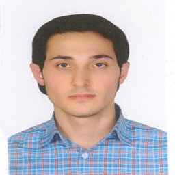 Yousef Rezaei合作论文数Seyyed-al-Shohada Heart Center, Urmia University of Medical Sciences45
 Niloufar Samiei合作论文数Shaheed Rajaie Cardiovascular Medical Center35
 Alireza Alizadeh Ghavidel合作论文数29
 Majid Maleki合作论文数Echocardiography Laboratory, Rajaei Heart Center22
 Mohamad Bashir合作论文数18
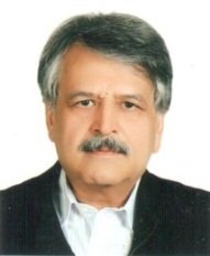 Peighambari Mohammad Mehdi合作论文数Rajaie Cardiovascular Medical &Research Center, Iran University of Medical Sciences;Rajaie Cardiovascular Medical & Research Center, Iran University of Medical Sciences18
 Mostafa Mousavizadeh合作论文数17
 Mohammed Idhrees合作论文数Department of Cardiovascular and Thoracic Surgery, Sree Chitra Thirunal Institute for Medical sciences and Technology16
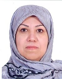 Fahimeh Kashfi合作论文数Department of Cardiac Surgery & Cardiology Shahid Rajaee;Heart Centre Tehran;Laleh General Hospital15
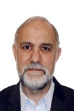 Feridoun Noohi合作论文数Shaheed Rajaei Cardiovascular Medical and Research Center;Sina General Hospital;Tehran University of Medical Sciences;Sina General Hospital, Tehran University of Medical Sciences15
 Ahmad Amin合作论文数Departments of Heart Failure, Rajaee Cardiovascular, Medical and Research Center13
 Pouraliakbar Hamidreza合作论文数Rajaie Cardiovascular Medical and Research Center, Iran University of Medical Sciences11
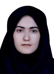 Parsaee Mozhgan合作论文数Department of Echocardiography, Shaheed Rajaie Cardiovascular Medical and Research Center;Department of Echocardiography, Shaheed Rajaie Cardiovascular Medical and Research Center10
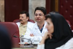 Bakhshandeh Hooman合作论文数Rajaie Cardiovasc Med & Res Ctr, Iran Univ Med Sci9
 Maryam Esmaeilzadeh合作论文数Echocardiography Lab., Rajaei Cardiovascular Medical and Research Center9
 Carlos A. Mestres合作论文数9
 Sepideh Banar合作论文数Rajaie Cardiovasc Med & Res Ctr, Iran Univ Med Sci8
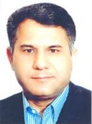 Ahmad Mohebbi合作论文数Shaheed Rajaei Cardiovascular Medical and Research Center;Sina General Hospital;Tehran University of Medical Sciences;Sina General Hospital, Tehran University of Medical Sciences8
 Avisa Tabib合作论文数Heart Center, Rajaee Hospital8
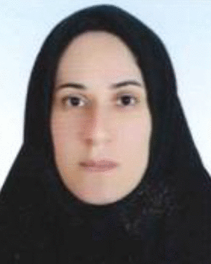 Maryam Shojaeifard合作论文数Rajaie Cardiovascular Medical and Research Center, Iran University of Medical Sciences7
 Naderi Nasim合作论文数7
 Yalda Mirmesdagh合作论文数7
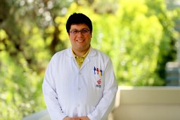 Parham Sadeghipour合作论文数Heshmat Cardiovascular Medical Center, Guilan University of Medical Sciences7
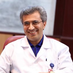 Majid Haghjoo合作论文数School of Medicine, Iran University of Medical Sciences7
 Azin Alizadehasl合作论文数Cardio-Oncology Department, Rajaie Cardiovascular, Medical and Research Center7Mahdi Daliri合作论文数6
 Hadi Abo Aljadayel合作论文数6a j khamoushi合作论文数5s m b tabatabaei合作论文数5Raheleh Kaviani合作论文数5Soheila Salari合作论文数5Sven Z C P Tan合作论文数5
 Akbar Nikpajouh合作论文数Rajaie Cardiovasc Med & Res Ctr, Iran Univ Med Sci5
 Matti Jubouri合作论文数Hull York Medical School, University of York5Taghavi Sepideh合作论文数5
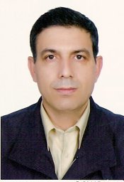 Alavi Seyed Mostafa合作论文数Rajaie Cardiovascular Medical and Research Center, Iran University of Medical Sciences5
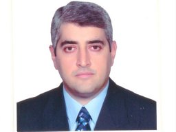 Dehaki Maziar Gholampour合作论文数Rajaee Heart Center, Iran University of Medical Sciences and Health Services5Touraj Babaee合作论文数Rajaie Cardiovascular Medical and Research Center, Tehran University of Medical Sciences5
 Zargham Hossein Ahmadi合作论文数Lung Transplantation Research Center, Shahid Beheshti University of Medical Sciences5
 Bahram Mohebbi合作论文数School of Medicine, Iran University of Medical Sciences5
 Mohammad Rafie Khorgami合作论文数5Anita Sadeghpour合作论文数Rajaie Cardiovascular Medical and Research Center, Iran University of Medical Sciences5Sedigheh Saedi合作论文数5Zahra Emkanjoo合作论文数Department of Pacemaker and Electrophysiology, Rajaie Cardiovascular Medical Center5
 Ali Rafati合作论文数4Yeganeh Pasebani合作论文数4
 Gholamreza Omrani合作论文数Rajaie Cardiovasc Med & Res Ctr, Iran Univ Med Sci4Mozaffari Kambiz合作论文数Shaheed Rajaie Hospital, Tehran University of Medical Sciences4
 Tafti Seyed Hossein Ahmadi合作论文数4




