IntroductionRespiratory syncytial virus A (RSVA) and respiratory syncytial virus B (RSVB) may induce distinct airway host responses. We compared them with influenza A virus (IAV) and human metapneumovirus (hMPV) in airway organ tissue equivalents (OTEs) and developed the Relaxed Magnitude Altitude Score with Hub-Centric Network Analysis framework (RMAS-HCNA).MethodsOTEs were infected with RSVA-GFP5 or RSVB-GFP3 at low multiplicity of infection and profiled at 24 and 72 h using the NanoString nCounter Host Response Panel. Previous IAV and hMPV profiles were integrated. Analyses included source-matched differential expression, cross-virus ranking, network-based module selection, nested cross-validation, and Gene Ontology Biological Process enrichment.ResultsRSVA showed the strongest temporal expansion and developed a late response centered on intracellular transport, localization, and organelle organization. RSVB remained the most restricted response, shifting from early inflammatory and signaling-associated activity to a later interferon-stimulated profile. IAV maintained a broad antiviral response, whereas hMPV combined limited induction with extensive late downregulation of stress, catabolic, metabolic, and transport-related processes. A shared antiviral component was present across infections, while downregulated responses were largely virus dependent. Under five-fold outer and four-fold inner nested cross-validation, the selected genes achieved a mean outer-test Macro F1 of 0.859 and balanced accuracy of 0.888, compared with 0.604 and 0.616 for the all-gene comparator.DiscussionThe four viruses shared an antiviral foundation but followed distinct temporal and functional trajectories. The framework identified compact, biologically representative panels that generalized better than the complete measured gene set, although independent genome-wide and in vivo validation remains necessary.
There is a critical need to understand pathophysiological mechanisms involved in injury from acute chlorine gas (Cl2) exposure. Limited information is available regarding the time course and mechanisms of injury after acute Cl2 exposure due to a lack of human clinical data and limited fidelity of pre-clinical animal models. We designed and integrated a Cl2 exposure platform to generate and deliver precise concentrations of Cl2 to a microfluidic human airway-on-a-chip microphysiological system. Chemical, biological, structural, and functional airway-on-a-chip responses to Cl2 exposure were characterized across multiple concentrations, exposure times, and post-exposure timepoints. Transcriptomics and metabolomics analyses delineated key molecular, cellular, and physiological pathways involved in the acute response to Cl2 exposure. This work represents a significant advancement towards high-throughput, human-relevant characterization of pulmonary toxicants and medical countermeasure development, addressing critical gaps in toxicology modeling while reducing reliance on animal studies.
BackgroundRadiation-induced renal injury (RRI) is characterized by a prolonged latent phase from radiation exposure to clinical dysfunction. Conventional markers such as serum creatinine and blood urea nitrogen (BUN) may only increase after substantial nephron loss, thereby missing a critical therapeutic window during which renal injury may remain amenable to mitigation.ObjectiveThis review synthesizes emerging strategies for the early, pre-fibrotic detection of RRI, emphasizing molecular damage biomarkers, genomic and epigenetic indicators, and advanced functional imaging techniques that may identify subclinical abnormalities before irreversible fibrosis develops.ContentTubular damage biomarkers such as kidney injury molecule-1 (KIM-1) and neutrophil gelatinase-associated lipocalin (NGAL) provide sensitive signals that precede functional decline. Filtration markers such as cystatin C may improve early risk stratification compared with creatinine alone. Genomic indicators, including γ-H2AX foci and circulating or urinary microRNAs such as miR-21 and miR-29 capture DNA damage and fibrotic signaling transitions. Advanced imaging modalities—particularly non-contrast multiparametric MRI (diffusion-weighted imaging/intravoxel incoherent motion, arterial spin labeling, blood oxygen level–dependent MRI, and T1 mapping)—demonstrate strong correlations with biopsy-proven fibrosis and microvascular dysfunction. Tc-99m MAG3 renography further reveals split renal function and asymmetrical injury masked by preserved contralateral reserve.ConclusionEvidence supporting these candidate approaches in human radiation-induced renal injury remains limited. Tubular biomarkers, cystatin C, genomic indicators, urinary extracellular-vesicle signatures, and multiparametric MRI have demonstrated potential in other renal disorders or preclinical radiation models, but their RRI-specific kinetics, thresholds, sensitivity, and specificity remain insufficiently established. We therefore propose a multimodal research framework integrating radiation exposure and renal dosimetry, serial renal function assessment, candidate biofluid biomarkers, and functional imaging. Prospective longitudinal validation is required before this framework can be used for routine clinical diagnosis or treatment selection.
The effectiveness of radiotherapy for lung cancer is constrained by the inherent radiosensitivity of healthy lung tissue, presenting a significant clinical challenge. To improve our understanding of radiation exposure, there is a need for accurate representation of the native human lung response to radiation exposure as well as the effects of radioprotectant treatments. Here, we employed primary human bronchial epithelial cells in vitro to quantitatively assess damage and protection in response to ionizing radiation from two clinically relevant sources-gamma rays and X-rays-across doses ranging from 0 to 10 Gy. Subconfluent human bronchial epithelial cultures were exposed to 0-10 Gy of gamma radiation or X-rays, and cellular responses were assessed through morphological analysis, DNA damage quantification, and growth dynamics. Distinct biological effects were observed between radiation types, underscoring differences in damage profiles and cell sensitivity. We also investigated the protective effects of WR-1065, the active metabolite of Amifostine, as a model radioprotectant compound. Pretreatment reduced radiation-induced injury, with differential efficacy depending on radiation type, reflecting the mechanisms of damage involved. Our findings demonstrate that primary human bronchial epithelial cells can report on radiation source-specific differences in cellular responses and radioprotectant efficacy. This system offers a physiologically-relevant and sensitive platform for evaluating the interplay of radiation exposure and radioprotectants with human airway cells in vitro.
Cystic fibrosis (CF), a life-shortening genetic disease, is hallmarked by mucus obstruction, respiratory deficiency, and chronic bacterial infections. Pseudomonas aeruginosa is the most common virulent respiratory pathogen that is detrimental to the overall survival of CF patients. Here we evaluate the efficacy of farnesol emulsion, a broad-spectrum agent recently used to combat P. aeruginosa biofilm infections, for reducing P. aeruginosa infections in CF using a three-dimensional (3D) airway "organ tissue equivalent" (OTE) model. OTEs are fabricated using cells derived from human primary cells sourced from CF donors (CF-OTEs), which accurately recapitulate multiple key traits of human CF airways, including increased mucin accumulation and lower cilium beating frequency, compared to OTEs derived from normal donors (N-OTEs). The OTE model closely approximates the native CF condition to provide a platform where both mucoid and nonmucoid P. aeruginosa establish biofilms. Luminescence quantification and viable bacterial enumeration demonstrated that more P. aeruginosa biofilm mass developed upon CF-OTEs compared to non-CF (normal) OTEs. The capability to establish infection and biofilm formation, without acute tissue toxicity, allows for rapid discrimination of therapeutic efficacy in an accurate, human in vitro model. Farnesol emulsion disrupted P. aeruginosa biofilms in situ and also protected OTE lung cell viability. We propose that the 3D airway OTE infection model is a reliable preclinical tool for CF drug screening, with farnesol emulsion being a prospective drug candidate to treat P. aeruginosa biofilm infections in CF.
The COVID-19 pandemic, driven by the Severe Acute Respiratory Syndrome Coronavirus-2 (SARS-CoV-2), has underscored the need to understand the virus's evolution due to its global health impact. This study employed RNA sequencing (RNA-Seq) to analyze gene expression differences across multiple SARS-CoV-2 variants. We used publicly available datasets from the Gene Expression Omnibus (GEO) with IDs GSE157103, GSE171110, GSE189039, and GSE201530, which contain RNA-Seq data extracted from white blood cells, whole blood, or PBMCs of individuals infected with the Original Wuhan variant (both hospitalized and non-hospitalized), the French variant (hospitalized), the Beta variant (hospitalized), and the Omicron variant (moderate and mild cases), along with COVID-negative controls. Our first objective was to examine differences in gene expression dynamics using Generalized Linear Models with Quasi-Likelihood F-tests and the Magnitude-Altitude Scoring (GLMQL-MAS) technique, followed by Gene Ontology (GO) and pathway analyses. Our second objective was to employ Cross-MAS to identify a robust set of genes indicative of SARS-CoV-2 infection regardless of the variant and to assess their classification performance. GO and pathway analyses revealed a significant evolutionary shift in how SARS-CoV-2 interacts with the host. Early variants such as the Original Wuhan and French cases primarily affected pathways related to viral replication, including Eukaryotic Translation Elongation and Viral mRNA Translation. In contrast, later variants like Beta and Omicron showed a strategic shift toward modulating and evading the host immune response, engaging immune-related pathways such as Interferon Alpha/Beta signaling and Cytokine signaling in the immune system. To evaluate the classification potential of the identified genes, we tested them on held-out datasets GSE152418, PMC8202013, GSE161731, and GSE166190, which contain RNA-Seq data from whole blood or PBMCs of COVID-positive and healthy individuals. Using top-ranked genes such as IFI27, CDC20, RRM2, HJURP, and CDC45 in linear models including logistic regression and linear SVM, we achieved 97.31% accuracy, with precision and recall rates of 0.97 and 0.99, respectively. These signatures also achieved perfect classification (100% accuracy, precision, and recall) in two additional datasets: GSE294888, which includes blood-derived plasmacytoid dendritic cells (pDCs) and type 2 conventional dendritic cells (DC2s) stimulated with Delta or Omicron variants, and GSE239595, which features Omicron-infected nasopharyngeal tissue. These findings demonstrate the potential of transcriptomic signatures for variant-agnostic COVID-19 detection and provide a foundation for flexible diagnostic and therapeutic approaches in response to SARS-CoV-2 evolution.
Mpox re-emerged globally in 2022 with atypical clinical features and efficient human-to-human transmission, which underscores the need to better understand host immune responses. We conducted an integrative transcriptomic analysis of mpox virus (MPXV) infection in nonhuman primates by leveraging RNA-Seq datasets from whole-blood and skin. We applied the Generalized Linear Model with Quasi-Likelihood F-test and Magnitude-Altitude Score (GLMQL-MAS), a method that combines rigorous statistical testing with a ranking metric to identify and prioritize differentially expressed genes (DEGs), and the Cross-Magnitude-Altitude Score (Cross-MAS) gene selection strategy, which integrates results across multiple time points to identify reproducible signatures, to define compact and robust markers of infection. Longitudinal analysis of whole-blood revealed a staged immune trajectory: early induction of interferon-stimulated genes (IFI27, ISG15) was followed by proliferative and hematopoietic programs and later enrichment of adaptive immune signatures, including B cell activation and TNF signaling. A minimal six-gene panel (IFI27, ISG15, MYO7B, HEY1, VASH1, CNTD2) distinguished infected from control animals with 100% accuracy in both training and independent held-out test sets. In contrast, skin transcriptomes showed upregulation of keratinocyte-related genes (KRT10, KRT1, SOSTDC1) and downregulation of immune mediators (IL1B, CXCL8, S100A8, ISG15), which suggests epithelial remodeling with limited local immune activation. Cross-tissue comparisons revealed that under stringent criteria (BH-adjusted p-value < 0.05 and |log₂FC|> 1), only 10 genes were commonly significant between whole-blood and skin, and all (100%) exhibited opposite regulation. When thresholds were relaxed for exploratory purposes (unadjusted p-value < 0.05 and |log₂FC|> 1), 58 common significant immune-related genes were identified from the curated nCounter® Host Response Panel, of which 74.1% were oppositely regulated. Shared immune genes such as ISG15, MX1, IFIT2, and OAS2 were upregulated in blood but suppressed in skin, and Reactome enrichment of discordant genes highlighted interferon and cytokine signaling pathways. These contrasts suggest that MPXV may trigger systemic interferon activation while suppressing local antiviral responses in lesions; however, because they are derived from independent secondary datasets in different species, they should be regarded as hypothesis-generating signals that require confirmation in matched, longitudinal studies.
Despite the well-known risks of tobacco use, tobacco exposure remains a major contributor to morbidity and mortality worldwide. Although cigarette use has declined, the popularity of novel tobacco products (NTPs), such as electronic cigarettes (ECs) and heated tobacco products (HTPs), has increased. Given the evolving landscape of the tobacco industry, robust in vitro models are needed to evaluate the potential for harm of novel products on the airways. We applied a 3D in vitro human airway organ tissue equivalent (OTE) model to evaluate its ability to characterize the acute effects of aerosol exposure from a combustible cigarette, an HTP, and two ECs using a VITROCELL VC1 Smoking Machine. Each product was tested using a nicotine-matched single exposure dose, providing a standardized benchmark relevant to real-world use. To deliver comparable amounts of nicotine (~35-38 μg), exposures ranged from 20 to 64 min depending on the product. Following exposure, OTEs were evaluated for cytotoxicity, oxidative stress, epithelial barrier function, ciliary function, inflammatory cytokine release, and inflammatory gene expression changes. Compared to cigarettes, NTP exposures resulted in reduced OTE cytotoxicity and inflammation. HTP exposure resulted in moderate cytotoxicity and oxidative stress, an increased inflammatory response, reduced epithelial barrier function, and temporary impairment of ciliary function. For the selected nicotine-matched dose, neither EC notably induced cytotoxicity nor inflammation or disrupted epithelial barrier or ciliary function. This work establishes a methodology for comparing NTPs using a physiologically relevant human in vitro model and supports further examination of NTPs using delivered nicotine as a benchmark.
IntroductionDeveloping robust predictive models from multi-omics data is challenging because sample sizes are typically small (often fewer than 100) while the feature space is vast (over 20,000 molecular features such as genes, transcripts, and proteins), which increases the risk of overfitting and limits generalizability. To address this challenge, this study introduces the Magnitude-Altitude Score Analysis for Tracking Infection and Time-Dependent Genes (MASIT), a novel method adept at filtering out irrelevant features/genes while focusing on important ones.MethodsApplied to the 3D airway organ tissue equivalent model that mimics human airway physiology, MASIT employed both RNA-Seq and NanoString technologies for a comprehensive analysis. RNA-Seq offered a transcriptomic overview of 19,671 protein coding genes, whereas NanoString targeted 773 specific genes. We used MASIT to analyze gene expression changes in the airway tissue equivalent after exposure to Influenza A virus, Human metapneumovirus, and Parainfluenza virus type 3 at 24- and 72-hour post-infection. MASIT was trained and validated on NanoString data, tested on the held-out RNA-Seq test set, and benchmarked against widely used feature selection approaches, including Fisher score, minimum Redundancy Maximum Relevance, embedded Lasso regression, and Boruta feature importance.ResultsMASIT achieved a 92% accuracy in differentiating eight groups of infected samples. Our findings showed that MASIT outperformed models using the full gene set, notably in algorithms like Random Forest, XGBoost, and AdaBoost. Selected genes such as IFIT1, IFIT2, IFIT3, OASL, IFI44, and OAS3 were particularly effective in categorizing samples by viral type and infection stage. Benchmarking further demonstrated that MASIT not only exceeded the performance of existing feature selection methods within NanoString data but also uniquely maintained high accuracy and stability when applied to held-out RNA-Seq data.DiscussionThese results provide insights into the host’s molecular response to viral infections and highlight MASIT as a robust tool for analyzing high-dimensional, small-sample multi-omics datasets.
Tissues on a chip are sophisticated three-dimensional (3D) in vitro microphysiological systems designed to replicate human tissue conditions within dynamic physicochemical environments. However, the current fabrication methods for tissue spheroids on a chip require multiple parts and manual processing steps, including the deposition of spheroids onto prefabricated "chips." These challenges also lead to limitations regarding scalability and reproducibility. To overcome these challenges, we employed 3D printing techniques to automate the fabrication process of tissue spheroids on a chip. This allowed the simultaneous high-throughput printing of human liver spheroids and their surrounding polymeric flow chamber "chips" containing inner channels in a single step. The fabricated liver tissue spheroids on a liver-on-a-chip (LOC) were subsequently subjected to dynamic culturing by a peristaltic pump, enabling assessment of cell viability and metabolic activities. The 3D printed liver spheroids within the printed chips demonstrated high cell viability (>80%), increased spheroid size, and consistent adenosine triphosphate (ATP) activity and albumin production for up to 14 days. Furthermore, we conducted a study on the effects of acetaminophen (APAP), a nonsteroidal anti-inflammatory drug, on the LOC. Comparative analysis revealed a substantial decline in cell viability (<40%), diminished ATP activity, and reduced spheroid size after 7 days of culture within the APAP-treated LOC group, compared to the nontreated groups. These results underscore the potential of 3D bioprinted tissue chips as an advanced in vitro model that holds promise for accurately studying in vivo biological processes, including the assessment of tissue response to administered drugs, in a high-throughput manner.
Exosome therapy holds great promise as a novel approach to improve acute skin wound healing. This review provides a comprehensive overview of the current understanding of exosome biology and its potential applications in acute skin wound healing and beyond. Exosomes, small extracellular vesicles secreted by various stem cells, have emerged as potent mediators of intercellular communication and tissue repair. One advantage of exosome therapy is its ability to avoid potential risks associated with stem cell therapy, such as immune rejection or stem cells differentiating into unwanted cell types. However, further research is necessary to optimize exosome therapy, not only in the areas of exosome isolation, characterization, and engineering, but also in determining the optimal dose, timing, administration, and frequency of exosome therapy. Thus, optimization of exosome therapy is critical for the development of more effective and safer exosome-based therapies for acute skin wound healing and other diseases induced by cancer, ischemia, or inflammation. This review provides valuable insights into the potential of exosome therapy and highlights the need for further research to optimize exosome therapy for clinical use.
Bioprinting is a promising alternative method to generate skin substitutes because it can replicate the structural organization of the skin into biomimetic layers in vitro. In this study, six primary human skin cell types were used to bioprint a trilayer skin construct consisting of epidermis, dermis, and hypodermis. Transplantation of the bioprinted skin with human cells onto full-thickness wounds of nu/nu mice promoted rapid vascularization and formation of epidermal rete ridges analogous to the native human epidermis, with a normal-looking extracellular matrix. Cell-specific staining confirmed the integration of the implanted cells into the regenerated skin. Using a similar approach, a 5 centimeter–by–5 centimeter bioprinted autologous porcine skin graft was transplanted onto full-thickness wounds in a porcine excisional wound model. The bioprinted skin graft improved epithelialization, reduced skin contraction, and supported normal collagen organization with reduced fibrosis. Differential gene expression demonstrated pro-remodeling protease activity in wounds transplanted with bioprinted autologous skin grafts. These results demonstrate that bioprinted skin can support skin regeneration to allow for nonfibrotic wound healing and suggest that the skin bioprinting technology may be applicable for human clinical use.
The hallmark of severe COVID-19 involves systemic cytokine storm and multi-organ injury including testicular inflammation, reduced testosterone, and germ cell depletion. The ACE2 receptor is also expressed in the resident testicular cells, however, SARS-CoV-2 infection and mechanisms of testicular injury are not fully understood. The testicular injury could be initiated by direct virus infection or exposure to systemic inflammatory mediators or viral antigens. We characterized SARS-CoV-2 infection in different human testicular 2D and 3D culture systems including primary Sertoli cells, Leydig cells, mixed seminiferous tubule cells (STC), and 3D human testicular organoids (HTO). Data shows that SARS-CoV-2 does not productively infect any testicular cell type. However, exposure of STC and HTO to inflammatory supernatant from infected airway epithelial cells and COVID-19 plasma decreased cell viability and resulted in the death of undifferentiated spermatogonia. Further, exposure to only SARS-CoV-2 Envelope protein caused inflammatory response and cytopathic effects dependent on TLR2, while Spike 1 or Nucleocapsid proteins did not. A similar trend was observed in the K18-hACE2 transgenic mice which demonstrated a disrupted tissue architecture with no evidence of virus replication in the testis that correlated with peak lung inflammation. Virus antigens including Spike 1 and Envelope proteins were also detected in the serum during the acute stage of the disease. Collectively, these data strongly suggest that testicular injury associated with SARS-CoV-2 infection is likely an indirect effect of exposure to systemic inflammation and/or SARS-CoV-2 antigens. Data also provide novel insights into the mechanism of testicular injury and could explain the clinical manifestation of testicular symptoms associated with severe COVID-19.
Sulfur mustard (SM) is a highly toxic chemical agent that causes severe tissue damage, particularly to the eyes, lungs, and skin. Despite advances in treatment, there is a need for more effective therapies for SM-induced tissue injury. Stem cell and exosome therapies are emerging as promising approaches for tissue repair and regeneration. Stem cells can differentiate into multiple cell types and promote tissue regeneration, while exosomes are small vesicles that can deliver therapeutic cargo to target cells. Several preclinical studies demonstrated the potential of stem cell, exosome, or combination therapy for various tissue injury, showing improvements in tissue repairing, inflammation, and fibrosis. However, there are also challenges associated with these therapies, such as the requirement for standardized methods for exosome isolation and characterization, the long-term safety and efficacy and reduced SM-induced tissue injury of these therapies. Stem cell or exosome therapy was used for SM-induced eye and lung injury. Despite the limited data on the use for SM-induced skin injury, this therapy is a promising area of research and may offer new treatment options in the future. In this review, we focused on optimizing these therapies, evaluating their safety and efficacy, and comparing their efficacy to other emerging therapeutic approaches potentially for SM-induced tissue injury in the eye, lung, and skin.
The human airways are complex structures with important interactions between cells, extracellular matrix (ECM) proteins and the biomechanical microenvironment. A robust, well-differentiated in vitro culture system that accurately models these interactions would provide a useful tool for studying normal and pathological airway biology. Here, we report the development and characterization of a physiologically relevant air–liquid interface (ALI) 3D airway ‘organ tissue equivalent’ (OTE) model with three novel features: native pulmonary fibroblasts, solubilized lung ECM, and hydrogel substrate with tunable stiffness and porosity. We demonstrate the versatility of the OTE model by evaluating the impact of these features on human bronchial epithelial (HBE) cell phenotype. Variations of this model were analyzed during 28 days of ALI culture by evaluating epithelial confluence, trans-epithelial electrical resistance, and epithelial phenotype via multispectral immuno-histochemistry and next-generation sequencing. Cultures that included both solubilized lung ECM and native pulmonary fibroblasts within the hydrogel substrate formed well-differentiated ALI cultures that maintained a barrier function and expressed mature epithelial markers relating to goblet, club, and ciliated cells. Modulation of hydrogel stiffness did not negatively impact HBE differentiation and could be a valuable variable to alter epithelial phenotype. This study highlights the feasibility and versatility of a 3D airway OTE model to model the multiple components of the human airway 3D microenvironment.
A traction inverter converts battery energy into power that controls motor torque and speed, giving it the most influence over an EV's range, performance and driving experience. The typical system consists of safety MCU, DSP for real time motor control processing along with hardware resolver for position sensing. The speed for traction motor is pushed to higher e.g. 10-30K rpm to reduce cost by miniaturizing motor and passive components, which necessitates the real time control loop performance in uSec range. The paper proposed a single chip traction Inverter System using Texas Instruments (TI) AM263 MCU, which is designed for real-time and high performance for optimal system cost for traction inverter. The paper proposes innovative techniques namely optimized Field Oriented Control (FOC) control algorithm with custom math algorithms, Software based resolver and double PWM update for better control performance. The overall solution is prototyped and demoed on hardware design and measured for overall performance. The real-time performance of control loop is 3.9 usec, which is less than 1% for overall processing power for 10K rpm. The proposed single chip solution has 99% headroom for running AUTOSAR operating system and customer's applications for differentiation.
There are an estimated 500,000 patients treated with full-thickness wounds in the United States every year. Fire-related burn injuries are among the most common and devastating types of wounds that require advanced clinical treatment. Autologous split-thickness skin grafting is the clinical gold standard for the treatment of large burn wounds. However, skin grafting has several limitations, particularly in large burn wounds, where there may be a limited area of non-wounded skin to use for grafting. Non-cellular dermal substitutes have been developed but have their own challenges; they are expensive to produce, may require immunosuppression depending on design and allogenic cell inclusion. There is a need for more advanced treatments for devastating burns and wounds. This manuscript provides a brief overview of some recent advances in wound care, including the use of advanced biomaterials, cell-based therapies for wound healing, biological skin substitutes, biological scaffolds, spray on skin and skin bioprinting. Finally, we provide insight into the future of wound care and technological areas that need to be addressed to support the development and incorporation of these technologies.
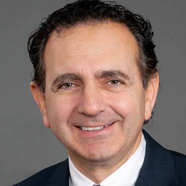 Anthony Atala合作论文数Wake Forest Institute for Regenerative Medicine;Department of Urology, Wake Forest School of Medicine;Brenner Children's Hospital, Atrium Health Levine Children's44
 Aleksander Skardal合作论文数Department of Bioengineering, The University of Utah17
 A. Atala合作论文数16Jackson John D合作论文数14
 Shay Soker合作论文数Wake Forest Institute for Regenerative Medicine, School of Medicine, Wake Forest University13
 James J. Yoo合作论文数Wake Forest Institute for Regenerative Medicine, School of Medicine, Wake Forest University13
 Khoury Oula合作论文数School of Arts and Sciences, Lebanese American University11
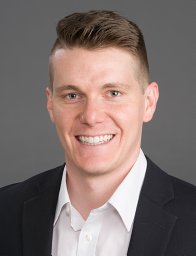 T. Leach合作论文数Wake Forest School of Medicine, Wake Forest Institute for Regenerative Medicine9
 Rebecca Lim合作论文数Monash Institute of Medical Research, Monash University9
 Sangjin Lee合作论文数Wake Forest Institute for Regenerative Medicine;International Society for Biofabrication;Korea University;School of Medicine, Wake Forest University9
 Euan Wallace合作论文数Victorian Government8Adam Jorgensen合作论文数7
 Graham Jenkin合作论文数Monash Immunology and Stem Cell Laboratories, Monash University7Cara Clouse合作论文数6Kidiyoor Amritha合作论文数6
 Victor E. Ortega合作论文数Department of Pulmonary Medicine, Mayo Clinic6
 Hooman Sadri-Ardekani合作论文数5S Walker合作论文数5
 Goodwell Nzou合作论文数Wake Forest Institute for Regenerative Medicine, Wake Forest School of Medicine5
 Colin Edward Bishop合作论文数Wake Forest Institute for Regenerative Medicine, Wake Forest University School of Medicine;Center for Redox Biology and Medicine, Wake Forest University School of Medicine5
 Adam R. Hall合作论文数Delft University of Technology5U. Gandhi合作论文数4N. Mahajan合作论文数Wake Forest Inst Regenerat Med4
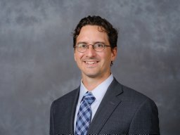 Carlos Kengla合作论文数Wake Forest Baptist Medical Center4
 Julio Aleman合作论文数Swanson School of Engineering, University of Pittsburgh4
 Dongxu Ke (柯东旭)合作论文数School of Biomedical Engineering, Nanjing University4
 Devarasetty Mahesh合作论文数4
 Anastasiya Alexeyevna Gorkun合作论文数4
 Mohammad Z. Albanna合作论文数4
 Thomas D Shupe合作论文数College of Medicine, University of Florida4Weixin Zhao合作论文数4
 Yuanyuan Zhang合作论文数Wake Forest Institute for Regenerative Medicine, Wake Forest University;Bowman Gray Center for Medical Education, School of Medicine, Wake Forest University4Martha L. Simmons合作论文数4C. Clouse合作论文数3
 Giuseppe Orlando合作论文数3Steven T. Leung合作论文数3Kelsey Willson合作论文数3Khiry Sutton合作论文数3
 Mostafa Rezapour合作论文数Dept Math, Wake Forest Univ3
 Nima Pourhabibi Zarandi合作论文数Department of General Surgery, Atrium Health Wake Forest Baptist Comprehensive Cancer Center3A. M. Jorgensen合作论文数3S. Rajan合作论文数Wake Forest Inst Regenerat Med3Robert Wicks合作论文数Department of Neurosurgery, Johns Hopkins University Hospital;Department of Neurosurgery, Johns Hopkins University Hospital3
 Hayley Dickinson合作论文数Monash Immunology and Stem Cell Laboratories (MISCL), Melbourne, VIC, Australia, hayley.dickinson@monash.edu.3Reid Tanya合作论文数3Chan Siow Teng合作论文数3
 Tan Jean L合作论文数3Gretchen Bruner合作论文数3
 Joshua Tan合作论文数3




