Nephrolithiasis, commonly termed urolithiasis, is characterized by the development of calculi anywhere within the urinary tract. The occurrence of this condition varies across different regions and is influenced by multiple factors, including climate, age, gender, dietary patterns, and genetic predisposition. The highest incidence is typically reported during the second and third decades of life, with males being affected more frequently than females in an approximate ratio of 2:1. Stone formation results from supersaturation of urinary solutes such as calcium, oxalate, and uric acid, along with low levels of citrate. This physicochemical imbalance promotes crystal aggregation and subsequent calculus formation. From the perspective of Unani medicine, calculi (Ḥaṣā) are believed to develop due to the retention and accumulation of dense, morbid matter (Mādda Ghalīẓ) within the organs, coupled with impaired expulsion. Progressive deposition of this material eventually leads to stone formation. The concept of Hararat (heat) contributes to the evaporation of Ratubat (moisture), resulting in desiccation (Tajfīf), condensation, and hardening of the retained matter into calculi.
Background: To assess the effectiveness of hyperbaric oxygen therapy (HBOT) as an adjunct to standard treatment in early-stage oral submucous fibrosis (OSMF), focusing on mouth opening, burning sensation, pain, and systemic inflammation.Methods: A randomized comparative study was conducted on early-stage OSMF patients divided into two groups. Group 1 received HBOT along with intralesional triamcinolone, hyaluronidase, and oral antioxidants. Group 2 received only the standard intralesional therapy with antioxidants. Clinical parameters—interincisal distance, burning sensation, and pain—were recorded at baseline, first, and second followup. Serum C-reactive protein (CRP) levels were measured pre- and post-treatment. Statistical analysis included unpaired t-tests and chisquare tests.Results: Both groups improved clinically, but Group 1 showed significantly greater reductions in burning sensation (p= 0.020), pain, and CRP levels (p= 0.033). Although intergroup differences in mouth opening were not statistically significant, Group 1 showed greater improvement over time. Fibrotic band distribution remained unchanged in both groups. No complications were reported with HBOT.Conclusion: HBOT provides enhanced symptomatic relief and anti-inflammatory effects in early-stage OSMF when used alongside conventional therapy. While it may not reverse fibrosis, its safety and noninvasive nature support its role in multimodal OSMF management.
Purpose of the Study:Reconstruction of post-mucor oral cavity defects (ranging from simple alveolectomy defects to complete maxillectomy defects) will improve the function, esthetics, and general health of an individual. Various surgical and prosthetic options are available for the successful reconstruction of the defects. The nasolabial flap has versatile role in the reconstruction of oral cavity defects because of its proximity to the defect area, optimal esthetic results, and less surgical morbidity unlike distant flaps. In this study, we assessed the efficiency of the nasolabial flap in reconstruction of post-mucor infrastructure maxillectomy defects in 42 individuals by comparing the pre-surgical and post-surgical abilities to speak and swallow. Patients and Methods:Patients with post-mucor infrastructure maxillectomy defects were involved in this prospective study. Fifty-one patients who met the inclusion criteria were selected. All the study parameters (swallowing ability, hypernasality, speech intelligibility, and subjective satisfaction of an individual) were assessed preoperatively. Reconstruction of the defect was done with a superiorly based nasolabial flap. Nine patients were lost to follow up, and 42 patients were followed up for six months to evaluate the study parameters. Results:Pre-surgical and post-surgical statistical data analysis shows that there is a significant improvement in the functional outcomes of an individual. 76% of the patients showed no signs of nasal regurgitation, dribbling of saliva, or coughing upon swallowing after reconstruction of the defect with nasolabial flap. Significant improvement in speech after the closure of maxillary defect was observed as 93% of patients had socially acceptable speech and 64.5% had normal intelligible speech postoperatively. Upon subjective satisfaction assessment, 64% of the patients were satisfied with the treatment procedure. Conclusion:Closure of oro-antral communication and creation of a seal between oral and nasal cavities using the nasolabial flap leads to improvement in the function of an individual in terms of speech intelligibility and swallowing ability. The nasolabial flap can be considered as an optimal option for managing post-mucor infrastructure maxillectomy defects by restoring functional and psychosocial well-being of a person.
Introduction:Oral submucous fibrosis (OSMF) is a prevalent disease in India, mainly caused by tobacco chewing (areca nut, pan masala, and betel quid). Its treatment is still under debate, as none of the treatment modalities have been statistically superior to one another, may it be medical or surgical. Aim and Objective:Clinical and interleukin-6 (IL-6) assessment in patients of submucous fibrosis at baseline and 3 months of follow-up, after intervening their usual treatment with the herbal extract. Methodology:Sixty patients were randomly divided into two groups. Group 1 was given the standard treatment, while group 2 was given herbal extract along with the standard treatment. Results:A significant reduction (P < 0.05) in burning sensation and pain upon mouth opening was observed in group 2 patients. Also, comparatively more decrease in the level of IL-6 was noted in group 1. Discussion:Sphaeranthus, Ocimum, and honey are already being used, separately or together, in many diseases as anti-inflammatory, antimicrobial, and antioxidant. Conclusion:The herbal extract, used in this study, can be used as an adjunct in OSMF for alleviating the painful symptoms at an early stage.
Aims and Objective:To evaluate lingual split technique vs buccal guttering in the extraction of lingually placed impacted mandibular third molar and to assess the clinical outcome of the two techniques in relation to pain, swelling, mouth opening, intra-operative time, dry socket, paraesthesia due to injury to the lingual and inferior alveolar nerve and hemorrhage. Materials and Methods:The present clinical study comprised of 36 lingually impacted mandibular third molars. Patients were divided into two groups, and bone covering the third molar was removed by the lingual split technique using chisel and mallet and buccal guttering approach technique using rotary instruments. Results:The conventional buccal guttering technique took longer time with higher incidence of dry socket than the lingual split technique. Significant findings were also recorded in the lingual split technique in terms of pain, swelling, and trismus. Postoperative nerve injury was significantly higher in lingual split technique. Conclusion:The study concluded that lingual split technique using chisel and mallet is found to be better than the buccal guttering approach technique using rotary instruments.
BACKGROUND:Maxillofacial trauma presents with a myriad of complications, including functional disability and aesthetic compromise. Optimal treatment includes management of bone and soft tissue injuries along with dental rehabilitation. Prompt management, including open reduction and internal fixation (ORIF) combined with immediate dental implant rehabilitation, facilitates both functional recovery and aesthetic restoration, thereby leading to improved quality of life. METHODS:A total of 36 patients with maxillofacial bony trauma, along with non-restorable fractured, avulsed, or missing teeth, were managed with ORIF with dental rehabilitation. Eighteen patients underwent delayed implant placement after 6 months of ORIF, and the other 18 underwent immediate implant rehabilitation during ORIF. The outcomes measured included implant stability, crestal bone loss, and evaluation of pain, swelling, and patient satisfaction. All follow-up visits were scheduled with reference to the post-implant placement period in both groups. RESULTS:Implant stability was significantly increased in the immediate implant placement group of patients at the third- and sixth month follow-up post-implant placement (p< 0.001). Pain assessment revealed that the immediate implant group experienced significantly higher pain scores than the delayed implant group on the first day (p< 0.001), seventh day (p< 0.001), and 28th day (p< 0.001) following implant placement. Significant differences in facial swelling were observed at the third and sixth months (p= 0.009), with the immediate implant group exhibiting lower swelling. Swelling was significantly higher along the tragal-commissure line in the immediate implant group on the first and seventh days (p< 0.001). Patient satisfaction was initially higher in the delayed implant group on the first day (p= 0.047) and remained so by the third month (p< 0.001). However, by the sixth month, both groups demonstrated similar levels of satisfaction after implant placement. CONCLUSION:Immediate implant rehabilitation during ORIF in patients sustaining maxillofacial trauma improves implant success rates, reduces morbidity, and ultimately enhances patients' overall quality of life.
Background:Head and neck tumors often result in extensive maxillofacial defects requiring complex reconstructive procedures. Free tissue transfer has become the gold standard for reconstruction due to its ability to provide optimal tissue matching, functional restoration, and aesthetic outcomes. The success of free flap reconstruction depends on effective microvascular anastomosis, particularly venous anastomosis. Purpose:This study evaluates the efficacy of coupler devices for venous anastomosis in free flap reconstruction of oral defects. These devices, which use a pin-based connection system, are compared with traditional hand-sewn techniques regarding patency, operative time, and complication rates. Methods:A prospective analysis was conducted on patients undergoing free flap reconstruction for oral defects. The study assessed the use of coupler devices in anterolateral thigh, fibular, and radial forearm flaps. Primary outcomes included venous patency rates, intraoperative time reduction, and postoperative complications such as thrombosis or flap failure. Results:Preliminary findings suggest that coupler devices reduce venous anastomosis time and provide comparable or improved patency rates compared to traditional sutured anastomosis. Additionally, a lower incidence of thrombosis was observed in coupler-assisted anastomoses. Conclusion:Coupler devices offer a reliable and efficient alternative for venous anastomosis in free flap reconstruction of oral defects. Their use may contribute to reduced surgical time and improved anastomotic outcomes, making them a valuable tool in microvascular reconstruction.
Introduction The 'progression model' of oral carcinogenesis emphasizes that initial irreversible genotypic damage produces tissue disorganization and dysmaturation which, if allowed to progress, leads invariably towards invasive oral squamous cell carcinoma. Recognition of these genotypic changes is fundamental for a successful management strategy. Genetic studies have confirmed the role of pro-carcinogens present in tobacco and alcohol contributing to DNA damage and genotoxicity. Many molecular epidemiological studies have evaluated the association of head and neck cancer with polymorphisms in DNA repair genes. Our study aimed to investigate the association between single nucleotide polymorphisms (SNPs) of DNA repair genes OGG1 Ser326Cys, APE1-Asp148Glu, XRCC1- Arg194Trp, XRCC1-Arg280His, XRCC1-Arg399Gln, ADPRT-V762A, APEX1-D148E, LIG3-R780H, MUTYH-Tyr165Cys and the risk of oral squamous cell carcinoma and oral precancerous lesions. Materials and method Our study included two patient groups. Group I (n = 58) included patients with leukoplakia, oral submucous fibrosis and biopsy-proven oral squamous cell carcinoma. Group II (n = 58) included patients who had no clinically detectable lesions. Patients in both groups had tobacco chewing and smoking history. Blood samples were collected, and gene polymorphisms were identified using PCR-RFLP technique. Results and discussion Our study results revealed a statistically significant association between gene polymorphisms OGG1-Ser326Cys (p = 0.008), XRCC1-Arg194Trp (p = 0.009), XRCC1-Arg399Gln (p < 0.001), APEX1-Asp148Glu (p < 0.001) and the occurrence of leukoplakia, OSMF and OSCC.
Background & Objective:Telomeres consist of repetitive G-rich nucleotides located at the end of each chromosome, acting as protein binding sites. The aim of this study was to examine the differences in telomere length in blood, saliva, and tissue samples at various stages of oral precancerous and cancerous lesions. Methods:Samples of blood, tissue, and saliva were collected from patients with oral precancerous and cancerous lesions. DNA extraction was performed. Then, a TRAP assay was conducted to assess and compare the telomere length and telomerase expression. Results:The levels of telomerase activity (TA) in the DNA samples ranged from 0.19 to 6.91 (2.05+1.37) among oral squamous cell carcinoma (OSCC) patients and from 0.17 to 4.5 (0.28+4.25) among precancerous patients. A significant difference was observed in TA levels between OSCC and precancerous samples (t=3.9691, P= 0.0000). Conclusion:Assessing the telomerase activity is crucial for studying the behavior of carcinoma in the clinical setting. The augmented telomerase expression and the length of telomere contribute to OSCC progression. Hence, this study adds a diagnostic tool that can serve as a biomarker for the early detection and prognosis of OSCC.
Introduction: Fractures of orbital walls especially floor are complex and are difficult to address clinically due to extreme fragility of the same. Fractures of the floor can be classified in to pure or impure blow out fractures and can lead to various complications such as enophthalmos, extraocular muscle entrapment, severe shift of orbital content, emphysema and space occupying lesions. Depending upon size or surface area of defects and nature of the fracture, various treatment options or materials are considered for reconstruction of these defects.
ABSTRACT Background and Aim: As oral submucous fibrosis (OSMF) is a chronic progressive disorder, the treatment is based on the severity of the disease. Surgical treatment is the only choice for grade III and grade IV OSMF cases because the patient can neither clean his/her mouth nor properly chew. The resulting soft tissue defect requires resurfacing with various well-vascularized tissues such as extraoral flaps, intraoral flaps, microvascular flaps, and allografts that have been used. Reconstruction of the resultant defects proved to be challenging. Till date, none of the flaps has been proven to be effective and is universally accepted for the treatment of OSMF because of various drawbacks of the available techniques. This study was conducted to know whether an endoscopic-assisted platysma flap is associated with better outcomes in terms of ease of operation and postoperative function than the conventional approach. Materials and Methods: This study included 40 patients of grade III and grade IV OSMF reporting to the outpatient department of oral and maxillofacial surgery in a tertiary center of North India. These patients were divided randomly into two groups. Group I and Group II had 20 patients each, undergoing endoscopic-assisted platysma flap and non–endoscopic-assisted platysma flap for reconstruction after resection of OSMF bands, respectively. Data were analyzed for the mouth opening, operating time, flap viability, congestion of neck and oral cavity, signs of inflammation, neurologic assessment, and measurement of the drain. Results: The results showed significant increase in mouth opening from the preoperative value to the values immediately after surgery and at 24 h, 1 week, 15 days, 1 month, 3 months, and 6 months after surgery in both the study groups. Reduced bleeding incidence was found in group I compared to group II, with better postoperative outcomes noted during follow-up. But the mean intraoperative time of the subjects in group I was 130.80 ± 5.5.908 min and in group II was 105.74 ± 2. 491 min. Increased time taken in group I may be due to the long learning curve. Conclusion: The present study concluded that the Endoscope-assisted technique has a key role during supra and subplatysmal dissection to allow for better accessibility, handling, and visibility of the flap and its orientation in relation to the underlying structures to avoid postoperative complications and to overcome the drawback of platysma myocutaneous flap in reconstruction of OSMF defects.
ObjectiveThis study aimed to explore genetic variations associated with DNA repair mechanisms to enhance the management of both oral cancer (OC) and oral precancer (OPC).MethodsA cohort of 380 patients diagnosed with OC and OPC, comprising 220 males and 160 females, was analyzed. Participants were categorized based on their tobacco-chewing habits, with corresponding control groups established. Key genetic markers investigated for polymorphisms included OGG1, APE1, and XRCC1.ResultsThe XRCC1 Arg280H variant demonstrated significant associations with the susceptibility to both OC and OPC across various models. Further analyses, incorporating factors such as tobacco and alcohol consumption, unveiled a correlation between the XRCC1 Arg194Trp variant and an elevated risk of developing head and neck cancer. Stratified analyses also revealed an increased risk of OC or OPC based on the specific site of the cancer.ConclusionThe study underscores the importance of XRCC1 polymorphisms, particularly XRCC1 Arg280H and XRCC1 Arg194Trp, within the genetic framework of OC and OPC. Understanding these genetic associations provides valuable insights for the potential development of targeted interventions aimed at individuals predisposed to these conditions.
Introduction:Auricular reconstruction is a technically challenging and aesthetically demanding procedure as the ear has a complex anatomy. Anthropometry aids in achieving aesthetic ear reconstruction. We considered that implication of stereophotogrammetric technology will lead to a better understanding of human ear morphology.Material and methods:A cross-sectional study was conducted in our institutional OPD in a tertiary health care centre in the Northern part of India.400 people were chosen based on selection criteria. facial scans were done for 3D pictures using Canfield VECTRA® H2 3D imaging device. Study variables were assessed after marking landmarks on the 3D-generated auricular image of an individual.Discussion:This study consisted of 55.5% males and 44.5% females belonging to the age group of 5-25 years (30.3%), 26-40 years (38.8%) and>40 yr (31.0%). Out of 400 cases, the majority had; oval shaped auricle, normally rolled helix, square earlobe; knob shaped tragus. The attached type of earlobe attachment was more in the right auricle (37%) and the partial attachment ear lobe was more in the left side auricle (35.5%). Darwin's tubercle showed more proportion in the case of males. The mean length and width of the auricle & attachment length are higher in males compared to females. Ear Angulation is highest among females.Conclusion:Assessment of ear morphology using technologically sound methods like stereophotogrammetry paves the way for a more quick, reliable and easy-to-use method for understanding ear morphology. Precise assessment of ear morphology using stereophotogrammetry helps in providing more cosmetic and acceptable ear restoration.
Background:The use of dental implants has become a very predictive method of rehabilitation for patients with partial or complete edentulism. It is more challenging to treat the posterior quadrants of the maxillary ridges using dental implants due to their anatomical and physiological characteristics. So to overcome the limitations of other techniques, short implants were introduced recently as a new approach to simplify implant placement in compromised alveolar bone and to prevent possible damage to vital structures.Purpose:This study aims to compare the clinical outcomes of dental implants placed using the osteotomized sinus floor elevation (OSFE) technique side engaging the bony floor of the maxillary sinus (bicortical anchorage) on one side and the conventional technique by split mouth on the other side.Materials and Method:This study included 15 patients. Study participants had dental implants placed on both sides of the mouth at the same time, so one side was implanted according to the test method, while the other side used the control method. Randomization determined which side would be implanted.Conclusion:The OSFE technique provides greater stability to the implant via bicortical anchorage than conventional techniques, which only provide unicortical anchorage.
Purpose: The behavior of trigeminocardiac reflex (TCR) is limited to few case reports only in maxillofacial surgery, especially for temporomandibular joint (TMJ) ankylosis cases. The present study aims to find out the incidence of bradycardia due to TCR during intraoperative forceful mouth opening in TMJ ankylosis patients. Materials and Methods: A prospective, unicentric observational study was conducted selecting those patients who were planned for osteoarthrectomy with interpositional gap arthroplasty under general anesthesia. Sixty cases of TMJ ankylosis were randomly selected from December 2018 to-03-2020. Predictor variables were patient age, gender, and type of ankylosis, and outcome variables were pulse rate, mean arterial blood pressure (MABP), and oxygen saturation level (SPO2). Data were recorded at baseline and during intraoperative mouth opening via heister jaw stretcher. A Chi-square test was used for testing the association between variables. P values were considered statistically significant at <0.05. Results: The sample size of 60 subjects has been divided into two age groups (10–19) years and (20–40) years. The mean standard deviation age of the patients was 18.32 ± 6.81 years. About 63.3% were male. Out of 60 cases, 14 patients developed bradycardia. The frequency of bradycardia was found 23.3%. According to logistic regression analysis, age and type of ankylosis were the significant predictors of bradycardia. Conclusion: We conclude that bradycardia due to TCR in TMJ ankylosis patients is not an uncommon entity. Incidences are more prevalent in the age group of 10–19 years and type IV bony ankylosis. Male had a high incidence of bradycardia though P value was nonsignificant.
The origin of ameloblastic fibrosarcoma (AFS) or ameloblastic dentinosarcoma in the background of ameloblastic fibroma has been widely reported in literature. To the best of our knowledge, the malignant transformation of a benign ameloblastoma to an aggressive variety of AFS has never been reported. Here, we report a rare case of follicular ameloblastoma of mandible in a 16-year-old female, who initially presented with a painless slow growing swelling, gradually transformed into a painful, aggressive variety of AFS requiring extensive surgery and chemoradiation therapy.
Edentulism, a common problem can occur either as a congenital defect or acquired later due to dental caries, periodontitis, as a consequence of aging, maxillofacial trauma or post-ablation in tumor resections. The rehabilitation of the missing teeth can be done using dental implants. To overcome the deficiency of available bone, processes like sinus augmentation with substituted bone graft and Le Fort I osteotomy with interpositional bone graft have been described in the literature. In order to overcome the associated limitations with these procedures, implants were designed that can be placed in specific anatomical areas like zygoma. This study aims to compare two different types of surgical approaches (Intrasinus vs Extrasinus) for the placement of zygomatic implants to treat atrophic maxilla. The placement of zygomatic implant through both extrasinus and intrasinus approaches were evaluated on the basis of different parameters and it was observed that postoperative pain and swelling was significantly found in intra sinus approach as compared to extra sinus approach. However, in the intranasal approach, poor patient compliance or low satisfaction rate was observed as compared to extra sinus approach. On the basis of the results of the study and post operative evaluation based on various criteria, it was concluded that extra sinus approach has got an edge over intra sinus approach.
A field experiment was conducted during kharif season of 2021 at the Crop Research Farm, Department of Agronomy, Naini Agricultural Institute, SHUATS, Prayagraj (U.P.) India. The soil of experimental field was sandy loam in texture, nearly neutral in soil reaction (pH 7.2), low in organic carbon (0.35%), available N (108.0 kg/ha), available P2O5 (22.15 kg/ha) and available K2O (280.0 kg/ha). The experiment was carried out to find the performance of 10 hybrids, which laid out in Randomized Block Design (RBD) with three replications. To find the performance of 10 Rice hybrids. The result of experimentation revealed that the Rice hybrid UR-35 recorded significantly higher growth attributes viz. plant height (120.58 cm), number of tillers/hill (15 no.), plant dry weight/plant (55.91 g), CGR (40.50 g/m2/day), yield attributes viz., effective tillers/m2 (387 no.), panicle length (28.41 cm), test weight (26.45 g), and yields viz., grain yield per hill (28.37 g), grain yield (6.90 t/ha), straw yield (12.77 t/ha).
ABSTRACT On January 30, 2020, the World Health Organization (WHO) declared a severe respiratory disorder syndrome which originated in Wuhan city as a global public health emergency, and the pandemic declaration by the WHO was made on March 11, 2020. Persons infected with SARS-CoV-2 are frequently asymptomatic, yet they have high respiratory viral loads, and they are major purveyors of viral spread. These factors have led to the current explosion of COVID-19 hospitalizations and deaths. Vaccines could play an important role by preventing severe diseases and increasing population immunity and reducing the ongoing health crisis. There is wealth of information for the review available since it is a current topic of interest. Initially, Google Scholar was utilized to take an initial sample of what types of articles are available. We searched other databases such as PubMed, EMBASE, and COCHRANE LIBRARY for research articles published up to March 2021, with no language restrictions. We found seven peer-reviewed publications available on the efficacy of SARS-CoV-2 vaccines: AZD1222 (AstraZeneca/University of Oxford), a ChAdOx1-based vaccine with a reported efficacy of 70.4% and two mRNA-based vaccines: BNT162b2 (Pfizer/BioNTech) with a reported efficacy of 95% and mRNA-1273 (Moderna/NIAID) with a reported efficacy of 94.1%. Internet was used as a source because of its limitless networking of resources. Sources used from the internet were written by professionals in their fields and published on reliable sites, in referred publications, or on professional organization sites. The cited references were within the last 2 years.
Introduction:Blunt trauma to the orbital rim is a frequent cause of both orbital fractures and damage to the surrounding facial bones and soft tissues. The inception of endoscopy and minimal invasive intervention has revolutionized the surgical treatment of blow out fracture of orbital walls. Thus, the present study was conducted to compare the outcome of conventional open reduction approach with endoscopic approach for orbital floor repair.Materials and method:The study included 10 patients with clinical and radiographic evidence of orbital floor fracture, divided randomly into two groups: Group I-operated using endoscopic approach; and Group II- operated using conventional external approach. All the patients were evaluated clinically preoperatively, immediate post-operative and after 1 month for Diplopia, Extrinsic ocular motility, Enophthalmos and Hypoglobus.Results:The study consisted of 40% females and 60% males, with mean age of Endoscopic and Conventional group being 27.20 ± 6.14 years and 27.60 ± 7.23 years respectively. In relation to diplopia, an insignificant difference was observed statistically. The mean change in enophthalmos and hypoglobus in endoscopic and conventional group showed a significant difference statistically (p < 0.001). Unrestricted ocular motility was present only in one case of each group and significantly increased to 5 cases in both the groups (p = 0.048). The mean duration of surgery in endoscopic and conventional group showed a significant difference statistically.Conclusion:Diplopia, hypoglobus and Enophthalmos correction was better achieved in endoscopic group as compared to conventional group. But duration of surgery was more in endoscopic group as compared to conventional group.
 Vibha Singh合作论文数Department of Oral and Maxillofacial Surgery, King George's Medical University30
 Uma shanker Pal合作论文数Associate Professor, Department of Oral &Maxillofacial Surgery, King George's Medical University;Department of Oral & Maxillofacial Surgery, King George's Medical University27
 Hari Ram合作论文数CSM Medical University, Erstwhile King George’s Medical University24
 Geeta Singh合作论文数Division of Microbiology, Indian Agricultural Research Institute21
 Divya Mehrotra合作论文数Department of Oral and Maxillofacial Surgery, CSMMU (Formerly King George’s Medical University),21
 Rk Singh合作论文数16
 Nimisha Singh合作论文数15Malkunje Laxman R合作论文数11R. Pradhan合作论文数8
 Shalini Gupta合作论文数8Sana Farooqui合作论文数7
 Saurabh Srivastava合作论文数Department of Electronics Engineering, Indian Institute of Technology7
 Agrawal A合作论文数7Somdipto Das合作论文数6
 Nuzhat Husain合作论文数6
 Fahad. M. Samadi合作论文数5Prem Narayan Gupta合作论文数4aslam mohammad合作论文数4zuber ahmad合作论文数4
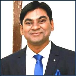 Deepak Passi合作论文数4Mehta Gagan合作论文数Department of Oral and Maxillofacial Surgery, King George's Medical University4
 Gupta Hemant合作论文数Department of Oral and Maxillofacial Surgery, BBD College of Dental Sciences4Agrawal Dhirendra Kumar合作论文数4
 Arunesh Kumar Tiwari合作论文数4
 Ravi Katrolia合作论文数3Stuti Singh合作论文数3
 Gaurav Singh合作论文数3
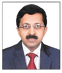 Shaleen Chandra合作论文数Dept Oral Pathol & Microbiol, King Georges Med Univ3
 Najmul Islam合作论文数3
 Ranjana Singh合作论文数Department of Biochemistry, King George's Medical University3
 Debraj Howlader合作论文数3
 Saumyendra V Singh合作论文数Dental Faculty, CSM Medical University3
 Kumar Sumit合作论文数3
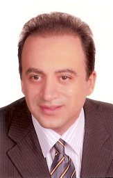 Abdolreza Jamilian合作论文数Department of Orthodontics, Islamic Azad University3
 Kumar Vijay合作论文数3
 Mehrangiz Sepide Ghassemi合作论文数3
 Sumit Gupta合作论文数3Alireza Ghassemi合作论文数3
 Abbas Ali Mahdi合作论文数Era University;Department of Biochemistry, King George's Medical University3
 M L B Bhatt合作论文数Department of Radiotherapy, King George's Medical University;Department of Radio Therapy, All India Institute of Medical Sciences;Ram Manohar Lohia Hospital3Ramesh K Pandey合作论文数2Seema Gupta合作论文数2n m kalariya合作论文数2Harsha Pradhan合作论文数2Showkatbakhsh R合作论文数2Khushboo Arif合作论文数2
 Fauzia Khan合作论文数2Roop Ganguly合作论文数2Harshita Maurya合作论文数2




