According to research, an increase in chronic diseases, including asthma, has been identified. The number of asthma patients in Saudi Arabia is a cause for concern due to the weather conditions and lifestyle, especially in the post-pandemic era. It demands a solution to reduce infections by developing an intelligent system to detect asthma at an early stage, thereby preventing the disease or enabling early treatment. In this study, machine learning has been utilized to develop tools to track asthma symptoms at an early stage. Although there have been various prior attempts to apply machine learning to predict the occurrence of asthma. Nevertheless, focusing on the identification of the disease at the pre-symptom stage, particularly in the Saudi Arabian context, is a relatively neglected area. The dataset for the current study was obtained from King Fahad University Hospital, Dammam, Saudi Arabia, and included standard tests performed on patients, including blood tests, viral tests, and biochemistry tests. The dataset contains 17 significant attributes and includes information for 328 asthma patients: 165 are positive, and 163 are negative. The methods selected for application here are random forests (RF), artificial neural networks (ANN), support vector machines (SVM), and naive Bayes (NB). Each of these methods has been chosen based on its distinctive features. The experimental outcome revealed that the RF, SVM, and ANN approaches yielded 94%, which is the highest accuracy and improved upon the state of the art by 3.9%. It is worth noting that nine of the seventeen possible features were used to achieve the above accuracy. Despite RF, SVM, and ANN achieving the same accuracy, ANN has a higher error cost. Therefore, RF and SVM are superior based on the pattern of results, and hence they are suggested for this problem.
Sentiment analysis is a natural language processing (NLP) based computational approach, helps machines understand and categorize human emotions expressed in unstructured textual format. Traditional models, such as long short term memory (LSTM) networks, are good at recognizing patterns in text but often lack interpretability and struggle to include domain specific sentiment knowledge. These issues been addressed in this study, proposes a new hybrid model called attention based LSTM with VADER driven domain specific feature integration for sentiment analysis which uses an attention mechanism to focus on the most relevant words in the text and VADER sentiment scores as extra features related to specific domains, improving the model's ability to capture emotional sentiment. Sentiment analysis becomes more accurate and stable with the combination of both the LSTM's contextual understanding with VADER's sentiment knowledge. The experimental results claims, this proposed ensemble method improves on traditional LSTM models in terms of accuracy and adaptability, especially when handling specific domain specific sentiment analysis activities, to create more intelligent and context aware sentiment analysis model.
Glaucoma is a leading global cause of blindness, making early detection essential. This paper introduces GlaucoXAI (Glaucoma Explainable AI), an advanced computer-aided diagnosis (CAD) model that integrates machine learning and explainable AI for glaucoma detection using retinal fundus images. The proposed model consists of four stages, including preprocessing, feature extraction, dimensionality reduction, and classification. Initially, features are extracted using the fast discrete curvelet transform with wrapping (FDCT-WRP) to obtain curve-type features. During the next stage, principal component analysis (PCA) and linear discriminant analysis (LDA) are combined to reduce the dimensionality of the feature matrix, followed by a classification stage employing an improved grey wolf optimization (IMGWO) with an extreme learning machine (ELM) to optimize the weight and bias to reduce the overfitting of the model. The model has been experimented with two publicly available datasets named G1020 and ORIGA. The model has achieved 93.87% accuracy on G1020 and 95.38% on ORIGA, outperforming existing methods. The 10 × 5-fold stratified cross-validation (SCV) with explainable AI enhances the interpretability of models and improves clinician trust. Overall, the proposed approach offers accurate, efficient, and explainable glaucoma diagnosis, potentially supporting ophthalmologists in early disease detection.
Congenital heart diseases (CHDs), including Ventricular Septal Defect (VSD), Atrial Septal Defect (ASD), and Patent Ductus Arteriosus (PDA), are among the most prevalent birth-related anomalies in children, often requiring early diagnosis to prevent severe complications. Similarly, respiratory diseases such as tuberculosis, pneumonia, and COVID-19 continue to pose significant public health challenges globally. Early and precise identification of these conditions through chest X-ray analysis plays a crucial role in supporting accurate diagnosis and guiding treatment strategies. This research presents an automated deep learning framework designed to classify congenital heart diseases (CHDs) and respiratory disorders by utilizing two distinct chest X-ray datasets. To capture and learn discriminative image features, advanced models, including Vision Transformer (ViT), ConvMixer, and VGG-19, were applied. The models were trained and evaluated on labeled datasets containing paediatric chest X-rays for CHD detection and mixed adult/paediatric chest X-rays for respiratory disease classification. The performance of each model was assessed using metrics such as accuracy(acc), precision(pr), recall(re), F1-score, Area under the curve (AUC), and Precision Recall(P-R) curves. Experimental results demonstrate that the Vision Transformer outperforms traditional convolution-based models, achieving superior classification accuracy across all disease categories. Based on our findings, the ViT model outperformed the other two models across both datasets. For Dataset-1, the ViT model achieved an average acc of 0.86, pr of 0.83, re of 0.71, an F1-score of 0.76, and an AUC of 0.80. For Dataset-2, the model achieved an acc of 0.93, a pr of 0.89, a re of 0.88, an F1-score of 0.87, and an AUC of 0.89. This work emphasizes the effectiveness of transformer-based models in the field of healthcare imaging analysis tasks and their applicability in real-time healthcare environments through integration with IoT and cloud platforms.
Precision agriculture requires accurate discrimination between winter crops and weeds, but there is a lack of annotated image data for winter cropping systems. This paper investigates a two-stage deep learning approach that integrates self-supervised feature learning with supervised fine-tuning for winter crop and weed image classification. A new winter crop and weed image dataset, WinterCropWeedDB, is proposed and used in this paper, which contains 1,136 high-resolution images of six winter crop species and four weed species collected from agricultural fields in central India. In the first stage of self-supervised learning, an EfficientNet-B3 model is pre-trained using a SimCLR-style self-supervised learning approach with an InfoNCE loss function (temperature τ = 0.5) on the images. The average contrastive loss value reduces from 2.0712 in the first iteration to 1.6835 at the end of pretraining. In the second stage of supervised fine-tuning, the pre-trained EfficientNet-B3 model is fine-tuned with a supervised classifier head on the images and tested on a single internal validation split (30%) of the dataset. The fine-tuned model reaches a maximum validation accuracy of 98.27%, with a macro-averaged F1-score of 0.98. Gradient-weighted class activation mapping (Grad-CAM) and Grad-CAM++ are used on the fine-tuned model to provide a qualitative visualization of the image regions that contribute to class predictions. The experiment outcomes demonstrate the viability of using self-supervised pretraining and supervised fine-tuning for the classification of winter crop and weed images on a region-specific dataset, while also emphasizing the importance of additional testing on independent test sets.
Diabetes remains a major public health challenge, contributing to complications such as kidney disease, cardiovascular disorders, and diabetic retinopathy. Early detection is essential for timely intervention, yet prediction from structured biomedical data is often hindered by limited sample size and feature diversity. This study investigates a deep learning framework that combines tabular-to-image transformation, pre-trained Convolutional Neural Networks, and Long Short-Term Memory (LSTM) networks to enhance diabetes prediction. Using the Pima Indians Diabetes Dataset, numerical features were transformed into 2D image representations based on correlation patterns and feature importance scores. Conditional Generative Adversarial Networks generated additional synthetic samples for training. Feature extraction was performed with DenseNet201, ResNet152, Xception, and EfficientNetB4, followed by classification using LSTM networks optimised via Bayesian search. In five-fold cross-validation, the deep learning pipeline achieved 94% accuracy and 98% AUC on the augmented PIMA dataset, showing improved performance compared to commonly reported benchmarks; however, these results may partially reflect the influence of synthetic data. When evaluated on the Frankfurt Diabetes Dataset, the model exhibited comparable performance, although the limited number of samples indicates that additional studies are required to firmly establish its generalizability. The proposed framework demonstrates promising performance for diabetes prediction from structured data. While the results suggest potential applicability to broader biomedical classification tasks, further validation on large, demographically diverse, and multi-institutional datasets is essential before considering any clinical translation.
Hypothyroidism is one of the most common yet underdiagnosed medical conditions in Saudi Arabia. It is more prevalent in aged and pregnant women as well as in patients with diabetes and sleep apnea. Hypothyroidism is characterized by the thyroid gland producing inadequate thyroid hormones, which might result in other chronic illnesses if left untreated. For this reason, this study proposes machine learning techniques to preemptively diagnose this disease using a straightforward clinical dataset from Saudi Arabia. Given the data size, this work serves as proof of concept. Algorithms such as KNN, SVM, Gradient boosting, and soft voting ensemble classifier were chosen for their promising performance in the proactive diagnosis of hypothyroidism and associated diseases compared to other algorithms in literature. The best performing model was the soft voting ensemble classifier, which achieved an accuracy of 94.7%. SVM, KNN, and XGBoost achieved 94%, 93.42%, and 92.1% accuracies, respectively. These results were obtained using 10 fold cross validation and forward sequential feature selection.
In recent years, the integration of deep learning and bionic algorithms has opened a new horizon for the development of high-end chatbots. This article explores the design and implementation of a chatbot that uses deep learning (specifically neural networks) and bionic algorithms (such as genetic algorithms and Ant colony optimization) to well enhance communication and interaction. Chatbot architecture integrates deep learning models to generate natural and intuitive messages for target users. At the same time, bionic algorithms are used to optimize the chatbot's responses and improve its adaptation to a dynamic conversation environment. For example, genetic algorithms facilitate the development of response strategies by selecting and displaying the most efficient communication patterns, while the development of ants helps find the shortest path and stay relevant in the discussion tree. The combination method outperforms legitimate and deep learning chatbots in terms of correct answers, user satisfaction, and adaptability. The integration of biomimetic algorithms not only improves the chatbot's functionality but also supports its ability to learn and evolve over time by optimally following natural selection and activation in biological systems. The results demonstrate the potential of combining deep learning with bionic algorithms to create stronger, smarter and more user-friendly chatbots. This article lays the groundwork for future research to further explore the use of bionic technology in artificial intelligence and natural language processing.
Diabetes Mellitus is a long-term health disorder that interferes with the body's ability to regulate blood sugar, and it has emerged as a major life-threatening disease. As Artificial Intelligence (AI) advancement is rising continuously, it plays a vital role in diagnosing a disease in the healthcare sector. Early detection of Diabetes may help many people from different complications. Numerous researchers are actively engaged in using AI to detect diabetes early. In this study, feature-relevant weight-based feature selection with the Random Forest has been proposed for the prediction of Diabetes disease. Mutual Information helps to find the feature interaction between two or more input features. For this study, the Kaggle Diabetes Dataset obtained from Frankfurt Hospital, Germany, is employed and it includes missing data. Missing data is filled using the interpolation approach The Boxplot method is used to find the outliers and cleaned up by the Capping method. The imbalanced data may reduce the performance of the Classification. Therefore, the Synthetic Minority Oversampling Technique (SMOTE) is employed to upsample the minority class and create a balanced dataset. Selecting hyperparameter values is the essential task for a prediction model to enhance performance. So, Bayesian optimization is applied for hyperparameter tuning. The proposed method outperforms existing techniques, with an accuracy of 99.94% on training data, 99% on testing data, and an average of 99% in 10-fold cross-validation.
Epilepsy is a neurological disorder that causes repeated seizures. One of the biggest problems in accurately classifying epilepsy is that there are not enough samples in the minority class, which makes the dataset unbalanced. This can lead to biased model performance, particularly in predicting the minority class. To address the issue of imbalanced datasets, we propose a framework utilizing an Extreme Learning Machine (ELM) enhanced with Principal Component Analysis (PCA) and Synthetic Minority Over-sampling Technique (SMOTE) to improve classification performance for epilepsy. PCA reduces the dataset's dimensionality while retaining essential data, to improve the discriminative ability of the features used by the classification model. SMOTE ensures that the minority class samples are properly represented, allowing the model to learn their distinguishing features effectively. We compare the performance of ELM on the imbalanced dataset with and without balancing techniques and PCA. We evaluate the classification accuracy, precision, recall, and F1-score to assess the effectiveness of our proposed approach. Our experiments demonstrate that the PCA-ELM model, when combined with SMOTE, outperforms other models such as Support Vector Machine (SVM) and ELM kernel based Support Vector Machine (SVM-ELM). The optimized hyperparameters obtained through the grid-search optimization algorithm further enhance the performance of the algorithms. The findings of this study have important implications for developing more effective diagnostic tools and personalized treatment strategies for epilepsy patients.
Brain tumors lead to a severe medical concern characterized by their heterogeneity and complex behavior. Timely diagnosis is crucial for effective treatment of brain tumor before it brings deadly consequences. In recent time, use of deep learning techniques are ubiquitous in healthcare industries for developing automated system for disease prediction and diagnosis. This study highlights the use of different image augmentation techniques augmented with pre-trained models like VGG-16, ResNet-50, Inception V3, and MobileNet V2 for the identification and prevention of brain tumor. The proposed architecture uses brain MRI images which are classified as tumor or no tumor. Results confirm that the performance of pre-trained model architectures is enhanced significantly by applying image augmentation and balancing technique. Leveraging image augmentation technique, ResNet-50 model outperformed all other pre-trained models. It achieved accuracy, recall, precision, and f1 score of 99.12
Glaucoma is a prominent threat to vision and ranks as the third leading cause of blindness in India. Early detection is crucial to limit its progression. Retinal image analysis, particularly computer-aided diagnosis (CAD), has gained significant attention due to its potential in effectively screening for and managing glaucoma. Nowadays, Artificial intelligence (AI) has achieved significant progress has been made in medical image analysis across various applications. However, the main challenge hindering the widespread adoption of deep neural models in clinical settings is their lack of interpretability. In response to this need, we propose an enhanced CAD model with four key components: image pre-processing, feature extraction, feature dimensionality reduction, and classification. Utilizing the G1020 and ORIGA datasets, we employ a fast discrete curvelet transform with wrapping (FDCT-WRP) for curve-like feature extraction. A combined feature reduction technique, principal component analysis (PCA) and linear discriminant analysis (LDA), is applied to generate relevant features and reduce the feature dimensionality. Incorporating an improved grey wolf Optimization (IMGWO) with an Extreme Learning Machine (ELM) for classification. Then, IMGWO optimizes parameters, enhancing the efficiency of single-hidden-layer feedforward neural networks. Through a 10 × 5-fold stratified cross-validation (SCV) test on standard datasets, our CAD model demonstrates superior performance. The proposed CAD model achieved better classification result i.e., 93.87% and 95.38% on the G1020 and ORIGA datasets respectively. Additionally, we have proposed a framework named as GlaucoXAI (Glaucoma explainable artificial intelligence), which utilizes seven advanced explanation methods to improve the interpretability of deep learning models for medical experts’ trust. GlaucoXAI has been employed on glaucoma detection using fundus images. GlaucoXAI’s adaptable design could aid ophthalmologists and medical professionals in detection of glaucoma. The experimental results demonstrate that the proposed model surpasses other existing models in classification accuracy, while significantly reducing the number of features. ### Competing Interest Statement The authors have declared no competing interest.
One of the most common types of cancer globally is skin cancer. Quick identification of this cancer plays a significant role in patient treatment and successful recovery. It is a condition characterized by the uncontrolled growth of skin cells. Skin cancer consists of two broad categories: melanoma and non-melanoma, each with distinct characteristics and treatment approaches. Recently, deep convolutional techniques have contributed high-quality models to the automation system for the segmentation, visualization, and detection of skin cancer. A deep convolutional neural network architecture, known as U-shaped encoder-decoder network (U-net), is used in various fields, mainly in medical image segmentation. It is also used for various tasks like image processing and computer vision. We proposed a customized extended deep U-net architecture with configured layer dissemination for the classification and segmentation of each image with an affected area and visually explainable visibility of skin cancer consisting of two categorical data samples: melanoma and non-melanoma. The model achieved high accuracy.
Early identification of brain tumors is indeed pivotal in cancer diagnosis. Timely detection significantly enhances the prospects of successful treatment and improved survival rates for patients. Gliomas, originating from glial cells, represent a prevalent form of brain tumors in adults. Despite significant strides in medical image analysis and glioma research, achieving precise diagnoses remains a persistent challenge. Gliomas are broadly categorized into High Grade (HG) and Low Grade (LG), pivotal in assessing disease progression and devising treatment strategies. While Convolutional Neural Networks (CNNs) have shown impressive success in medical image classification, accurately classifying 3D medical images remains arduous. The inherent difficulty lies in optimizing CNNs for 3D volumetric classification. In our current study, we tackle this challenge by introducing a cascaded approach, combining CNNs with Long Short-Term Memory (LSTM) Networks, aimed at classifying 3D brain tumor MR images into HG and LG gliomas. Leveraging features from the pre-trained VGG-16 model, we fed these into the LSTM network to learn high-level feature representations for the classification of 3D brain tumor volumes. Our findings reveal that features extracted from VGG-16 outperformed those from AlexNet and ResNet, showcasing superior classification accuracy. This cascaded CNN-LSTM framework demonstrates promising potential in enhancing the precise classification of gliomas, marking a substantial step forward in accurate diagnostic methodologies for brain tumors.
Early detection of Oral Squamous Cell Carcinoma (OSCC) remains a critical challenge in clinical oncology. Traditional diagnostic techniques such as biopsies are invasive, time-intensive, and often fail to detect the disease in early stages. In this study, we propose a gene expressionbased predictive framework for oral cancer detection using ensemble machine learning models. Gene expression data from the TCGA-HNSC cohort is preprocessed through normalization, feature selection, and batch correction techniques. Models including Random Forest, XGBoost, and CatBoost were trained and evaluated using stratified crossvalidation and independent testing. The proposed system achieved a test accuracy of 99.52% and an F1-score of 1.00, demonstrating exceptional predictive capability. Furthermore, model explainability was incorporated via feature importance analysis to highlight biologically relevant gene markers. This research highlights the potential of ensemble learning models in transforming genomic data into actionable diagnostic tools for early and interpretable oral cancer detection. The code is available at: https://github.com/Bhupeshdk/OralGen
 Atta Rahman合作论文数Imam Abdulrahman Bin Faisal Univ, Coll Comp Sci & Informat Technol, Dept Comp Sci, POB 1982, Dammam 31441, Saudi Arabia17
 Saurav Mallik合作论文数16
 Sourav Kumar Giri合作论文数Dept CSE, Srinix Coll Engn12
 Ganapati Panda合作论文数School of Electrical Sciences, Indian Institute of Technology Bhubaneswar;National Institute of Technology Meghalaya12
 Dr. Subhendu Kumar Pani合作论文数9
 Bichitrananda Patra合作论文数8
 Sitanath Biswas合作论文数8
 Adyasha Rath合作论文数Department of Computer Science and Engineering, Siksha O Anusandhan (Deemed to be) University7
 Debendra Muduli合作论文数Pattern Recognit Lab, NIT Rourkela7
 Bibhuprasad Sahu合作论文数7
 Madhumita Panda合作论文数Sch Comp Applicat, Seemanta Engn Coll6
 Mohd Asif Shah合作论文数5
 Singh Kashmir合作论文数Biotechnology Division, Institute of Himalayan Bioresource Technology5
 Rabi Narayan Behera合作论文数National Institute of Technology Rourkela4
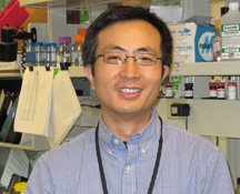 Hong Qin合作论文数Department of Biology, Spelman College, Atlanta, Georgia, United States4
 Ashish Kr. Luhach合作论文数4
 Sanjay Kumar Sen合作论文数3Dilip k. Choudhury合作论文数3
 B Mohammed Imran合作论文数College of Computer Science and Information Technology, Imam Abdulrahman Bin Faisal University3
 Madhumita Pal合作论文数Department of Computer Science and Engineering, Siksha O Anusandhan (Deemed to be) University3
 Ruppa (Tulsi) Thulasiram合作论文数Department of Computer Science, University of Manitoba3Parimala Thulasiraman合作论文数Department of Computer Science
University of Manitoba3
Ruppa (Tulsi) Thulasiram合作论文数Department of Computer Science, University of Manitoba3Parimala Thulasiraman合作论文数Department of Computer Science
University of Manitoba3 Madasu Hanmandlu合作论文数CSE Department, Maturi Venkata Subba Rao Engineering College3
 Babita Majhi合作论文数ECE Department, National Institute of Technology Rourkela3
 Bhupesh Deka合作论文数2g k mahanti合作论文数2Vijay Agrawal合作论文数2
 Nehad Mohammed Ibrahim合作论文数2
 Chinmay Chakraborty合作论文数2
 Ratnakar Dash合作论文数2
 Haipeng Liu合作论文数2Padma Charan Sahu合作论文数Godavari Institute of Engineering and Technology2Munir Ahmad合作论文数2
 Dhiaa Musleh合作论文数Dept Comp Sci CS, Imam Abdulrahman Bin Faisal Univ IAU2avinash sinha合作论文数2
 Achyut Shankar合作论文数Amity Univ, Dept CSE, Noida, India2
 Prasad, B.合作论文数2Subudhi B N R合作论文数2
 Ajith Abraham合作论文数School of Artificial Intelligence, Sai University2
 Rohit Sharma合作论文数2
 Joel Rodrigues合作论文数Instituto de Telecomunicações2
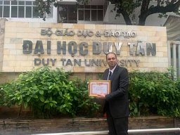 Anand Nayyar合作论文数2
 Ananta Charan Ojha合作论文数ICFAI University2Kiran Sultan合作论文数2
 B.K. Tripathy合作论文数School of Computing Sciences, VIT University2
 Nazar Abbas Saqib合作论文数Department of Computer Science, College of Computer Science and Information Technology, Imam Abdulrahman Bin Faisal University2Debasish Pradhan合作论文数1T. C. Panda合作论文数1Smita Rani Parija合作论文数1




