The Deep Brain Stimulation (DBS) Think Tank XIII was held September 2-4th, 2025, in Gainesville, Florida, at the Norman Fixel Institute for Neurological Diseases at the University of Florida. The theme was "The Evolving Landscape of DBS: New Indications, New Goals." This theme was a continuation of the DBS Think Tank XI and XII, which were focused on emerging technology and pushing the horizon of indications. Since its founding in 2012, the DBS Think Tank has provided a global forum for leading clinicians, engineers, and researchers in both in industry and academia to present, discuss, and debate the current state of DBS technologies as well as to consider important logistics and ethical challenges. Over the course of three days, members of each panel presented and facilitated discussions on the cutting edge of DBS research. The keynote speaker was Dr. Kamil Uğurbil of the University of Minnesota, who led the first group of researchers to demonstrate the feasibility of imaging the human brain using fMRI technology and who was a pioneer in the development of high-field human MRI scanning. Nobel laureate Dr. Stanley Prusiner, from the University of California, San Francisco, used the story of the discovery of prions to demonstrate the power of pursuing a finding even when the idea conflicted with the prevailing state of the field. The think tank was divided into sections, including: Next Generation Neuromodulation for Gait, Brain Networks and Neuromodulation, Neuroscience & Society, Interventional Psychiatry & Behavior, Devices for Closing the Loop, Physiology & Closing the Loop, and A Roadmap for Genetics & Neuromodulation.
The NIH Common Fund's SPARC (Stimulating Peripheral Activity to Relieve Conditions) program was launched in 2015 to catalyze bioelectronic medicine by advancing foundational knowledge of the autonomic nervous system (ANS) and enabling clinical translation of novel bioelectronic medicines. While the program officially ended in 2025, activities related to the program are still ongoing through no-cost extensions, data sharing, and derivative efforts. We provide here an overview of the major outcomes of SPARC under its four focus areas: Anatomical and Functional Mapping, Technology Development, Translational Research, and Data/Modeling Infrastructure. In the Anatomical and Functional Mapping focus areas, teams generated cross-species, cross-organ ANS connectivity datasets, detailed nerve anatomy, along with detailed maps and connectivity resources to support query, visualization, and hypothesis-generation. Under the Technology Development focus area, teams developed new tools for recording and stimulating autonomic pathways and built computational pipelines for realistic neuromodulation simulations. Within the Translational Research, teams conducted pre-clinical and clinical proof-of-concept studies and used prize-driven efforts to accelerate clinically ready neuromodulation solutions. Data/Modeling Infrastructure teams built a durable open-science ecosystem enabling FAIR sharing, exploration, and reuse of SPARC datasets, models, and workflows by the broader community. Together, SPARC's integrated approach has lowered barriers to precision neuromodulation research and established reusable resources to sustain the field beyond the program.
This protocol describes acute experiments involving sacral stimulation of minipigs while monitoring distal colon motility. Stimulation at various amplitudes and frequencies is applied to the S2 or S3 sacral roots using a bipolar cuff. Colon motility is monitored using multiple sensing modalities including electromyography (EMG), colon wall strain sensing, intraluminal pressure manometry, and electrochemical dopamine sensing.
We introduce OpenNerve, an open-source modular clinical-grade neuromodulation platform developed by the Center for Autonomic Nerve Recording and Stimulation Systems (CARSS) with Med-Ally LLC, Medipace Inc., and the University of Southern California (USC). The system combines an Implantable Pulse Generator (IPG) with 8 stimulation and 8 sensing channels, plus a library of interchangeable leads and sensors for diverse bioelectronic-medicine applications. All implantable components including sensors, stimulation electrodes, and IPG are verified to relevant electrical safety and biocompatibility standards. The architecture supports userconfigurable open- and closed-loop control, onboard signal processing, wireless charging, and Bluetooth Low Energy (BLE) telemetry for communication with patient and clinician interfaces. Pilot in-vivo animal studies are underway to demonstrate safe chronic use and efficacy. All hardware, firmware, software, and related test protocols are released under permissive open-source licenses, making OpenNerve the first openly shared, end-to-end platform for accelerating clinical translation of bioelectronic therapies.
Bioelectronic medicine is a growing field which involves directly interfacing with the vagus, sacral, enteric, and other autonomic nerves to treat conditions. Therapies based on bioelectronic medicine could address previously intractable diseases and provide an alternative to pharmaceuticals. However, translating a bioelectronic medicine therapy to the clinic requires overcoming several challenges, including titrating stimulation parameters to an individual’s physiology, selectively stimulating target nerves without inducing off-target activation or block, and improving accessibility to clinically approved devices. This review describes recent progress towards solving these problems, including advances in mapping and characterizing the human autonomic nervous system, new sensor technology and signal processing techniques to enable closed-loop therapies, new methods for selectively stimulating autonomic nerves without inducing off-target effects, and efforts to develop open-source implantable devices. Recent commercial successes in bringing bioelectronic medicine therapies to the clinic are highlighted showing how addressing these challenges can lead to novel therapies.
The open-source development model has been successfully applied to consumer and enterprise software, and recently to consumer hardware. Medical devices may become a beneficiary of this trend, as open-source medical device development has the potential to reduce costs, democratize patient access, and provide continued support to abandoned devices from failed companies. Unlike the consumer device market, the medical device market is highly regulated and involves considerable manufacturer liability that may limit the use of open-source technology. This review of open-source medical device development explores the current state of development in research and clinical products and suggests best practices for creating sustainable and effective open-source medical devices.
BACKGROUND AND AIMS:Sacral nerve stimulation (SNS) showed anti-inflammatory properties in animal models of inflammatory bowel disease. We aimed to evaluate the effectiveness and safety of SNS in patients with ulcerative colitis (UC).MATERIALS AND METHODS:Twenty-six patients with mild and moderate disease were randomized into two groups: SNS (delivered at S3 and S4 sacral foramina) and sham-SNS (delivered 8-10 mm away from sacral foramina), with the therapy applied once daily for one hour, for two weeks. We evaluated the Mayo score and several exploratory biomarkers, including C-reactive protein in the plasma, pro-inflammatory cytokines and norepinephrine in the serum, assessment of autonomic activity, and diversity and abundance of fecal microbiota species.RESULTS:After two weeks, 73% of the subjects in the SNS group achieved clinical response, compared with 27% in the sham-SNS group. Levels of C-reactive protein, pro-inflammatory cytokines in the serum, and autonomic activity were significantly improved toward a healthy profile in the SNS group but not in the sham-SNS group. Absolute abundance of fecal microbiota species and one of the metabolic pathways were changed in the SNS group but not in the sham-SNS group. Significant correlations were observed between pro-inflammatory cytokines and norepinephrine in the serum on the one side and fecal microbiota phyla on the other side.CONCLUSIONS:Patients with mild and moderate UC were responsive to a two-week SNS therapy. After performing further studies to evaluate its efficacy and safety, temporary SNS delivered through acupuncture needles may become a useful screening tool for identifying SNS therapy responders before considering long-term implantation of the implantable pulse generator and SNS leads for performing long-term SNS therapy.
Background and objectives:In this systematic review, we assessed the efficacy, potential mechanisms, and safety of two neuromodulation therapies in patients with inflammatory bowel disease (IBD), including Crohn's disease and ulcerative colitis. The first therapy is vagus nerve stimulation (VNS) utilizing implantable or transcutaneous electrodes, and the second is sacral nerve stimulation (SNS) using implantable or percutaneous electrodes.Methods:We conducted a systematic literature review following the Preferred Reporting Items for Systematic Reviews and Meta-Analyses (PRISMA) guidelines. The PubMed database was comprehensively searched, and studies were rigorously assessed for inclusion and exclusion criteria.Results:Our analysis encompassed five clinical studies, three on VNS and two on SNS. Most investigated studies demonstrated significant beneficial effects on IBD symptoms, including disease activity, severity of intestinal lesions, and intestinal pain. When evaluating the impact on key IBD pathophysiologies, both VNS and SNS exhibited trends toward reducing biomarkers of intestinal mucosal inflammation and mitigating sympathetic dominance. Importantly, none of the evaluated neuromodulation methods resulted in long-term adverse effects.Conclusions:Cumulative evidence from the evaluated studies indicates that VNS and SNS therapies effectively alleviate IBD symptoms and may hold promise in addressing the underlying pathophysiologies of IBD, including intestinal mucosal inflammation and sympathetic dominance. Consequently, they represent valuable options for individualized IBD treatment.
The aim of this mini-review is to introduce most prevalent autoimmune diseases, emphasize the importance of sympathoparasympathetic imbalance in these autoimmune diseases, demonstrate how such imbalance can be effectively treated using the bioelectronic medicine, and describe potential mechanisms of bioelectronic medicine effects on the autoimmuneactivity at the cellular and molecular levels.
Background and objectives:In this systematic review, we evaluated the efficacy, mechanisms and safety of three neuromodulation therapies in patients with gastroesophageal reflux disease (GERD), including the effect of neuromodulation therapies on symptoms and key GERD pathophysiologies, lower esophageal sphincter (LES) pressure, esophageal motility, gastric motility, and parasympathetic activity. The first therapy is LES electrical stimulation using an implantable electrical stimulator, the second is transcutaneous electrical acustimulation, and the third is manual acupuncture.Methods:A systematic review of literature according to the PRISMA guidelines was performed. Online databases searched include Medline (Ovid), Embase, and PubMed. Studies were assessed for inclusion and exclusion criteria with Covidence, a systematic review software.Results:The analysis included thirteen clinical studies. Four papers included were registered under two open-label trials on ClinicalTrials.gov for LES electrical stimulation; Five randomized trials with sham-treated controls were analyzed for transcutaneous electrical acustimulation; Four studies, including three involving standard therapy controls and one involving shamtreated controls were included for manual acupuncture. All evaluated studies demonstrated significant beneficial effects on GERD symptoms, using patient-completed questionnaires, objective 24-h measurement of esophageal pH, and patient-reported use of proton pump inhibitors. In evaluating the effect on key GERD pathophysiologies, electrical stimulation significantly increased LES pressure, and transcutaneous electrical acustimulation significantly improved esophageal motility, gastric motility, and parasympathetic activity. None of the evaluated neuromodulation methods produced severe adverse effects.Conclusions:Cumulative evidence from the evaluated studies indicates that neuromodulation therapies were effective in treating the GERD symptoms and key underlying GERD pathophysiologies. They are thus valuable options for individualized GERD treatment.
Purpose: To investigate how modulating ocular sympathetic activity affects progression of choroidal neovascularization (CNV), a hallmark feature of wet age-related macular degeneration (AMD). Methods: In the first of two studies, Brown Norway rats underwent laser-induced CNV and were assigned to one of the following groups: daily eye drops of artificial tears (n = 10; control group); daily eye drops of the β-adrenoreceptor agonist isoproterenol (n = 10); daily eye drops of the β-adrenoreceptor antagonist propranolol (n = 10); sympathetic internal carotid nerve (ICN) transection 6 weeks prior to laser-induced CNV (n = 10). In the second study, rats underwent laser-induced CNV followed by ICN transection at different time points: immediately after the laser injury (n = 6), 7 days after the laser injury (n = 6), and sham surgery 7 days after the laser injury (n = 6; control group). All animals were euthanized 14 days after laser application. CNV development was quantified with fluorescein angiography and optical coherence tomography (in vivo), as well as lesion volume analysis using 3D confocal reconstruction (postmortem). Angiogenic growth factor protein levels in the choroid were measured with ELISA. Results: In the first study, blocking ocular sympathetic activity through pharmacological or surgical manipulation led to a 75% or 70% reduction in CNV lesion volume versus the control group, respectively (P < 0.001). Stimulating ocular sympathetic activity with isoproterenol also led to a reduction in lesion volume, but only by 27% versus controls (P < 0.05). VEGF protein levels in the choroid were elevated in the three treatment groups (P < 0.01). In the second study, fluorescein angiography and CNV lesion volume analysis indicated that surgically removing the ocular sympathetic supply inhibited progression of laser-induced CNV, regardless of whether ICN transection was performed on the same day or 7 days after the laser injury. Conclusion: Surgical and pharmacological block of ocular sympathetic activity can inhibit progression of CNV in a rat model. Therefore, electrical block of ICN activity could be a potential bioelectronic medicine strategy for treating wet AMD.
Background Previous studies have shown that neurons of the cerebral cortex can be injured by implantation of, and stimulation with, implanted microelectrodes. Objectives Objective 1 was to determine parameters of microstimulation delivered through multisite intracortical microelectrode arrays that will activate neurons of the feline cerebral cortex without causing loss of neurons. Objective 2 was to determine if the stimulus parameters that induced loss of cortical neurons differed for all cortical neurons vs. the subset of inhibitory neurons expressing parvalbumin. Methods The intracortical microstimulation was applied for 7 h/day for 20 days (140 h). Microelectrode site areas were 2000 and 4000 μm2, Q was 2–8 nanocoulombs (nC) at 50 Hz, and QD was 50–400 μcoulombs/cm2. Results Neuron loss due to stimulation was minimal at Q = 2 Ncp, but at 8 Ncp, 20%–50% of neurons within 250 μm of the stimulated microelectrodes were lost, compared to unstimulated microelectrodes. Loss was greatest in tissue facing electrode sites. Stimulation-induced loss was similar for neurons labeled for NeuN and for inhibitory neurons expressing parvalbumin. Correlation between neuron loss and QD was not significant. Electrodes in the medullary pyramidal tract recorded neuronal activity evoked by stimulation in the cerebral cortex. The pyramidal neurons were activated by intracortical stimulation of 2 nC/phase. 140 h of microstimulation at 2 nC/phase and 50 Hz induced minimal neuron loss.
Objective. Bladder dysfunction is a significant and largely unaddressed problem for people living with spinal cord injury (SCI). Intermittent catheterization does not provide volitional control of micturition and has numerous side effects. Targeted electrical microstimulation of the spinal cord has been previously explored for restoring such volitional control in the animal model of experimental SCI. Here, we continue the development of the intraspinal microstimulation array technology to evaluate its ability to provide more focused and reliable bladder control in the feline animal model. Approach. For the first time, a mechanically robust intraspinal multisite silicon array was built using novel microfabrication processes to provide custom-designed tip geometry and 3D electrode distribution. Long-term implantation was performed in eight spinally intact animals for a period up to 6 months, targeting the dorsal gray commissure area in the S2 sacral cord that is known to be involved in the coordination between the bladder detrusor and the external urethral sphincter. Main results. About one third of the electrode sites in the that area produced micturition-related responses. The effectiveness of stimulation was further evaluated in one of eight animals after spinal cord transection (SCT). We observed increased bladder responsiveness to stimulation starting at 1 month post-transection, possibly due to supraspinal disinhibition of the spinal circuitry and/or hypertrophy and hyperexcitability of the spinal bladder afferents. Significance. 3D intraspinal microstimulation arrays can be chronically implanted and provide a beneficial effect on the bladder voiding in the intact spinal cord and after SCT. However, further studies are required to assess longer-term reliability and safety of the developed intraspinal microstimulation array prior to eventual human translation.
Purpose:To investigate specific effects of denervation and stimulation of the internal carotid nerve (ICN) on the choroid and retina.Methods:Female Sprague Dawley rats underwent unilateral ICN transection (n = 20) or acute ICN electrical stimulation (n = 7). Rats in the denervation group were euthanized 6 weeks after nerve transection, and eyes were analyzed for changes in choroidal vascularity (via histomorphometry) or angiogenic growth factors and inflammatory markers (via ELISA). Rats in the stimulation group received acute ICN electrical stimulation with a bipolar cuff electrode over a range of stimulus amplitudes, frequencies, and pulse widths. Choroidal blood flow and pupil diameter were monitored before, during, and after stimulation.Results:Six weeks after unilateral ICN transection, sympathectomized choroids exhibited increased vascularity, defined as the percentage of choroidal surface area occupied by blood vessel lumina. Vascular endothelial growth factor (VEGF) and VEGF receptor-2 (VEGFR-2) protein levels in denervated choroids were 61% and 124% higher than in contralateral choroids, respectively. TNF-α levels in denervated retinas increased by 3.3-fold relative to levels in contralateral retinas. In animals undergoing acute ICN electrical stimulation, mydriasis and reduced choroidal blood flow were observed in the ipsilateral eye. The magnitude of the reduction in blood flow correlated positively with stimulus frequency.Conclusions:Modulation of ICN activity reveals a potential role of the ocular sympathetic system in regulating endpoints related to neovascular diseases of the eye.
A new class of treatments termed bioelectronic medicines are now emerging that aim to target individual nerve fibres or specific brain circuits in pathological conditions to repair lost function and reinstate a healthy balance. Carotid sinus nerve (CSN) denervation has been shown to improve glucose homeostasis in insulin-resistant and glucose-intolerant rats; however, these positive effects from surgery appear to diminish over time and are heavily caveated by the severe adverse effects associated with permanent loss of chemosensory function. Herein we characterise the ability of a novel bioelectronic application, classified as kilohertz frequency alternating current (KHFAC) modulation, to suppress neural signals within the CSN of rodents.
The polycystic ovary syndrome (PCOS) is the most prevalent ovarian pathology in women, with excessive sympathetic activity in the superior ovarian nerve (SON) playing an important role in inducing the PCOS symptoms in the rats and humans. Our previous studies have shown that surgical transection of the SON can reverse the disease progression, prompting us to explore the effect of the kilohertz frequency alternating current (KHFAC) modulation as a method of reversible non-surgical suppression of the nerve activity in the rodent model of PCOS. 56 animals were randomly allocated to three groups: the Control group (n = 18), the PCOS group (n = 15), and the PCOS + KHFAC group (n = 23). The physiological, anatomical, and biochemical parameters of ovarian function were evaluated during the progression of the experimentally-induced PCOS and during long-term KHFAC modulation applied for 2–3 weeks. The KHFAC modulation has been able to reverse the pathological changes in assessed PCOS parameters, namely the irregular or absent estrous cycling, formation of ovarian cysts, reduction in the number of corpora lutea, and ovarian norepinephrine concentration. The fertility capacity was similar in the PCOS and the PCOS + KHFAC groups, indicating the safety of KHFAC modulation approach. In summary, these results suggest that the KHFAC modulation approach of suppressing the SON activity could become a useful treatment modality for PCOS and potentially other pathological ovarian conditions.
The external anal sphincter (EAS) is important for the maintenance of bowel continence and may be compromised by a variety of neuropathic conditions. However, large animal models for the study of EAS functions have been sparse. The EAS guarding reflex was examined by electromyography (EMG) in neurologically intact rhesus macaques ( n = 6) and at 4-6 wk after a unilateral EAS denervation from an L6-S3 ventral root avulsion (VRA) injury ( n = 6). Baseline EAS EMG recordings were quiescent in all subjects, and evoked responses showed an initial large-amplitude EMG activity, which gradually returned to baseline within 1-2 min. At 4-6 wk postoperatively, the EAS guarding reflex showed a significantly reduced EMG response duration of 47 ± 15 s and area under the curve (AUC) of 0.198 ± 0.097 mV·s compared with the corresponding evoked EAS EMG duration of 102 ± 19 s and AUC of 0.803 ± 0.225 mV·s ( P < 0.05) in the control group. Detailed time- and frequency-domain analysis of the evoked EAS EMG responses for the first 40 s showed no difference between groups for the maximum amplitude but a significant decrease for the mean amplitude across the study period and an early AUC reduction for the first 10 s in the VRA injury group. Time-frequency analysis and power spectrum plots indicated decreased intensity and a narrower midrange of frequencies in the VRA injury group. We conclude that the EAS guarding reflex in rhesus macaques shows characteristic EMG features in control subjects and signs of partial target denervation after a unilateral L6-S3 VRA injury. NEW & NOTEWORTHY The external anal sphincter guarding reflex showed initial large-amplitude peaks and a gradual return to a quiescent baseline after a rectal probe stimulus in rhesus macaques. At 4-6 wk after a unilateral ventral root avulsion (VRA) injury, the electromyography duration, mean amplitude, and area under the curve measurements were decreased. Time-frequency analysis and power spectrum plots indicated decreased intensity and a narrowed midrange of frequencies in the VRA injury cohort.
A novel Parylene C-based peripheral nerve interface that combines both electrodes and microfluidic channels in an adjustable cuff was designed, fabricated, and characterized. This minimally invasive interface incorporates a drug delivery system for targeted delivery of lysing agents and neurotrophic factors to the nerve surface to locally disrupt the epineurium and allow fascicular selectivity. Multiple platinum (Pt) electrodes were embedded in the microfluidic channels for neural stimulation and recording. The lyse-and-attract cuff electrode (LACE) uses a simple locking mechanism that is adjustable for close contact with nerves of varying diameters. Devices were fabricated using standard Parylene microfabrication techniques resulting in low variability and high device yield. A procedure for the implantation of the LACE was developed and successfully demonstrated in vivo around rat sciatic nerves. The adjustable locking mechanism demonstrated adequate holding strength and fit around the nerves. Benchtop electrochemical characterization of the thin-film Pt electrodes showed that the electrodes possessed high charge storage capacity (>1 mC/cm2) and low impedances ( $< 2~\text{k}\Omega $ at 1 kHz) suitable for neural stimulation and recording. As expected, embedded electrodes demonstrated higher impedance values. Acute neural recording from the rat sciatic nerve verified the capability of the LACE to record evoked neural activity (compound action potentials). Controlled and localized microfluidic infusions were achieved at low flow rates ( $< 1~\mu \text{L}$ /min). Finally, infusion experiments in vivo demonstrated targeted drug delivery to the sciatic nerve fascicles. This multifunctional peripheral nerve interface has the potential to enhance implant-tissue integration in vivo and provide reliable chronic performance not available in the existing extraneural or intraneural interfaces. [2018-0192]
 Peter H. Siegel合作论文数Division of Biology, California Institute of Technology;Jet Propulsion Laboratory, California Institute of Technology;THz Global19
 Douglas B. McCreery合作论文数Huntington Medical Research Institutes14
 Ellis Fan-Chuin Meng合作论文数Alfred E. Mann Department of Biomedical Engineering, Viterbi School of Engineering, University of Southern California;Fluid Synchrony LLC;Senseer LLC11
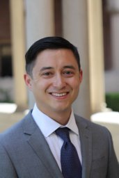 Seth a Hara合作论文数6
 Kuo, J.T.W.合作论文数6
 Curtis Lee合作论文数Department of Metallurgical Engineering, The University of Utah6
 Tuan Hoang合作论文数Dept Intervent Radiol, Univ Texas MD Anderson Canc Ctr5
 Martin Han合作论文数5
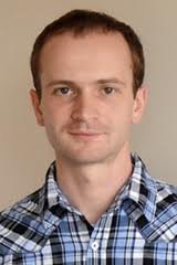 Sergii V Romanenko合作论文数California Inst. of Technol.;c;California Inst. of Technol.4
 Juyong Brian Kim合作论文数Department of Medicine, Stanford University4
 Arun Sridhar合作论文数3
 Sahar Elyahoodayan合作论文数Dept Biomed Engn, Univ Southern Calif3
 Lawrence Yu合作论文数Department of Pharmaceutical Sciences, College of Pharmacy, University of Michigan-Ann Arbor3
 Albert S Lossinsky合作论文数3
 Jean R. Wrathall合作论文数Department of Neuroscience, Georgetown University Medical Center, Georgetown University3
 Mark Humayun合作论文数Dr. Allen and Charlotte Ginsburg Institute for Biomedical Therapeutics, University of Southern California3Andrew C. Weitz合作论文数2B. J. Kim合作论文数2Anthony Rodriguez合作论文数2Alessandra Giarola合作论文数2
 Christian Gutierrez合作论文数2C. A. Gutierrez合作论文数2
 Matteo Donega合作论文数Neuromodulation, Galvani Bioelectronics2
 Alex Baldwin合作论文数Dept Biomed Engn, Univ Southern Calif2
 Christopher Larson合作论文数Department of Biomedical Engineering, University of Southern California2
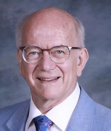 Michael G Harrington合作论文数Dept Neurol, Univ Southern Calif2
 Kee Scholten合作论文数Applied Physics Program, University of Michigan;c;Applied Physics Program, University of Michigan2
 Silvia V Conde合作论文数CEDOC, Univ Nova Lisboa2Angelica Cobo合作论文数2Famm Kristoffer合作论文数2
 Joana F. Sacramento合作论文数NOVA Medical School, Universidade Nova de Lisboa2
 X. Liu合作论文数2
 A. Tang合作论文数Ministry of Defence of Singapore2
 David E. Hinton合作论文数Duke University2
 Mau-Chung Frank Chang合作论文数Hong Kong Institute for Advanced Study, City University of Hong Kong;University of California Los Angeles2Xianghong Arakaki合作论文数Molecular Neurology Program, Huntington Medical Research Institutes2
 Daniel Wagenaar合作论文数Division of Biology and Biological Engineering, California Institute of Technology2
 Leo A. Bullara合作论文数2Leif A. Havton合作论文数2
 Gabriel Virbila合作论文数2Christine Spee合作论文数2LA BULLARA合作论文数1Jesus Prieto-Lloret合作论文数1Juan-Carlos Martinez-Camarillo合作论文数1SCHÜTTLER MARTIN合作论文数1
 Bryan McLaughlin合作论文数1Wesley Dopson合作论文数1Thao X. Hoang合作论文数1MA Qiyao (麻琦瑶)合作论文数1




