Augmentation can corrupt a training example when an image and its annotations receive different random changes. A crop must use the same coordinates for the image, mask, boxes, keypoints, stereo views, video frames, or volume. Code paths that choose these values separately can silently misalign the data. AlbumentationsX keeps the transform list, probabilities, annotation settings, and random seed in one Compose object. Each call chooses random values once and applies them to every supported part of the training example. The library keeps each object's mask, box, and label together and lets projects add their own transforms. It can also save the pipeline definition, show what happened in one call, and run that call again. The examples place Compose after files have been decoded into arrays and before PyTorch groups examples into a batch. AlbumentationsX executes the declared transforms. Practitioners still decide whether a flip, crop, color change, or other operation preserves the correct label for their task.
Data augmentation is a commonly used technique for increasing both the size and the diversity of labeled training sets by leveraging input transformations that preserve corresponding output labels. In computer vision, image augmentations have become a common implicit regularization technique to combat overfitting in deep learning models and are ubiquitously used to improve performance. While most deep learning frameworks implement basic image transformations, the list is typically limited to some variations of flipping, rotating, scaling, and cropping. Moreover, image processing speed varies in existing image augmentation libraries. We present Albumentations, a fast and flexible open source library for image augmentation with many various image transform operations available that is also an easy-to-use wrapper around other augmentation libraries. We discuss the design principles that drove the implementation of Albumentations and give an overview of the key features and distinct capabilities. Finally, we provide examples of image augmentations for different computer vision tasks and demonstrate that Albumentations is faster than other commonly used image augmentation tools on most image transform operations.
With the growth of popularityKalinin, Alexandr A. of deep neural networks for image analysis, segmentation is the most common subject of studies applying deep learning to medical imaging and establishing state-of-the-art performance resultsIglovikov, Vladimir I. in many applications. However, it still remains a challenging problem, for which performance improvements can potentiallyRakhlin, Alexander benefit diagnosis and other clinical practice outcomes. In this chapter, we consider two applications of multiple deep convolutional neural networks to medical image segmentation. First, we describe angiodysplasia lesion segmentation from wireless capsule endoscopy videos. Angiodysplasia is the most common vascular lesion of the gastrointestinal tract in the general population andShvets, Alexey A. is important to detect as it may indicate the possibility of gastrointestinal bleeding and/or anemia. As a baseline, we consider the U-Net model and then we demonstrate further performance improvements by using different deep architectures with ImageNet pre-trained encoders. In the second example, we apply these models to semantic segmentation of robotic instruments in surgical videos. Segmentation of instruments in the vicinity of surgical scenes is a challenging problem that is important for intraoperative guidance that can help the decision-making process. We achieve highly competitive performance for binary as well as for multi-class instrument segmentation. In both applications, we demonstrate that networks that employ ImageNet pre-trained encoders consistently outperform the U-Net architecture trained from scratch.
In 2015 we began a sub-challenge at the EndoVis workshop at MICCAI in Munich using endoscope images of ex-vivo tissue with automatically generated annotations from robot forward kinematics and instrument CAD models. However, the limited background variation and simple motion rendered the dataset uninformative in learning about which techniques would be suitable for segmentation in real surgery. In 2017, at the same workshop in Quebec we introduced the robotic instrument segmentation dataset with 10 teams participating in the challenge to perform binary, articulating parts and type segmentation of da Vinci instruments. This challenge included realistic instrument motion and more complex porcine tissue as background and was widely addressed with modifications on U-Nets and other popular CNN architectures. In 2018 we added to the complexity by introducing a set of anatomical objects and medical devices to the segmented classes. To avoid over-complicating the challenge, we continued with porcine data which is dramatically simpler than human tissue due to the lack of fatty tissue occluding many organs.
Motivated by the impact of large-scale datasets on ML systems we present the largest self-driving dataset for motion prediction to date, containing over 1,000 hours of data. This was collected by a fleet of 20 autonomous vehicles along a fixed route in Palo Alto, California, over a four-month period. It consists of 170,000 scenes, where each scene is 25 seconds long and captures the perception output of the self-driving system, which encodes the precise positions and motions of nearby vehicles, cyclists, and pedestrians over time. On top of this, the dataset contains a high-definition semantic map with 15,242 labelled elements and a high-definition aerial view over the area. We show that using a dataset of this size dramatically improves performance for key self-driving problems. Combined with the provided software kit, this collection forms the largest and most detailed dataset to date for the development of self-driving machine learning tasks, such as motion forecasting, motion planning and simulation. The full dataset is available at http://level5.lyft.com/.
Breast cancer is one of the main causes of death worldwide. Histopathological cellularity assessment of residual tumors in post-surgical tissues is used to analyze a tumor's response to a therapy. Correct cellularity assessment increases the chances of getting an appropriate treatment and facilitates the patient's survival. In current clinical practice, tumor cellularity is manually estimated by pathologists; this process is tedious and prone to errors or low agreement rates between assessors. In this work, we evaluated three strong novel Deep Learning-based approaches for automatic assessment of tumor cellularity from post-treated breast surgical specimens stained with hematoxylin and eosin. We validated the proposed methods on the BreastPathQ SPIE challenge dataset that consisted of 2395 image patches selected from whole slide images acquired from 64 patients. Compared to expert pathologist scoring, our best performing method yielded the Cohen's kappa coefficient of 0.69 (vs. 0.42 previously known in literature) and the intra-class correlation coefficient of 0.89 (vs. 0.83). Our results suggest that Deep Learning-based methods have a significant potential to alleviate the burden on pathologists, enhance the diagnostic workflow, and, thereby, facilitate better clinical outcomes in breast cancer treatment.
In mainstream computer vision and machine learning, public datasets such as ImageNet, COCO and KITTI have helped drive enormous improvements by enabling researchers to understand the strengths and limitations of different algorithms via performance comparison. However, this type of approach has had limited translation to problems in robotic assisted surgery as this field has never established the same level of common datasets and benchmarking methods. In 2015 a sub-challenge was introduced at the EndoVis workshop where a set of robotic images were provided with automatically generated annotations from robot forward kinematics. However, there were issues with this dataset due to the limited background variation, lack of complex motion and inaccuracies in the annotation. In this work we present the results of the 2017 challenge on robotic instrument segmentation which involved 10 teams participating in binary, parts and type based segmentation of articulated da Vinci robotic instruments.
Breast cancer is one of the main causes of cancer death worldwide. Early diagnostics significantly increases the chances of correct treatment and survival, but this process is tedious and often leads to a disagreement between pathologists. Computer-aided diagnosis systems showed potential for improving the diagnostic accuracy. In this work, we develop the computational approach based on deep convolution neural networks for breast cancer histology image classification. Hematoxylin and eosin stained breast histology microscopy image dataset is provided as a part of the ICIAR 2018 Grand Challenge on Breast Cancer Histology Images. Our approach utilizes several deep neural network architectures and gradient boosted trees classifier. For 4-class classification task, we report 87.2% accuracy. For 2-class classification task to detect carcinomas we report 93.8% accuracy, AUC 97.3%, and sensitivity/specificity 96.5/88.0% at the high-sensitivity operating point. To our knowledge, this approach outperforms other common methods in automated histopathological image classification. The source code for our approach is made publicly available at https://github.com/alexander-rakhlin/ICIAR2018
Accurate detection and localization for angiodysplasia lesions is an important problem in early stage diagnostics of gastrointestinal bleeding and anemia. Gold-standard for angiodysplasia detection and localization is performed using wireless capsule endoscopy. This pill-like device is able to produce thousand of high enough resolution images during one passage through gastrointestinal tract. In this paper we present our winning solution for MICCAI 2017 Endoscopic Vision SubChallenge: Angiodysplasia Detection and Localization its further improvements over the state-of-the-art results using several novel deep neural network architectures. It address the binary segmentation problem, where every pixel in an image is labeled as an angiodysplasia lesions or background. Then, we analyze connected component of each predicted mask. Based on the analysis we developed a classifier that predict angiodysplasia lesions (binary variable) and a detector for their localization (center of a component). In this setting, our approach outperforms other methods in every task subcategory for angiodysplasia detection and localization thereby providing state-of-the-art results for these problems. The source code for our solution is made publicly available at https://github.com/ternaus/angiodysplasia-segmentation
The sign problem is the fundamental limitation to quantum Monte Carlo simulations of the statistical mechanics of interacting fermions. Determinant quantum Monte Carlo (DQMC) is one of the leading methods to study lattice models such as the Hubbard Hamiltonian, which describe strongly correlated phenomena including magnetism, metal-insulator transitions, and (possibly) exotic superconductivity. Here, we provide a comprehensive dataset on the geometry dependence of the DQMC sign problem for different densities, interaction strengths, temperatures, and spatial lattice sizes. We supplement these data with several observations concerning general trends in the data, including the dependence on spatial volume and how this can be probed by examining decoupled clusters, the scaling of the sign in the vicinity of a particle-hole symmetric point, and the correlation between the total sign and the signs for the individual spin species.
The most common approaches to instance segmentation are complex and use two-stage networks with object proposals, conditional random-fields, template matching or recurrent neural networks. In this work we present Ternaus-NetV2 - a simple fully convolutional network that allows extracting objects from a high-resolution satellite imagery on an instance level. The network has popular encoder-decoder type of architecture with skip connections but has a few essential modifications that allows using for semantic as well as for instance segmentation tasks. This approach is universal and allows to extend any network that has been successfully applied for semantic segmentation to perform instance segmentation task. In addition, we generalize network encoder that was pre-trained for RGB images to use additional input channels. It makes possible to use transfer learning from visual to a wider spectral range. For DeepGlobe-CVPR 2018 building detection sub-challenge, based on public leaderboard score, our approach shows superior performance in comparison to other methods.
Analysis of high-resolution satellite images has been an important research topic for traffic management, city planning, and road monitoring. One of the problems here is automatic and precise road extraction. From an original image, it is difficult and computationally expensive to extract roads due to presences of other road-like features with straight edges. In this paper, we propose an approach for automatic road extraction based on a fully convolutional neural network of U-net family. This network consists of ResNet-34 pre-trained on ImageNet and decoder adapted from vanilla U-Net. Based on validation results, leaderboard and our own experience this network shows superior results for the DEEPGLOBE - CVPR 2018 road extraction sub-challenge. Moreover, this network uses moderate memory that allows using just one GTX 1080 or 1080ti video cards to perform whole training and makes pretty fast predictions.
Pixel-wise image segmentation is demanding task in computer vision. Classical U-Net architectures composed of encoders and decoders are very popular for segmentation of medical images, satellite images etc. Typically, neural network initialized with weights from a network pre-trained on a large data set like ImageNet shows better performance than those trained from scratch on a small dataset. In some practical applications, particularly in medicine and traffic safety, the accuracy of the models is of utmost importance. In this paper, we demonstrate how the U-Net type architecture can be improved by the use of the pre-trained encoder. Our code and corresponding pre-trained weights are publicly available at https://github.com/ternaus/TernausNet. We compare three weight initialization schemes: LeCun uniform, the encoder with weights from VGG11 and full network trained on the Carvana dataset. This network architecture was a part of the winning solution (1st out of 735) in the Kaggle: Carvana Image Masking Challenge.
Semantic segmentation is in-demand in satellite imagery processing. Because of the complex environment, automatic categorization and segmentation of land cover is a challenging problem. Solving it can help to overcome many obstacles in urban planning, environmental engineering or natural landscape monitoring. In this paper, we propose an approach for automatic multi-class land segmentation based on a fully convolutional neural network of feature pyramid network (FPN) family. This network is consisted of pre-trained on ImageNet Resnet50 encoder and neatly developed decoder. Based on validation results, leader-board score and our own experience this network shows reliable results for the DEEPGLOBE - CVPR 2018 land cover classification sub-challenge. Moreover, this network moderately uses memory that allows using GTX 1080 or 1080 TI video cards to perform whole training and makes pretty fast predictions.
Semantic segmentation of robotic instruments is an important problem for the robot-assisted surgery. One of the main challenges is to correctly detect an instrument's position for the tracking and pose estimation in the vicinity of surgical scenes. Accurate pixel-wise instrument segmentation is needed to address this challenge. In this paper we describe our deep learning-based approach for robotic instrument segmentation. Our approach demonstrates an improvement over the state-of-the-art results using several deep neural network architectures. It addressed the binary segmentation problem, where every pixel in an image is labeled as an instrument or background from the surgery video feed. In addition, we address a multi-class segmentation problem, in which we distinguish between different instruments or different parts of an instrument from the background. In this setting, our approach outperforms other methods for automatic instrument segmentation thereby providing state-of-theart results for these problems. The source code for our solution is made publicly available at https://github.com/ternaus/robotsurgery-segmentation.
Source camera identification is the process of determining which camera or model has been used to capture an image. In recent years, there has been a rapid growth of research interest in the domain of forensics. In the current work, we describe our Deep Learning approach to the camera detection task of 10 cameras as a part of the Camera Model Identification Challenge hosted by Kaggle.com where our team finished 2nd out of 582 teams with the accuracy on the unseen data of 98%. Augmentations that allowed a stay robust against transformations. A number of experiments are carried out on datasets collected by organizers and scraped from the web.
HER2/neu status of breast cancer is important for treatment strategy choice, but nowadays it is evaluated manually by pathologist. The automation of this procedure is an urgent task, because it will allow to free a pathologist from routine work. But the problem of cells segmentation is difficult for classical methods of computer vision due to the frequent presence of erythrocytic background and non-cellular elements at specimen' image. In this work we propose segmentation algorithm, based on deep convolutional neural networks. Is it has been shown that using this approach, it's possible to get better results, than using classical computer vision algorithms.
This paper describes our approach to the DSTL Satellite Imagery Feature Detection challenge run by Kaggle. The primary goal of this challenge is accurate semantic segmentation of different classes in satellite imagery. Our approach is based on an adaptation of fully convolutional neural network for multispectral data processing. In addition, we defined several modifications to the training objective and overall training pipeline, e.g. boundary effect estimation, also we discuss usage of data augmentation strategies and reflectance indices. Our solution scored third place out of 419 entries. Its accuracy is comparable to the first two places, but unlike those solutions, it doesn't rely on complex ensembling techniques and thus can be easily scaled for deployment in production as a part of automatic feature labeling systems for satellite imagery analysis.
Skeletal bone age assessment is a common clinical practice to diagnose endocrine and metabolic disorders in child development. In this paper, we describe a deep learning approach to the problem of bone age assessment using data from the 2017 Pediatric Bone Age Challenge organized by the Radiological Society of North America. This dataset consists of 12,600 radiological images. Each radiograph in the dataset is an image of a left hand labeled with bone age and sex of a patient. Our approach introduces a comprehensive preprocessing protocol based on the positive mining technique. We use images of whole hands as well as specific hand parts for both training and prediction. This allows us to measure the importance of specific hand bones for automated bone age analysis. We further evaluate the performance of the suggested methods in the context of skeletal development stages. Our approach outperforms other common methods for bone age assessment.
The sign problem is the fundamental limitation to quantum Monte Carlo simulations of the statistical mechanics of interacting fermions. Determinant quantum Monte Carlo (DQMC) is one of the leading methods to study lattice fermions, such as the Hubbard Hamiltonian, which describe strongly correlated phenomena including magnetism, metal-insulator transitions, and possibly exotic superconductivity. Here, we provide a comprehensive dataset on the geometry dependence of the DQMC sign problem for different densities, interaction strengths, temperatures, and spatial lattice sizes. We supplement these data with several observations concerning general trends in the data, including the dependence on spatial volume and how this can be probed by examining decoupled clusters, the scaling of the sign in the vicinity of a particle-hole symmetric point, and the correlation between the total sign and the signs for the individual spin species.
 Alexey A. Shvets合作论文数Institut Charles Sadron, Université de Strasbourg11
 Richard T. Scalettar合作论文数Department of Physics and Astronomy, University of California, Davis8
 Alexandr A. Kalinin合作论文数Chan Zuckerberg Biohub San Francisco7
 Alexander V Buslaev合作论文数Mapbox R D Ctr Minsk4
 Sasha Rakhlin合作论文数Statistics and Data Science Center, Department of Brain & Cognitive Science, Massachusetts Institute of Technology;Institute for Data, Systems, and Society, Massachusetts Institute of Technology4Selim S. Seferbekov合作论文数Veeva Syst, D-60314 Frankfurt, Germany3Francois Hebert合作论文数Institut Non-Lineaire de Nice;Universite de Nice-Sophia Antipolis;Institut Non-Lineaire de Nice, Universite de Nice-Sophia Antipolis3
 Lena Maier-Hein合作论文数German Cancer Research Center;Faculty of Medicine, University of Heidelberg2
 Rajiv Singh合作论文数Department of Physics and Astronomy, University of California, Davis2
 Khatami Ehsan合作论文数Dept Phys & Astron, San Jose State Univ2
 Azizian Mahdi合作论文数Intuitive Surgical Inc2
 Stefanie Speidel合作论文数Technische Universität Dresden;National Center for Tumor Diseases Dresden, German Cancer Research Center2
 Sebastian Bodenstedt合作论文数2
 Benoit Gremaud合作论文数Laboratoire Kastler Brossel;Université Pierre et Marie Curie;Laboratoire Kastler Brossel, Université Pierre et Marie Curie2
 Danail Stoyanov合作论文数Surgical Robot Vision Research Group, Department of Computer Science, Faculty of Engineering Sciences, University College London2
 Jaan Oitmaa合作论文数School of Physics, University of New South Wales2
 Max Allan合作论文数Intuitive Surgical Inc2
 George Batrouni合作论文数Université Côte d'Azur2V. N. Antonov合作论文数1Elena Slavnova合作论文数1Satoshi Kondo合作论文数1Eugene Khvedchenya合作论文数1
 Wenqi Li (李文琦)合作论文数NVIDIA1
 Guinther Saibro合作论文数1
 A. V. Fattakhov合作论文数1
 Vladimir Osin合作论文数Signify, Eindhoven, Netherlands1
 Denis Makhov合作论文数Moscow State Technical University1Ilya Kibardin合作论文数МФТИ1Guido C. A. Zuidhof合作论文数Radboud University1
 Anton Dobrenkii合作论文数Care Mentor AI, Research and Development Department1
 Madhu Reddiboina合作论文数RediMinds Inc1
 Andrey Samorodov合作论文数1Imanol Luengo合作论文数Medtronic Digital Surgery1
 Miguel Luna合作论文数Department of Robotics Engineering, Daegu Gyeongbuk Institute of Science and Technology, Daegu, South Korea1Stefan Leger合作论文数Tech Univ Dresden, Helmholtz Zentrum Dresden Rossendorf, OncoRay Natl Ctr Radiat Res Oncol, Fac Med, Dresden, Germany1David Rauber合作论文数1
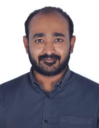 Alex Varghese合作论文数Indian Inst Technol, Madras, Tamil Nadu, India1Robert Mendel合作论文数Ostbayer TH Regensburg OTH Regensburg, Regensburg Med Image Comp ReMIC, Regensburg, Germany1Yawei Ye合作论文数Lyft Level 5 self-driving division1
 Avinash Kori合作论文数Blurgs Pvt. Ltd;Department of Computing, Imperial College London1
 Rahul Duggal合作论文数AWS-AI Computer Vision Group1
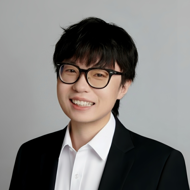 Long Chen合作论文数Xiaomi Technology1
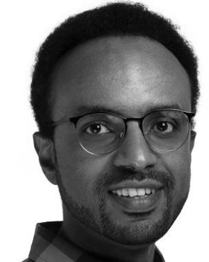 Ahmed Mohammed合作论文数Norwegian Univ Sci & Technol, Dept Comp Sci, N-2815 Gjovik, Norway1Evangello Flouty合作论文数1
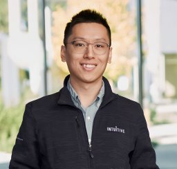 Cong Gao合作论文数Johns Hopkins Univ, Baltimore, MD 21218 USA1Thomas Kurmann合作论文数Univ Bern, ARTORG Ctr, Bern, Switzerland1
 Félix Fuentes-Hurtado合作论文数Univ Politecn Valencia, Inst Invest & Innovac Bioingn, Valencia, Spain1A. A. Kapustin合作论文数1
 Yun-Hsuan Su合作论文数1




