Pustular dermatoses represent a heterogeneous group of conditions frequently encountered in dermatologic practice. The differential diagnosis of pustular dermatoses is broad, encompassing an array of inflammatory, autoimmune, infectious, and genetic etiologies, each with characteristic clinical features and pathophysiology. This article, part of a continuing medical education series, dives into the evolving landscape of these conditions, delivering up-to-date insights into their epidemiology, pathogenesis, clinical presentations, diagnostic advances, and emerging management strategies. Part I will provide an overview of inflammatory and autoimmune etiologies. Part II will focus on infectious, genetic, and transient neonatal and infantile pustular eruptions. This continuing medical education series will help practitioners expand their differential diagnosis, highlight subtle clues that characterize the clinical presentations of these conditions, and spotlight cutting-edge developments that are reshaping management paradigms for some of these entities.
Figure S5: Evaluation of SETDB1 knockdown in human melanoma cells, related to Figure 4:
Pustular dermatoses represent a heterogeneous group of conditions frequently encountered in dermatologic practice. The differential diagnosis of pustular dermatoses is broad, encompassing an array of inflammatory, autoimmune, infectious, and genetic etiologies, each with characteristic clinical features and pathophysiology. This article, part of a continuing medical education series, dives into the evolving landscape of these conditions, delivering up-to-date insights into their epidemiology, pathogenesis, clinical presentations, diagnostic advances, and emerging management strategies. Part II will focus on infectious, genetic, and transient neonatal and infantile pustular eruptions. This continuing medical education series will help practitioners expand their differential diagnosis, highlight clues that characterize the clinical presentation of these conditions, and spotlight cutting-edge developments that are reshaping management paradigms for some of these entities.
Granuloma annulare (GA) is an inflammatory skin disease typically characterized by an erythematous eruption consisting of papules and annular plaques. GA can have a severe impact on quality of life, especially when widespread. GA can be difficult to treat and often recurs after treatment is stopped; there remain no Food and Drug Administration‑approved therapies. In recent years, there has been significant progress in understanding the epidemiology, disease associations, and molecular pathogenesis of GA. Patients with GA are more likely to have hyperlipidemia, diabetes mellitus, and autoimmune diseases including thyroiditis, rheumatoid arthritis, and systemic lupus erythematosus. These associations further underscore the importance of recognizing GA and evaluating for comorbid disease. Accurate diagnosis of GA requires distinguishing it from its clinical and histologic mimics, some of which share annular morphology or granulomatous inflammation. Molecular work has suggested a T cell‑mediated pathogenesis and identified key cytokines and other signals that drive macrophage accumulation and activation in tissue. Emerging therapies that block these cytokine signals are showing promise in the clinic. In this article, we provide an updated overview of the epidemiology, disease associations, clinical and histopathologic characteristics, molecular pathogenesis, and treatment of GA.
Figure S3: Validation and characterization of Setdb1-/- cell lines, tumors, and microenvironments, related to Figure 2.
Ranked CRISPR screen dropout hits, related to Figure 1. Analysis was performed using MaGeCK.
Figure S4: Evaluation of differences in SKO tumor gene expression and microenvironment profiles, related to Figure 3:
IFN-I signaling is a hallmark of discoid lupus erythematosus (DLE), but routine tissue-based assays to detect this pathway are limited. Re-analysis of public transcriptomic data identified ISG15 and IFI6 as candidate IFN markers, ranking among the top differentially expressed genes in DLE datasets. We performed RNA in situ hybridization for both markers on archival specimens from DLE (n =11), other inflammatory dermatoses (n = 11; atopic dermatitis, psoriasis, lichen planus), and controls (n = 8). A total of 81 slides were scored (0-4+) in epidermal and dermal compartments. RNA in situ hybridization showed strong staining in basal keratinocytes and dermis of DLE, with deep dermal positivity in 89% of specimens with sufficient depth for analysis. Deep staining was absent in controls and other dermatoses. DLE demonstrated significantly higher staining intensity than inflammatory controls (P < .001). RNA in situ hybridization for ISG15 and IFI6 identifies IFN activation in DLE, distinguishing it from other dermatoses and providing objective molecular evidence applicable to routine specimens.
Background Leukocytoclastic vasculitis (LCV) and microvascular occlusion (MVO) are distinct histopathologic patterns underlying dermatologic diagnoses of purpura. This study explores the potential of attention-based artificial intelligence (AI) models to enhance diagnostic accuracy and provide interpretable insights in differentiating these conditions, serving as a proof of concept for the application of explainable AI in dermatopathology.Methods We compared the performance of two attention-based AI models, clustering-constrained-attention multiple-instance learning (CLAM) and attention multiple instance learning (MIL), in analyzing whole slide images of LCV and MVO cases. The models were trained and evaluated using a cohort of 69 biopsies. Performance metrics included precision, recall, accuracy, AUROC, and F1 score. Attention-based heatmaps were generated to highlight diagnostic regions and reveal histopathologic patterns.Results The CLAM model outperformed the attention MIL model across all evaluation metrics. Generated heatmaps effectively highlighted key diagnostic regions, including subtle areas of occlusion in the superficial papillary dermis of MVO cases.Conclusions This study demonstrates the potential of attention-based AI models to improve diagnostic accuracy and provide interpretable insights in differentiating LCV and MVO. The use of explainable AI and heatmaps offers a valuable tool for pathologists, enhancing their ability to identify and understand subtle histopathologic patterns.
 Marcus Bosenberg合作论文数Laboratory for Medicine and Pediatrics, Yale School of Medicine58
 Brett King合作论文数Department of Dermatology, Yale School of Medicine, Yale University48
 Jeffrey Cohen合作论文数Department of Dermatology, Yale School of Medicine, Yale University39
 Harriet Kluger合作论文数Department of Dermatology, Yale School of Medicine, Yale University;Developmental Therapeutics, Yale School of Medicine, Yale University;Yale Cancer Center, Yale University33
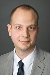 Micevic Goran合作论文数School of Medicine, Yale University30
 Mario Sznol合作论文数Department of Medical Oncology, School of Medicine, Yale University26
 Susan Kaech合作论文数Department of Immunobiology, Yale School of Medicine, Yale University;The Salk Institute for Biological Studies25
 Michael J Murphy合作论文数22
 Min Ding合作论文数Dept Med Cardiovasc Med, Yale Univ20
 Sandra Martinez Morilla合作论文数Boehringer Ingelheim20
 Akiko Iwasaki合作论文数Iwasaki Lab, School of Medicine, Yale University;Howard Hughes Medical Institute20
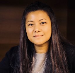 Thazin Nwe Aung合作论文数College of Natural Sciences, Pusan National University20
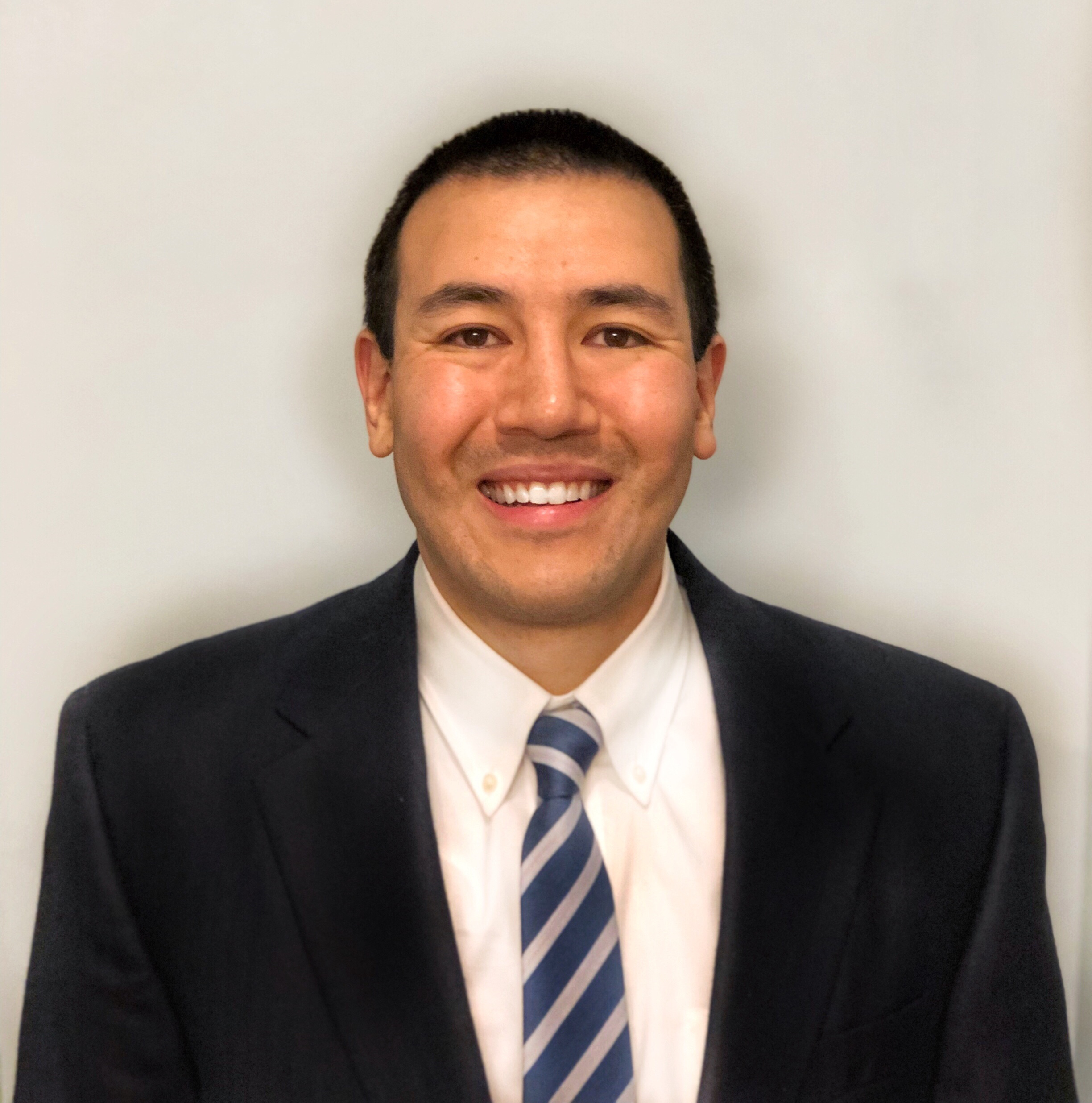 Jeffrey Ishizuka合作论文数Immunology, Biological & Biomedical Sciences, Yale University19
 David Rimm合作论文数Departments of Pathology, Yale University School of Medicine;Yale Cancer Center Tissue Microarray Facility, Yale University School of Medicine18
 Alice Wang合作论文数Dept Dermatol, Yale Sch Med17
 Audrey C Leasure合作论文数Department of Dermatology, Yale School of Medicine16Katelyn Singh合作论文数15
 Matthew Vesely合作论文数15Meaghan K McGeary合作论文数14
 Qin Yan (严钦)合作论文数Department of Pathology, School of Medicine, Yale University14
 Song Eric合作论文数Dept Immunobiol, Yale Sch Med14
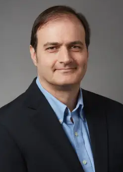 Benjamin Turk合作论文数Biochemistry, Quantitative Biology, Biophysics, and Structural Biology, Biological & Biomedical Sciences, Yale University13Thakral Durga合作论文数13Leventhal Jonathan Scott合作论文数13Lou Hua Jane合作论文数13Ryan Fan合作论文数12Danielle Peterson合作论文数12Huet-Calderwood Clotilde合作论文数12
 Jennifer Madison McNiff合作论文数Department of Dermatology, Yale School of Medicine, Yale University/Department of Pathology, Yale School of Medicine, Yale University11
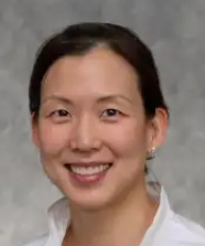 Christine J. Ko合作论文数Department of Dermatology, Yale School of Medicine, Yale University/Department of Pathology, Yale School of Medicine, Yale University11
 Nelson Caroline A合作论文数11Perry Curtis J合作论文数11David A. Calderwood合作论文数11Brooke Horowitch合作论文数10Feriel Ouerghi合作论文数10
 Misha Rosenbach合作论文数Dermatology, Perelman School of Medicine, University of Pennsylvania10Gehlhausen Jeffrey R合作论文数Department of Biochemistry, Herman B Wells Center for Pediatric Research10Bridget E Shields合作论文数9
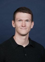 Ronan Talty合作论文数9
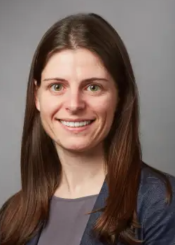 Sarah Weiss合作论文数Medical Oncology, Internal Medicine, Yale School of Medicine, Yale University9Irina Krykbaeva合作论文数8
 Mary M. Tomayko合作论文数Department of Dermatology and Pathology, Yale University School of Medicine8Michael Girardi合作论文数8A. Wang合作论文数7
 N. Turner合作论文数Yale Univ7
 Lucia Jilaveanu合作论文数Medical Oncology, Internal Medicine, Yale School of Medicine, Yale University7Gloria Chen合作论文数6Erica Hwang合作论文数6
 Anjela Galan合作论文数Department of Dermatology, Yale School of Medicine, Yale University/Department of Pathology, Yale School of Medicine, Yale University6




