Wave velocity is a key parameter for imaging complex media, but in vivo measurements are typically limited to reflection geometries, where only backscattered waves from short-scale heterogeneities are accessible. As a result, conventional reflection imaging fails to recover large-scale variations of the wave velocity landscape. Here we show that matrix imaging overcomes this limitation by exploiting the quality of wave focusing as an intrinsic guide star. We model wave propagation as a trainable multi-layer network that leverages optimization and deep learning tools to infer the wave velocity distribution. We validate this approach through ultrasound experiments on tissue-mimicking phantoms and human breast tissues, demonstrating its potential for tumour detection and characterization. Our method is broadly applicable to any kind of waves and media for which a reflection matrix can be measured.
This is the third article in a series of three dealing with the exploitation of speckle for imaging purposes. In complex media, a fundamental limit is the multiple scattering phenomenon that completely blurs the imaging process in depth. Matrix imaging can provide a relevant framework for solving this problem. As it proved to be an adequate tool for probing reverberations in speckle [E. Giraudat et al., Part I], we will show how it can be used to tailor complex spatio-temporal focusing laws to monitor the interference between the multiply-reflected paths and the ballistic component of the wave-field. To do so, we extend the distortion matrix concept to the frequency domain. An iterative phase reversal process operated from the space-time Fourier space is then used to compensate for reverberations and optimize both the axial and transverse resolution of the confocal image. Here, we first present an experimental proof-of-concept consisting in imaging a tissue-mimicking phantom through a reverberating plate before outlining the potential and the limits of this strategy for transcranial ultrasound and beyond.
This is the first article in a series of three dealing with the exploitation of speckle for imaging purposes. Speckle is the complex interference wave-field produced by a random distribution of un-resolved scatterers. In this paper, we show how these scatterers can be used as virtual microphones to monitor the spatio-temporal propagation of a wave-packet inside the medium. To do so, the concept of matrix imaging is particularly useful. It consists in decoupling the location of the transmitted and received focal spots in a standard beamforming process. By scanning the wave-field with the output focal spot that then acts as a virtual transducer, one can image the spatio-temporal evolution of the wave-packet inside the medium. This unique observable will allow us to highlight the imperfections of the focusing process, in particular the defocus and reverberations induced by a strong aberrating layer. As a proof-of-concept, we will consider ultrasound experiments on tissue-mimicking phantoms. In the next two papers, we will show how this observable can be leveraged to compensate for these phenomena that hamper wave focusing and imaging in all fields of wave physics. Our method is indeed broadly applicable to different types of waves beyond ultrasound for which multi-element technology allows a reflection matrix to be measured.
When waves propagate through a complex medium, they undergo several scattering events. This phenomenon is detrimental to imaging, as it causes full blurring of the image. Here we describe a method for detecting, localizing and characterizing any scattering target embedded in a complex medium. We introduce a fingerprint operator that contains the specific signature of the target with respect to its environment. When applied to the recorded reflection matrix, it provides a likelihood index of the target state. This state can be the position of the target for localization purposes, its shape for characterization or any other parameter that influences its response. We demonstrate the versatility of our method by performing proof-of-concept ultrasound experiments on elastic spheres buried inside a strongly scattering granular suspension and on lesion markers, which are commonly used to monitor breast tumours, embedded in a foam mimicking soft tissue. Furthermore, we show how the fingerprint operator can be leveraged to characterize the complex medium itself by mapping the fibre architecture within muscle tissue. Our method is broadly applicable to different types of waves beyond ultrasound for which multi-element technology allows a reflection matrix to be measured.
We present a physically intuitive matrix approach for quantitative imaging of complex media. While standard reflection imaging methods generally rely on confocal focusing operations, matrix imaging consists of decoupling the location of the incident and received focal spots [1]. Following this principle, a self-portrait of the focusing process can be obtained around each point of the medium. The Gouy phase shift exhibited by each focal spot can be leveraged to finely monitor the wave velocity distribution inside the medium [2]. A local multiple scattering rate can also be evaluated and its depth evolution can lead to a local measurement of the scattering mean free path, independently from absorption losses [3]. The approach is here demonstrated with ultrasound for a controllable phantom system before being applied in vivo to liver. The wave velocity and the scattering mean free path are quantitative markers for biomedical diagnosis but they are also important monitoring parameters for nondestructive testing and geophysical applications. This work thus opens important perspectives for quantitative imaging of heterogeneous media in all fields of acoustics. [1] W. Lambert etal., Phys. Rev. X 10, 021048 (2020). [2] F. Bureau et al., arXiv:2409.13901 (2024). [3] A. Goicoechea et al., Phys. Rev. Lett. 133, 176301 (2024).
As waves propagate through a complex medium, they undergo multiple scattering events. This phenomenon is detrimental to imaging, as it causes a full blurring of the image beyond a transport mean free path. Here, we show how to detect, localize, and characterize any scattering target through the reflection matrix of the complex medium in which this target is embedded and thus hidden from direct view. More precisely, we introduce a fingerprint operator that contains the specific signature of the target with respect to its environment. Applied to the recorded reflection matrix, this operator provides a likelihood index of the target in any given state, despite the scattering fog induced by the surrounding environment. This state can be the target position for localization purposes, its shape for characterization, or any other parameter that influences the target response. Our concept is versatile and broadly applicable to different type of waves for which multi-element technology allows a reflection matrix to be measured. We demonstrate this here explicitly by performing different proof-of-concept experiments with ultrasound on targets buried inside a strongly scattering granular suspension, on lesion markers for clinical applications, and on the architecture of muscle tissue.
This is the second article in a series of three dealing with the exploitation of speckle for aberration correction and reverberation compensation in reflection imaging. When probing heterogeneous media with waves, we have to cope with multi-scale fluctuations of the wave velocity. On the one hand, short-scale heterogeneities induce back-scattered echoes whose random interference generate a speckle pattern on the beamformed image. On the other hand, large-scale fluctuations of the wave-velocity can distort the focused wave-fronts, resulting in aberrations on the same image. In this paper, we show how the self-portrait of the wave evolves as a function of the speed-of-sound model. Strikingly, a Gouy phase shift is observed when the speed-of-sound model is optimal. This particularly sensitive feature enables: (i) an optimization of the speed-of-sound model for each pixel of the image; (ii) a local and fine compensation of defocus across the field-of-view, thereby compensating for most aberrations in the image. Experiment in a tissue-mimicking phantom and numerical simulations are first presented to validate our method. It is then applied to in-vivo liver data of a difficult-to-image patient. The speed-of-sound optimization allows an axial compensation of aberrations and a depth-reassignment of each singly-scattered echo to the actual position of the associated scatterer. As distance measurement is often critical for diagnosis, such a wave speed optimization can be crucial for ultrasound but also for any other imaging methods based on the principle of echo-location.
Multiple scattering of waves presents challenges for imaging complex media but offers potential for their characterization. Its onset is actually governed by the scattering mean free path ℓ_{s} that provides crucial information on the medium microarchitecture. Here, we introduce a reflection matrix method designed to estimate this parameter from the time decay of the single scattering rate. Our method is first validated by an ultrasound experiment on a tissue-mimicking phantom before being applied in vivo to a human liver. This Letter opens important perspectives for quantitative imaging of heterogeneous media with waves, whether it be for nondestructive testing, biomedical, or geophysical applications.
Matrix imaging paves the way towards a next revolution in wave physics. Based on the response matrix recorded between a set of sensors, it enables an optimized compensation of aberration phenomena and multiple scattering events that usually drastically hinder the focusing process in heterogeneous media. Although it gave rise to spectacular results in optical microscopy or seismic imaging, the success of matrix imaging has been so far relatively limited with ultrasonic waves because wave control is generally only performed with a linear array of transducers. In this paper, we extend ultrasound matrix imaging to a 3D geometry. Switching from a 1D to a 2D probe enables a much sharper estimation of the transmission matrix that links each transducer and each medium voxel. Here, we first present an experimental proof of concept on a tissue-mimicking phantom through ex-vivo tissues and then, show the potential of 3D matrix imaging for transcranial applications.
This is the first article in a series of two dealing with a matrix approach for aberration quantification and correction in ultrasound imaging. Advanced synthetic beamforming relies on a double focusing operation at transmission and reception on each point of the medium. Ultrasound matrix imaging (UMI) consists in decoupling the location of these transmitted and received focal spots. The response between those virtual transducers form the so-called focused reflection matrix that actually contains much more information than a confocal ultrasound image. In this paper, a time-frequency analysis of this matrix is performed, which highlights the single and multiple scattering contributions as well as the impact of aberrations in the monochromatic and broadband regimes. Interestingly, this analysis enables the measurement of the incoherent input-output point spread function at any pixel of this image. A fitting process enables the quantification of the single scattering, multiple scattering and noise components in the image. From the single scattering contribution, a focusing criterion is defined, and its evolution used to quantify the amount of aberration throughout the ultrasound image. In contrast to the state-of-the-art coherence factor, this new indicator is robust to multiple scattering and electronic noise, thereby providing a contrasted map of the focusing quality at a much better transverse resolution. After a validation of the proof-of-concept based on time-domain simulations, UMI is applied to the in-vivo study of a human calf. Beyond this specific example, UMI opens a new route for speed-of-sound and scattering quantification in ultrasound imaging.
This is the second article in a series of two which report on a matrix approach for ultrasound imaging in heterogeneous media. This article describes the quantification and correction of aberration, i.e. the distortion of an image caused by spatial variations in the medium speed-of-sound. Adaptive focusing can compensate for aberration, but is only effective over a restricted area called the isoplanatic patch. Here, we use an experimentally-recorded matrix of reflected acoustic signals tosynthesize a set of virtual transducers. We then examine wave propagation between these virtual transducers and an arbitrary correction plane. Such wave-fronts consist of two components: (i) An ideal geometric wave-front linked to diffraction and the input focusing point, and; (ii) Phase distortions induced by the speed-of-sound variations. These distortions are stored in a so-called distortion matrix, the singular value decomposition of which gives access to an optimized focusing law at any point. We show that, by decoupling the aberrations undergone by the outgoing and incoming waves and applying an iterative strategy, compensation for even high-order and spatially-distributed aberrations can be achieved. After a numerical validation of the process, ultrasound matrix imaging (UMI) is applied to the in-vivo imaging of a gallbladder. A map of isoplanatic modes is retrieved and is shown to be strongly correlated with the arrangement of tissues constituting the medium. The corresponding focusing laws yield an ultrasound image with drastically improved contrast and transverse resolution. UMI thus provides a flexible and powerful route towards computational ultrasound.
Synthetic beamforming for ultrasound imaging relies on a double focusing operation of back-scattered waves at transmission and reception on each point of the medium. Matrix imaging consists in decoupling the location of these transmitted and received focal spots .The response between those virtual transducers forms the focused reflection matrix that contains much more information than raw ultrasound images. Here, we aim to investigate the focused reflection matrix beyond the focusing time to fully leverage the available temporal information. In this time-dependent frame, each virtual source is associated with a focused incident wave while each virtual sensor enables to probe the propagation of this wave through the medium in thetime domain. . This approach is the matrix equivalent of iterative time-reversal methods developed in speckle noise(Montaldo, PRL 2011). However, the matrix formalism enables to perform this whole process in post-processing and provides a much greater flexibility and efficiency. It constitutes a promising tool for an optimized local spatio-temporal focusing of ultrasonic waves, even in presence of reverberation and multiple scattering phenomena. It also paves the way towards a quantitative characterization of random scattering media, by providing, for instance, a high resolution map of integrated speed-of-sounds in soft tissues.
A 5V-input, high-frequency, high-density (9A/mm2) buck converter featuring a low-voltage GaN power transistor (with 5-10× better FoM than Si) with on-die gate clamps, integrated with a CMOS companion die in 4mm × 4mm package, achieves 94.2% peak efficiency for 5Vin/1Vout at 3MHz switching frequency with a 40nH inductor.
HAL is a multi-disciplinary open access archive for the deposit and dissemination of scientific research documents, whether they are published or not. The documents may come from teaching and research institutions in France or abroad, or from public or private research centers. L’archive ouverte pluridisciplinaire HAL, est destinée au dépôt et à la diffusion de documents scientifiques de niveau recherche, publiés ou non, émanant des établissements d’enseignement et de recherche français ou étrangers, des laboratoires publics ou privés. Matrix approach of aberration correction in medical ultrasound imaging Flavien Bureau, William Lambert, Laura Cobus, Thomas Frappart, Mathias Fink, Alexandre Aubry
This article questions the linguistic mediation through the analysis of practices related to virtual technology tools in the field of users with visual and hearing impairment taking into consideration their sensitivity and civil rights. The issues of the inclusion of a person who does not speak the language of a conference as well as the inclusion of a deaf, hard of hearing, blind or partially sighted audience are discussed on the basis of a methodology that articulates the comparative approaches by comparison and / or distinctions of audiovisual translation platforms (with emphasis on Discord) and interview with users and trainers in audiovisual translation and linguistic mediation. The results highlight that digital inclusion is possible and effective depending on the devices suggested and tested, with usage protocols in place that favour free access.
Focusing waves inside inhomogeneous media is a fundamental problem for imaging. Spatial variations of wave velocity can strongly distort propagating wavefronts and degrade image quality. Adaptive focusing can compensate for such aberration, but is only effective over a restricted field of view. Here, we introduce a full-field approach to wave imaging based on the concept of the distortion matrix. This operator essentially connects any focal point inside the medium with the distortion that a wavefront, emitted from that point, experiences due to heterogeneities. A time-reversal analysis of the distortion matrix enables the estimation of the transmission matrix that links each sensor and image voxel. Phase aberrations can then be unscrambled for any point, providing a full-field image of the medium with diffraction-limited resolution. Importantly, this process is particularly efficient in random scattering media, where traditional approaches such as adaptive focusing fail. Here, we first present an experimental proof-of-concept on a tissue mimicking phantom, and then apply the method to in vivo imaging of human soft tissues. While introduced here in the context of acoustics, this approach can also be extended to optical microscopy, radar or seismic imaging.
Circuit area scaling driven by Moore's law coupled with the push to thinner packages for mobile microprocessors has reduced the volume available for Air Core Inductors (ACI). This translates to a reduction in the conversion efficiency of Fully Integrated Voltage Regulator (FIVR). Magnetic Inductor Array (MIA) modules are used for the first time on the 10 th generation Intel Core™ microprocessor in mobile segments to improve FIVR efficiency. In addition to improving the peak efficiency, it is also possible to realize large light load efficiency gains and minimize voltage ripple through the use of magnetic inductors. This paper covers the magnetic inductor array (MIA) design considerations for improving performance as well as meeting high volume manufacturability and reliability requirements.
We present a physically intuitive matrix approach for wave imaging and characterization in scattering media. The experimental proof of concept is performed with ultrasonic waves, but this approach can be applied to any field of wave physics for which multielement technology is available. The concept is that focused beam forming enables the synthesis, in transmit and receive, of an array of virtual transducers which map the entire medium to be imaged. The interelement responses of this virtual array form a focused reflection matrix from which spatial maps of various characteristics of the propagating wave can be retrieved. Here we demonstrate (i) a local focusing criterion that enables the image quality and the wave velocity to be evaluated everywhere inside the medium, including in random speckle, and (ii) a highly resolved spatial mapping of the prevalence of multiple scattering, which constitutes a new and unique contrast for ultrasonic imaging. The approach is demonstrated for a controllable phantom system and for in vivo imaging of the human abdomen. More generally, this matrix approach opens an original and powerful route for quantitative imaging in wave physics.
Fully Integrated Voltage Regulators (FIVR) with package-embedded air-core inductors [1] or on-die solenoid inductors with planar magnetic core [2] promise efficient power delivery and fine-grain wide-range DVFS in complex SoCs while providing fast transient response. The FIVR must provide high conversion efficiency across a wide operating range of output voltages and load currents, including light to medium loads, to maximize the overall energy efficiency of the SoC across different power states. Phase shedding and switch scaling have been used for high-frequency FIVR designs with pulse-width modulation (PWM) control in continuous conduction mode (CCM) to maintain high efficiency for large load currents [1–5], and pulse-frequency modulation (PFM) and hysteretic control have been used to achieve high efficiency across light to medium loads [3–5]. In this paper, we present an FIVR in 14nm CMOS with a 2.5nH air-core inductor embedded in an ultrathin coreless package $( 200 \mu m$ thick) (Fig. 8.5.7), featuring self-trimmed, soft-switched and digitally controlled variable ON-time DCM operation up to 70MHz to achieve high conversion efficiencies across light to medium load currents ranging from 5mA to 500mA and wide 0.7-1.2V output voltage range. The FIVR uses a cascoded thin-gate powertrain (Fig. 8.5.1) to support input voltages up to 2Vmax with the cascode bias rail set at $V_{in} /2$ which consumes $\lt/p\gt\lt1$ uA at light load. A small thick-gate device is connected across the inductor to dampen oscillations when the power stage is in a high-impedance state. The output voltage is monitored by a comparator with sub-ns response time which triggers an inductor current pulse when the output drops below the reference voltage. A resistor divider with a feedforward capacitor is used to achieve fast response time.
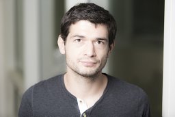 Alexandre Aubry合作论文数Institut Langevin;Universite Denis Diderot;Laboratoire Ondes et Acoustique;Laboratoire Ondes et Acoustique, Universite Denis Diderot15
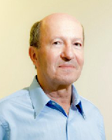 Mathias Fink合作论文数Langevin Institute, École Supérieure de Physique et de Chimie Industrielles14
 Flavien Bureau合作论文数5
 Radhakrishnan, K.合作论文数5
 Laura Cobus合作论文数Department of Physics, Faculty of Science, The University of Auckland5
 Arthur Le Ber合作论文数3
 Justine Robin合作论文数ESPCI Paris, PSL Res Univ3
 Sheldon Weng合作论文数3
 Christopher Schaef合作论文数Intel Corporation3
 Vivek De合作论文数Intel3
 Jim Tschanz合作论文数Circuit Research Lab, Intel Corporation3louie daniel garcia合作论文数2
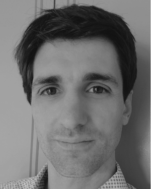 Thomas Frappart合作论文数2Jose Chavarria合作论文数2DANI ASHAY合作论文数2
 anne e augustine合作论文数2
 Krishnan Ravichandran合作论文数Intel2Arnaud Derode合作论文数2
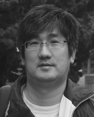 Yongki Min合作论文数2Leigh E. Wojewoda合作论文数2
 Claire Prada合作论文数Laboratoire Ondes et Acoustique2
 Arnaud Tourin合作论文数Laboratoire Ondes et Acoustique, Université Paris 7 — ESPCI2
 Stefan Rotter合作论文数Institute for Theoretical Physics, Vienna University of Technology2
 Harish K. Krishnamurthy合作论文数Intel2Vitor Spader合作论文数1Thomas T. Bauman合作论文数1Antton Goïcoechea合作论文数1Diane Doughty合作论文数1Johann Rode合作论文数1huong t do合作论文数1
 Jingshu Yu合作论文数1N. Nidhi合作论文数1Mark Chakravorti合作论文数Intel, Hillsboro, OR1Arnaud Amadjikpe合作论文数Intel, Hillsboro, OR1sell bernhard合作论文数1Divya Shree Vemparala合作论文数1
 Peter Sagazio合作论文数Intel, Hillsboro, OR1
 Michael J Hill合作论文数Intel1
 Naiara Korta Martiartu合作论文数Swiss Fed Inst Technol, Dept Earth Sci, CH-8092 Zurich, Switzerland1
 Nicolas Butzen合作论文数Department of Electrical Engineering, KU Leuven Heverlee1Robert Flory合作论文数1
 Beomseok Choi合作论文数1
 Somnath Kundu合作论文数Wireless Communication Research Intel Labs Hillsboro Oregon USA1
 Satoshi Suzuki合作论文数1
 Xiaoping Jia合作论文数School of Computer Science|Telecommunication and Information Systems|DePaul University1R. A. Vidal合作论文数1
 Abhishek Agrawal合作论文数Intel Labs1Glenn P. Jirka合作论文数1
 Christopher D. Hull合作论文数1




