Background: Primary hyperparathyroidism (PHPT) is typically a persistent disorder caused by a parathyroid adenoma requiring surgical intervention. Transient PHPT (tPHPT) refers to a rare phenomenon in which spontaneous normalization of hypercalcemia and PTH occurred without surgical or pharmacological intervention. The most common cause of tPHPT is spontaneous infarction or hemorrhage of adenoma. We report a unique case of tPHPT following upper respiratory infection (URI) in a patient without identifiable adenoma, suggesting a transient inflammatory 'parathyroiditis' mechanism. Case presentation: A 60-year-old woman was referred for evaluation of asymptomatic hypercalcemia (serum calcium 11.3–11.4 mg/dL) identified during preoperative testing for lingual tonsillectomy. Her ionized calcium was 5.9–6.1 mg/dL. She also developed inappropriately normal followed by elevated iPTH (57–110 pg/mL). The biochemical peak occurred after several episodes of URI. The patient had no history of renal disease or malignancy, and no use of thiazides, lithium, or vitamin D/calcium supplements. Laboratory evaluation, including creatinine, 25-hydroxyvitamin D, and CASR gene sequencing, was unremarkable. Neck ultrasound revealed multinodular goiter and mild hypervascularity but no parathyroid abnormalities. Without intervention, serum calcium, iPTH, and alkaline phosphatase spontaneously normalized (calcium 9.1–9.6 mg/dL, iPTH 37–38 pg/mL, ALP 105-117 IU/L) within 4, 8, and 11 months, respectively. Remission has persisted for over three years. Conclusion: Transient PHPT may occur via a post-viral, non-infarction mechanism. In PHPT patients with recent viral illness and no localized adenoma on imaging, a period of biochemical observation may be appropriate to prevent unnecessary surgery.
Currently, the total 25-hydroxyvitamin D (25(OH)D) is recognized as the indicator of vitamin D status that is used to define vitamin D sufficiency. Vitamin D binding protein (DBP) is the major carrier for circulating vitamin D and plays an important role in regulating circulating total and free vitamin D metabolites. Since the concentration of DBP and its affinity to vitamin D varies by different physiologic and clinical conditions, measuring total 25(OH)D may not accurately reflect functional vitamin D status. In addition, DBP is a potential prognostic indicator of clinical outcomes since it has other important functions beyond that as vitamin D carrier, including its role in actin scavenging after tissue injury and inflammation and immune modulation. It has been proposed that circulating DBP is altered in some clinical conditions, which affects the levels of total and free vitamin D metabolites and can explain clinical outcomes. Furthermore, in some clinical situations, total 25(OH)D levels are altered and knowing whether DBP is also changed may have diagnostic and therapeutic implications. The goal of this review is to assess clinical conditions altering DBP concentrations and then total 25(OH)D levels and their effects on prognosis. We suggest using the free 25(OH)D level as a better marker for vitamin D status in certain clinical conditions.
OBJECTIVE:Primary hyperparathyroidism (PHPT) is accompanied by a decreased 25-hydroxyvitamin D (25OHD) and vitamin D binding protein (DBP). High parathyroid hormone (PTH) is associated with elevated interleukin-6 (IL-6) and monocyte chemoattractant protein-1 (MCP-1), yet the effect of parathyroidectomy (PTX) on DBP and cytokines is not clear. This study aims to prospectively evaluate the effect of PTX on inflammatory profiles, total and free 25OHD, and DBP in patients with PHPT. METHODS:Newly diagnosed patients with PHPT were recruited for the study (n = 70). Twenty-eight patients returned after PTX, 3 months later. Biochemical markers were measured before and after PTX. A group of age and body mass index-matched healthy subjects were included as controls (n = 70). RESULTS:Before PTX, patients had lower serum DBP (37.5 ± 6.0 vs 41.5 ± 6.1 mg/dL, P < .001) and total 25OHD (30.1 ± 9.5 vs 33.3 ± 7.9 ng/mL, P < .05) but similar free 25OHD when compared to controls. Serum IL-6, C-reactive protein, and MCP-1 were higher in patients with PHPT (P < .05), whereas interleukin-10 was similar to that in controls. PTX increased total and free 25OHD and DBP (P < .001) and decreased serum IL-6 and MCP-1 (P < .05), but not C-reactive protein and interleukin-10. Multiple regression analysis indicated that the preoperative PTH explained a significant portion of the variance of IL-6 and MCP-1 (P < .05). CONCLUSION:These findings suggest that PTH may upregulate the production of MCP-1 and IL-6 and downregulate circulating DBP in patients with PHPT that are normalized by PTX. The exact mechanism of IL-6 and MCP-1 on DBP, vitamin D metabolites, and clinical outcomes in patients with PHPT is an area requiring further study.
Context Hip fracture is a serious injury that can lead to increased morbidity and mortality. Vitamin D binding protein (DBP) is a potential prognostic indicator of outcomes since it is important for actin scavenging and inflammation after tissue injury. Objective To determine whether circulating DBP is associated with mobility or mortality after hip fracture and its association with acute tissue injury markers. Methods Post hoc analysis of a multisite North American prospective study of 260 patients with hip fracture; mobility assessed at 30 and 60 days and mortality at 60 days after repair surgery. Biochemical markers were measured before, and 2 to 4 days after surgery. Tissue injury markers were measured in 100 randomly selected patients and controls. The primary outcome was mobility and mortality by DBP tertiles. Secondary outcomes were assessment of pre- and postoperative biomarkers. Results Among all patients (81 ± 9 years, BMI 25 ± 4 kg/m2; 72% female), the highest DBP tertile had greater mobility at 30 (OR: 2.66; 95% CI: 1.43, 4.92; P = .002) and 60 days (OR: 2.31; 95% CI: 1.17, 4.54; P = .014) and reduced mortality (OR: 0.18; 95% CI: 0.04, 0.86; P = .032) compared with the lowest DBP tertile (<28.0 mg/dL). Total 25-hydroxyvitamin-D did not differ between tertiles (22.0 ± 9.5 ng/mL). Circulating DBP and gelsolin were lower and interleukin-6, C-reactive protein, and F-actin were higher (P < .01) in patients vs controls, and worsened (P < .01) after surgery. Conclusion High circulating DBP concentrations are associated with better mobility and reduced mortality after hip fracture surgery. The role of DBP as an acute phase reactant to tissue injury and clinical outcomes should be addressed in future study.
EDITORIAL article Front. Endocrinol., 14 July 2023Sec. Translational Endocrinology Volume 14 - 2023 | https://doi.org/10.3389/fendo.2023.1238237
Background: Handgrip strength (HGS) and the appendicular lean mass index (ALMI) are important determinants of sarcopenia. Muscle quality (MQ) is a measure of muscle strength relative to muscle mass. We examined trends in handgrip strength, the appendicular lean mass index, and analyzed their relationship with age, anthropometry, and body composition in a sample of participants in the United States (US). Methods: This cross-sectional study analyzed data from 14,741 US males (49.7%) and females (50.3%) 6–80 years old who responded to the National Health and Nutrition Examination Survey (NHANES) from 2011 to 2014. Dual X-ray absorptiometry was used to measure appendicular skeletal muscle mass. HGS was evaluated using the Takei Digital Grip Strength Dynamometer. Smoothed normative curves for HGS and the ALMI were constructed using a generalized additive model. Multiple regression analyses were used to examine associations of HGS and the ALMI with age, nutrition-related factors, physical activity, and body composition. Results: Mean HGS and the ALMI declined with advancing age. While mean HGS increased with the ALMI, it decreased with the fat mass index. HGS increased in males with an increase in body mass index, energy intake, the ALMI, and vitamins; however, HGS in females increased with albumin, but it had a negative association with the fat mass index and age, but not with increasing adiposity. Conclusions: HGS and the ALMI change with age: HGS increases with age, then stabilizes and declines; the ALMI increases with age, then stabilizes. In addition, we provide evidence for the effect of anthropometry, nutrition, physical activity, and body composition on HGS and the ALMI in US population.
Abstract Disclosure: I.N. Farooqi: None. X. Wang: None. The familiar topic of vitamin D deficiency remains controversial in the context of bone metabolism and health, with findings supportive of differing thresholds for supplementation. While the Fifth International Workshop has not delineated exact cutoffs for 25(OH)D to rule out the secondary cause of vitamin D deficiency in evaluations of NPHPT, we believe higher thresholds will yield fewer rates of diagnosis. We postulate that a review of existing literature yields significant differences in the reported prevalence of NPHPT when comparing 25(OH)D cutoffs of ≥30ng/mL versus ≥20ng/mL. PubMed searches were conducted using the key words “normocalcemic primary hyperparathyroidism,” “prevalence,” “trial,” and 7400 studies were initially found. Excluding studies from before 2010, those using population sizes less than 100 subjects, and abstracts, we found a total of 14 articles for which full-text review was performed. We examined 9 studies using the 25(OH)D cutoff ≥20ng/mL and 5 studies using ≥30ng/mL for our analysis. Although research on NPHPT has been done worldwide, including Europe, East Asia, North and South Americas, none were found in Africa, South Asia, and Australia. Our review identified 9/14 studies in Canada, USA, UK, Sweden, Czech Republic, Italy, Belgium, and China which used 25(OH)D ≥20ng/mL to exclude vitamin D deficiency, with a prevalence range of 0.18-21%, averaging 5.81%. Conversely, the remaining 5/14 studies from Brazil, USA, Spain, and Italy using ≥30ng/mL as the lower limit of 25(OH)D for diagnosing NPHPT yielded prevalence rates ranging 0.02-8.9% with an average of 3.17%. The prevalence of NPHPT is 8.9% in the Brazilian study by Marques et al. in 2011 and 6% in the Spanish study by Garcia-Martin in 2012, while the remainder of North American and European studies using the ≥30ng/mL cutoff from 2015 onward reported prevalence rates of only 0.02-0.6%. Of the total number of publications reviewed since 2015, 30% of the studies implemented the higher threshold of ≥30ng/mL. Previously documented prevalence rates of NPHPT among healthy adults have used 25(OH)D ≥20ng/mL to define the exclusion of vitamin D deficiency; however, our current review of several studies since 2010 demonstrates a significantly lower prevalence of the disease when the cutoff is raised to ≥30ng/mL. Our findings suggest greater specificity in the exclusion of vitamin D deficiency in more recent publications. Recent research examining free 25(OH)D as a more effective marker for vitamin D status suggests total 25(OH)D ≥30ng/mL may still be insufficient to completely exclude vitamin D deficiency in NPHPT. Thus, the role of varying 25(OH)D cutoffs is interesting and requires further investigation on a geographic level, as it may provide more insight into interethnic variations, correlation with seasonality and sunlight exposure, and healthcare access in regions where such studies have yet to be conducted. Presentation: Saturday, June 17, 2023
The clinical presentation of primary hyperparathyroidism (PHPT) has evolved over the years from a symptomatic disorder to a predominantly asymptomatic condition. Altered vitamin D metabolism seems to play a role in the presentation of PHPT and may exacerbate the severity of disease. The epidemiology of PHPT differs in the developing versus the developed world, where more severe phenotypes occur in regions where vitamin D deficiency is common. Although it has been validated that patients with PHPT should be vitamin D sufficient, the threshold to supplement in relation to the severity of PHPT and the degree of vitamin D deficiency remains controversial. This review will highlight some of the controversy regarding vitamin D deficiency and the different phenotypes of PHPT.
Disease modifying therapies aiming to preserve β-cell function in patients with adult-onset autoimmune type 1 diabetes are lacking. Here, we conducted a multi-centre, randomized, controlled trial to assess the β-cell preservation effects of saxagliptin alone and saxagliptin combined with vitamin D as adjunctive therapies in adult-onset autoimmune type 1 diabetes. In this 3-arm trial, 301 participants were randomly assigned to a 24-month course of the conventional therapy (metformin with or without insulin) or adjunctive saxagliptin or adjunctive saxagliptin plus vitamin D to the conventional therapy. The primary endpoint was the change from baseline to 24 months in the fasting C-peptide. The secondary endpoints included the area under the concentration-time curve (AUC) for C-peptide level in a 2-h mixed-meal tolerance test, glycemic control, total daily insulin use and safety, respectively. The primary endpoint was not achieved in saxagliptin plus vitamin D group ( P = 0.18) and saxagliptin group ( P = 0.26). However, compared with the conventional therapy, 2-h C-peptide AUC from 24 months to baseline decreased less with saxagliptin plus vitamin D (-276 pmol/L vs. -419 pmol/L; P = 0.01), and not to the same degree with saxagliptin alone (-314 pmol/L; P = 0.14). Notably, for participants with higher glutamic acid decarboxylase antibody (GADA) levels, the decline of β-cell function was much lower in saxagliptin plus vitamin D group than in the conventional therapy group ( P = 0.001). Insulin dose was significantly reduced in both active treatment groups than in the conventional therapy group despite all groups having similar glycemic control. In conclusion, the combination of saxagliptin and vitamin D preserves pancreatic β-cell function in adult-onset autoimmune type 1 diabetes, an effect especially efficacious in individuals with higher GADA levels. Our results provide evidence for a novel adjunct to insulin and metformin as potential initial treatment for adult-onset type 1 diabetes. (ClinicalTrials.gov identifier: NCT02407899).
Background: Although SARS-CoV-2 virus infection has been reported to cause subacute thyroiditis, the mRNA vaccine for SARS-CoV-2 is suspected to induce thyroiditis with thyrotoxicosis. Case Report: : We describe 3 patients with no history of thyroid disease who presented with symptomatic, biochemical, and radiological evidence of thyroiditis with thyrotoxicosis, 10 to 20 days after receiving either Pfizer Bio-NTech or Moderna COVID-19 mRNA vaccines. All patients presented with thyrotoxicosis but with negative thyroid-stimulating immunoglobulins for Graves disease and no autonomous nodules. Two patients underwent thyroid uptake scans that confirmed thyroiditis. One patient had significantly increased erythrocyte sedimentation rate and interleukin-6. All patients showed improvement in symptoms with nonsteroidal anti-inflammatory drugs, and 1 patient eventually required steroids for symptom control. Discussion: The mRNA vaccine for SARS-CoV-2 was associated with thyroiditis and led to thyrotoxicosis. Elevated proinflammatory markers and cytokines after vaccines may play a major role. Conclusion: Our case series report highlights a possible relationship between the COVID-19 mRNA vaccine and thyroiditis with thyrotoxicosis, which has not been recognized by health providers.
Searchable abstracts of presentations at key conferences in endocrinology ISSN 1470-3947 (print) | ISSN 1479-6848 (online)
Primary Adrenal Lymphoma (PAL) is a very rare malignancy accounting for < 1% of non-Hodgkin's Lymphoma. We report, to our knowledge, the first case of PAL associated with adrenal insufficiency (AI) and hypercalcemia.
Purpose To evaluate total and free vitamin D metabolites and hormone-to-prohormone [1,25(OH)(2)D/25(OH)D] "activation ratio" in PHPT patients with low or insufficient vitamin D status. Methods Thirty female patients with primary hyperparathyroidism (PHPT) and 30 age and body mass index (BMI) matched healthy controls were enrolled. Serum levels of calcium, intact parathyroid hormone (iPTH), vitamin D binding protein (DBP), albumin, total 25(OH)D and 1,25(OH)(2)D were measured. The activation ratio of vitamin D was calculated as total 1,25(OH)(2)D/25(OH)D. Calculated serum-free 25(OH)D and 1,25(OH)(2)D levels were also reported. Results Compared to the control subject, patients with PHPT had a lower total 25(OH)D and DBP levels (p < 0.001). The serum concentration of free 25(OH)D and total 1,25(OH)(2)D were similar between the two groups; but free 1,25(OH)(2)D levels were about 26% higher in the PHPT patients compared to controls (p < 0.001). PHPT patients had a significantly higher activation ratio (p < 0.01), although their total 25(OH)D were lower than controls. The free (but not total) 1,25(OH)(2)D level was inversely correlated with DBP (p < 0.01). Both free 1,25(OH)(2)D levels and activation ratio were positively correlated with iPTH and calcium levels (p < 0.01). The activation ratio was highly correlated with levels of total vitamin D stores and free vitamin D metabolites (p < 0.001). Conclusion Patients with PHPT had significantly higher free 1,25(OH)(2)D levels and activation ratio compared to control subjects. We suggest that levels of free vitamin D metabolites and vitamin D activation ratio may provide additional values for the diagnosis and therapeutic choices in these patient populations with compromised vitamin D status.
Abstract Background: Vitamin D3 is metabolized to 25-hydroxyvitamin D [25(OH)D] in liver, and only after it goes to kidney is it converted to its biologically active form, 1,25-dihydroxyvitamin D [1,25(OH)2D]. Also, the majority of both total 25(OH)D and 1,25(OH)2D are tightly bound to vitamin D bind protein (DBP) and only a small portion remains in free form. In certain patient populations, like primary hyperparathyroidism (PHPT), concentrations of free vitamin D metabolites may be affected by altered levels of binding protein. Objective: To evaluate total and free 1,25(OH)2D levels in PHPT patients and healthy controls. Methods: Thirty female patients with PHPT and 30 healthy age and body mass index (BMI) matched controls were enrolled (57.1 ± 9.8 years and BMI of 32.2 ± 7.2 kg/m2). Serum levels of calcium, intact parathyroid hormone (iPTH), DBP, total 25(OH)D and 1,25(OH)2D levels were examined. Serum free 25(OH)D and 1,25(OH)2D levels were calculated using equations adapted from Bikle et al. Results: There were no significant differences in age and BMI between groups. Compared to controls, patients with PHPT had lower total 25(OH)D (25.2 ± 7.5 vs. 19.3 ± 6.4 ng/mL; p <0.001) and DBP levels (40.7± 3.1 vs. 36.5 ± 5.7 mg/dL; p <0.001). There were no significant differences in total 1,25(OH)2D levels or calculated free 25(OH)D levels between PHPT patients and controls; but the calculated free 1,25(OH)2D levels were 27% higher in the PHPT patients compared to controls (p<0.001). The calculated free (but not total) 1,25(OH)2D level was inversely correlated with DBP (r=-0.35, p<0.01) and positively correlated with iPTH levels (r=0.33, p<0.01). Conclusion: Postmenopausal patients with PHPT had lower serum total 25(OH)D, but similar free 25(OH)D levels. In contrast, total 1,25(OH)2D levels did not differ between patients and controls; however, patients had higher free 1,25(OH)2D. Because total 25(OH)D and 1,25(OH)2D levels do not reflect free levels, standard clinical measures of circulating vitamin D may not be an accurate estimate of true vitamin D status in patients with PHPT. References: Bikle et al. Serum Protein Binding of 1,25-Dihydroxyvitamin D: A Reevaluation by Direct Measurement of Free Metabolite Levels. JCEM 1985;61:969-75.
To investigate Klotho level and its association with biochemical indices of primary hyperparathyroidism (PHPT). Fifty PHPT patients and fifty-two age- and body mass index-(BMI) matched healthy control subjects were recruited. In addition, twenty-five PHPT patients underwent parathyroidectomy (PTX) and had 4-month follow-up visits. Intact parathyroid hormone (iPTH), 25-hydroxyvitamin D [25(OH)D], calcium, albumin, corrected calcium, and Klotho levels were determined. There was no significant difference in age and BMI between PHPT subjects and controls (p > 0.05). PHPT patients had Klotho levels (15.4 ± 1.2 ng/mL) about 23% higher compared with those of the controls (11.9 ± 0.8 ng/mL), but this difference was not significant (p = 0.063). However, postmenopausal PHPT patients had 45% higher Klotho levels (17.6 ± 1.5 ng/ml) compared with postmenopausal controls (12.1 ± 0.9 ng/mL, p = 0.008). For postmenopausal subjects, Klotho levels had positive correlation with levels of iPTH (r = 0.25, p = 0.026) and corrected calcium (r = 0.34, p = 0.003), but negative correlation with 25(OH)D (r = −0.23, p = 0.042). After PTX, levels of iPTH and corrected calcium decreased and 25(OH)D levels increased to normal range (p < 0.001). However, there was no significant change in Klotho levels after a 4-month follow-up. Serum Klotho levels are higher in postmenopausal PHPT patients than in healthy postmenopausal control subjects. The etiology of elevated Klotho level and its clinical significance requires further investigation.
We are writing to provide an update on the case that was published in the American Association of Clinical Endocrinologists Clinical Case Reports in 2016 by Mewani et al ( 1. Mewani V. Dhillon S. Cai X. Wang X. Transient primary hyperparathyroidism: a case report. AACE Clinical Case Rep. 2016; 2: e182-e185 Abstract Full Text Full Text PDF Google Scholar ).
Objective: We sought to determine the long-term bioavailability of single doses of intramuscular (IM) vitamin D-2 (D-2) in healthy adults. Methods: Forty healthy volunteers with hypovitaminosis D received a single dose of 200,000, 400,000, or 600,000 IU intramuscular D-2 or no treatment. Levels of 25-hydroxyvitamin D-2 (25[OH]D-2) and 25-hydroxyvitamin D-3 (25[OH]D-3) in serum were measured by liquid chromatography-tandem mass spectrometry. Vitamin D binding protein (DBP) and intact parathyroid hormone (iPTH), bone turnover markers (BTMs), and serum and urinary calcium were also measured. Results: After a single dose of D-2 injection, the level of 25(OH)D-2 increased slowly and reached a plateau at 8 weeks. The plateau remained stable for 12 weeks. The mean increase in 25(OH)D-2 was 6.8, 9.6, or 15.6 ng/mL after injection of 200,000 IU, 400,000 IU, or 600,000 IU D-2 . Although endogenous 25(OH)D levels were reduced by IM D-2, the total 25(OH)D levels increased by 5.0, 7.0, or 10.3 ng/mL in average after injection of 200,000 IU, 400,000 IU, or 600,000 IU D-2. The iPTH levels were also decreased by IM D-2. However, levels of serum calcium, BTMs, and DBP and urinary calcium were not altered by IM D-2. Conclusion: A single dose of 200,000 IU, 400,000 IU or 600,000 IU IM D-2 raises total 25-hydroxyvitamin D levels by 5.0, 7.0, or 10.3 ng/mL on average for at least 12 weeks and reduces iPTH and endogenous 25(OH)D-3 levels without affecting levels of serum calcium, BTMs, DBP, and urinary calcium.
 Ma Greer合作论文数36
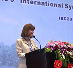 Sue Shapses合作论文数Dept Nutr Sci, Rutgers State Univ28N Sato合作论文数27Se Greer合作论文数20Lingqiong Meng合作论文数16S McAdams合作论文数15
 Zhiguang Zhou (周智广)合作论文数The Second Xiangya Hospital of Central South University10Stanley Z Trooskin合作论文数8T Inukai合作论文数7
 Chi Su合作论文数7
 Xinjiang Cai合作论文数Duke Univ, Med Ctr, Dept Med Cardiol, Durham, NC 27710 USA7Huang Cindy合作论文数7
 Gan Huang (黄干)合作论文数National Clinical Research Center for Endocrine and Metabolic Diseases, The Second Xiangya Hospital of Central South University7
 Sukumar Deeptha合作论文数Department of nutritional sciences, rutgers university6
 Xia Li (李霞)合作论文数The Second Xiangya Hospital of Central South University;Xiangya School of Nursing, Central South University6
 Yvette Schlussel合作论文数6
 Yufei Xiang (向宇飞)合作论文数Division of General Internal Medicine, The Ohio State University5Wei Sun合作论文数5Zhifeng Sheng合作论文数5Beatrice Wong合作论文数4Claudia Pop合作论文数4Yuling He合作论文数4Aseel Al Dayyeni合作论文数4Leslie Richard David合作论文数4Jian Lin合作论文数4Amy M. Toscano-Zukor合作论文数4
 Zhongjian Xie合作论文数Department of Metabolism and Endocrinology, Second Xiangya Hospital, Central South University4Hyon Kim合作论文数3Sita Chokhavatia合作论文数3vanessa mewani合作论文数3diane fresca合作论文数3Lin Yang合作论文数3
 Xiang Yan (颜湘)合作论文数3
 Jeffrey L. Carson合作论文数Department of Medicine, Robert Wood Johnson Medical School, Rutgers University3Zhao Yunjuan合作论文数3Padma Chamarthy合作论文数2
 Anna Ogilvie合作论文数2Hu Sophia合作论文数2Sudeep Dhillon合作论文数2Anupa Sharma合作论文数2dragana jokic合作论文数2Lingjiao LIU (刘玲娇)合作论文数2Ziwei Zhang合作论文数2Bingwen Liu合作论文数2Xiaohong Niu合作论文数2Liu ShuYing合作论文数2
 Nicola C.Partridge合作论文数Department of Molecular Pathobiology, College of Dentistry, New York University2
 Amar Mahdi合作论文数Graduate School of Biomedical Sciences, The State University of New Jersey2




