CD4+ T cells play a pivotal role in anti-tumor immunity by assisting CD8+ T cells in eliminating MHC I+ tumor cells and promoting immune responses against MHC-deficient tumors through myeloid-cell recruitment. Extracellular vesicles (EVs) are increasingly recognized as important mediators of intercellular immune communication; however, how CD4+ T helper (Th)-derived EVs modulate tumor immune cells, including CD8+ T cells and eosinophils, to orchestrate anti-tumor responses remains poorly understood. Here, we show that immune checkpoint molecules, including PD-1 and TIM-3, and the cytokine IL-5 are enriched on CD4+ Th cell microvilli and EVs. Activated CD4+ Th cells released both exosomes and ectosomes, which cooperatively suppressed tumor progression through two complementary mechanisms. First, CD4+ Th-derived exosomes and ectosomes displayed immune checkpoint receptors such as PD-1 and TIM-3 on their surface, thereby intercepting tumor-derived inhibitory signals, reversing T cell exhaustion, and enhancing cytotoxic T cell activity. Second, CD4+ Th-derived EVs carried IL-5 and other effector molecules that promoted JAK-STAT signaling, eosinophil recruitment to tumor sites, and eosinophil-mediated tumor cell killing through degranulation. Moreover, CD4+ Th-derived EVs remodeled the tumor microenvironment by enhancing CD8+ T cell and eosinophil infiltration and activation, suggesting their potential role in immune regulation and cancer therapy.
Plant-derived extracellular vesicles (EVs) have emerged as promising drug delivery vehicle due to their inherent biocompatibility, high stability and large-scale production. Chlorella vulgaris (C. vulgaris) has been increasing application in the food industry and biomedical fields. Nevertheless, the biogenesis and immunomodulatory functions of EVs from Chlorella species remain poorly understood. Herein, we found that the EVs in C. vulgaris originated from “vacuolar structures”, a process analogous to the biogenesis of exosomes which originates from endosomes in mammalian cells. Therefore, we further isolated and characterized EVs secreted by Chlorella vulgaris (C.V-EVs). Afterwards, we investigated the regulatory effects of C.V-EVs on immune cells, with a particular emphasis on their capacity to promote dendritic cell (DC) maturation, enhance antigen presentation, and improve T-cell priming efficiency. Furthermore, we developed C.V-EVs as multifunctional biomimetic nanocarriers for co-delivering gemcitabine (Gem) and a programmed cell death-1/ligand-1 checkpoint inhibitor (PD-1/PD-L1 checkpoint inhibitor, PD-1/L1i), and conjugated these drug-loaded C.V-EVs with platelet (Platelet-C.V-EVs-Gem&PD-1/L1i) to enhance tumor-targeted delivery. Simultaneously, the release of PD-1/L1i could block the PD-1/PD-L1 signaling axis, reinvigorating tumor specific-T cell activity, to trigger robust antitumor immune responses. In addition, C. vulgaris could generate oxygen in situ to alleviate hypoxia in the tumor microenvironment, synergistically with preloaded C. V-EVs to promote the infiltration and activation of immune cells to prevent the tumor relapse and metastasis.
Autoimmune destruction of pancreatic β-cells leads to impaired insulin production and onset of type 1 diabetes (T1D). Hence, immunomodulation of pancreas-infiltrated immune cells especially the β-cells autoreactive-T cells is a promising way to hinder and reverse the progress of T1D. Herein, megakaryocytes are primed with interferon-γ (IFN-γ) to produce platelets presenting high levels of immunosuppressive checkpoint ligands including programmed death-ligand 1 (PD-L1), Programmed Death-Ligand 2 (PD-L2), the B and T lymphocyte attenuator (BTLA) and Galectin-9 (Gal-9), termed as IFN-γ platelets. The IFN-γ platelets bound and interacted with T cells through immune checkpoint ligands and receptors, which efficaciously induced T cell exhaustion and apoptosis in vitro. Virtually, NOD diabetes mice received IFN-γ platelets treatments prominently preserved β-cell integrity and insulin production, ultimately hindering the progress to hyperglycemia. Intriguingly, both the amount and activity of the pancreas infiltrate-T cells intensively reduced, whereas the magnitude of regulatory T cells (Tregs) remarkably increased, which is attributed to IFN-γ platelets treatments. Moreover, IFN-γ platelets treatment instigated macrophage polarization toward an anti-inflammatory M2 phenotype that may stimulate pancreatic angiogenesis, and promote β-cell proliferation, consequently ameliorating the new-onset T1D.
Biomaterial-based implants encapsulating islets or R cells are desirable regimens for type 1 diabetes (T1D). However, current implants are restricted by poor durable R cell survival due to immune cell infiltration, graft fibrosis, and hypoxia. Programmed cell death-ligand 1 (PD-L1) induces T cell exhaustion, consequently protecting R cells from autoimmune attack. Herein, we report an implant encapsulating PD-L1-overexpressing R cell microspheres (PD-L1 R-MCSs) and Chlorella within alginate hydrogel to control hyperglycemia in T1D. Virtually, PD-L1-R cell-derived exosomes efficaciously induce T cell exhaustion and convert macrophages into M2 phenotype in vitro. The PD-L1 R-MCSs routinely secrete insulin in response to changes in glucose concentration. Furthermore, PD-L1-R cell microsphere artificial pancreas (PD-L1-R-MCSs aPancreas) prominently relieves hyperglycemia with fewer CD4+ T cells, CD8+ T cells, and M1 macrophage infiltration, inflammation, and fibrosis deposition. Intriguingly, Chlorella produces oxygen to relieve hypoxia and enables the PD-L1-R-MCSs aPancreas-transplanted mice to achieve sustained normoglycemia, which potentially benefits from both oxygen supplementation and exosomal PD-L1.
An important factor in the development of Type 1 diabetes (T1D) is the deficiency of inhibitory immune checkpoint ligands, specifically programmed cell death ligand 1 (PD-L1) and Galectin-9 (Gal-9), in β-cells. Hence, modulation of the pancreas infiltrated T lymphocytes by exogenous PD-L1 or Gal-9 is an ideal approach for treating the new-onset T1D. Herein, we genetic engineered the macrophage cells to generate artificial extracellular vesicles (aEVs) overexpressing PD-L1 and Gal-9, which could restrict the islets auto-reactive T lymphocytes and protect β-cells from destruction. Intriguingly, overexpressing Gal-9 spurred macrophage polarization to M2 phenotype with immune suppressive attribute. Alternatively, both of PD-L1 and Gal-9 presenting aEVs (PD-L1-Gal-9 aEVs) favorably adhere to T cells via the interaction of programmed cell death protein 1 (PD-1)/PD-L1 or T cell immunoglobulin mucin 3 (TIM-3)/Gal-9. Moreover, PD-L1-Gal-9 aEVs prominently promoted effector T cell apoptosis and splenic regulatory T cells (Treg) cells differentiation in vitro. Virtually, PD-L1-Gal-9 aEVs efficaciously reversed the new-onset hyperglycemia in the NOD mice, prevented T1D progress, and declined the proportion and activation of CD4+ and CD8+ T cells infiltrating the pancreas notably, which together contributed to preserving the residual β-cells survival and mitigating the hyperglycemia.
Neoantigens serve as ideal personalized cancer vaccines because of their high immunogenicity, ability to evade central thymic tolerance, and minimal risk of eliciting autoimmune responses. Herein, we describe a genetically engineered autophagosome-based neoantigen vaccine (APNV) in combination with an immune checkpoint inhibitor (anti-PD-1 antibody) for cancer immunotherapy. The APNV was derived from engineered NIH 3T3 cells, which co-express melanoma neoantigens and autophagosome maker microtubule-associated proteins 1 A/1B light chain 3B (LC3), from which the LC3-labeled neoantigen-autophagosomes were isolated. These purified autophagosomes, in conjunction with vaccine adjuvants high-mobility group box 1 (HMGB1) and granulocyte-macrophage colony-stimulating factor (GM-CSF), were integrated into a hydrogel to create an APNV. The APNV effectively activated dendritic cells both in vitro and in vivo. Moreover, APNV, in combination with checkpoint blockade therapy, significantly hampered post-surgical tumor recurrence in a subcutaneous melanoma tumor model and effectively impeded metastatic progression in a melanoma lung metastasis model. This APNV may be conducive to making personalized therapeutic neoantigen vaccines for cancer immunotherapy.
Immune-checkpoint blockade (ICB) reinvigorates T cells from exhaustion and potentiates T-cell responses to tumors. However, most patients do not respond to ICB therapy, and only a limited response can be achieved in a “cold” tumor with few infiltrated lymphocytes. Synthetic biology can be used to engineer bacteria as controllable bioreactors to synthesize biotherapeutics in situ. We engineered attenuated Salmonella VNP20009 with synthetic gene circuits to produce PD-1 and Tim-3 scFv to block immunosuppressive receptors on exhausted T cells to reinvigorate their antitumor response. Secreted PD-1 and Tim-3 scFv bound PD-1+ Tim-3+ T cells through their targeting receptors in vitro and potentiated the T-cell secretion of IFN-γ. Engineered bacteria colonized the hypoxic core of the tumor and synthesized PD-1 and Tim-3 scFv in situ, reviving CD4+ T cells and CD8+ T cells to execute an antitumor response. The bacteria also triggered a strong innate immune response, which stimulated the expansion of IFN-γ + CD4+ T cells within the tumors to induce direct and indirect antitumor immunity.
Chimeric antigen receptor (CAR) T cell therapy has implemented impressive advances in the treatment of B-cell lymphoma. However, the complex production process of CAR T cells and hindrance of solid tumor penetration remain substantial challenges. Intriguingly, cell-targeting delivery of messenger RNA (mRNA) with ionizable lipid nanoparticles (mRNA-LNPs) is able to efficiently and precisely engineer T cells and other immune cells in vivo to perform their functions. Herein, we harnessed the ionizable LNPs to encapsulate mRNA encoding antityrosinase related protein 1 (TRP1) CAR (CAR-LNPs) for in vivo generation of mRNA-CAR T cells to eliminate melanoma cells. Specifically, the anti-CD3 antibody (aCD3) armed mRNA-LNPs (CD3-mRNA-LNPs) selectively targeted T cells, resulting in the production of functional and therapeutic levels of CAR T cells both ex vivo and in vivo. These CD3-CAR-LNPs engineered CAR T cells were capable of infiltrating into the solid tumor and effectively eliminating melanoma cells with high TRP1 expression, significantly hindering tumor progression. Critically, CD3-7CAR-LNPs containing mRNA encoding both CAR and interleukin-7 (IL-7) generated 7CAR T cells that secreted IL-7, thereby enhancing the activity and proliferation of both CAR T cells and other intratumoral cytotoxic T cells. Alternatively, the employment of anti-programmed cell death protein 1 antibody (aPD-1) protected mRNA-CAR T cells from exhaustion, especially in combination with CD3-7CAR-LNPs, could significantly enhance the antitumor capability of CAR T cells without causing acute cytokine release syndrome (CRS).
Exosomes, minute vesicles ubiquitously released by diverse cell types, serve as critical mediators in intercellular communication. Their pathophysiological relevance, especially in malignancies, has garnered significant attention. A meticulous exploration of the exosomal impact on cancer development has unveiled avenues for innovative and clinically valuable techniques. The cargo conveyed by exosomes exerts transformative effects on both local and distant microenvironments, thereby influencing a broad spectrum of biological responses in recipient cells. These membrane-bound extracellular vesicles (EVs) play a pivotal role in delivering bioactive molecules among cells and organs. Cellular and biological processes in recipient cells, ranging from stromal cell reprogramming to immunological responses, extracellular matrix formation, and modulation of cancer cell activation, expansion, and metastasis, are subject to exosome-mediated cell-to-cell communication. Moreover, exosomes have been implicated in endowing cancer cells with resistance to treatment. Extensive research has explored the potential of exosomes as therapeutic targets and diagnostic indicators. This comprehensive review seeks to provide an in-depth understanding of the pivotal components and roles of exosomes in tumorigenesis, growth, progression, and therapeutic responses. The insights into the multifaceted involvement of exosomes in malignant cancers are essential for the scientific community, fostering the development of novel therapeutic and diagnostic strategies in the relentless pursuit of cancer.
Interleukin 15 (IL-15) is a pivotal immune cytokine that responses to cancer stress signals and maintains tissue-resident memory T (TRM) cells. It is therefore considered as a potent therapeutic immunomodulator agonist cancer. However, in addition to the aberrant expression of programmed cell death 1 ligand 1 (PD-L1), certain types of tumors lack surface expression of IL-15 and thereby leads to the deficiency of T cell-mediated anti-tumor response. Herein, we engineered cellular nanovesicles presenting IL-15/IL-15Rα (IL-15/IL-15Rα-NVs) complex. IL-15/IL-15Rα-NVs trans-present IL-15 to T cells and then boost the activation, proliferation and survival of tumor-infiltrated T cells to antagonize cancer cells. Small-molecule programmed cell death protein 1 (PD-1)/PD-L1 inhibitor 1 was loaded into the NVs and simultaneously prevented CD8+ T cell exhaustion mediated via PD-L1. Moreover, IL-15/IL-15Rα-NVs intensively evoked the activity and proliferation of CD8+ TRM cells, and could exert permanent anti-tumor response.
Immune intervention of B cell activation to blockade the production of autoantibodies provokes intense interest in the field of systemic lupus erythematosus (SLE) therapy development. Although the survival rate for SLE is improved, many patients die untimely. Engineered cell membrane vesicles manifest remarkable capacity of targeted drug delivery and immunomodulation of immune cells such as B cells. Herein, this work engineered cellular nanovesicles (NVs) presenting CD40 (CD40 NVs) that can blunt B cells and thus alleviate SLE. CD40 NVs disrupt the CD40/CD40 ligand (CD40L) costimulatory signal axis through the blockade of CD40L on CD4+ T cells. Therefore, the CD40 NVs restrain the generation of the germinal center structure and production of antibodies from B cells. Furthermore, immunosuppressive drug mycophenolate mofetil (MMF) is also encapsulated in the vesicles (MMF-CD40 NVs), which is employed to deplete immunocytes including B cells, T cells, and dendritic cells. Together, CD40 NVs are promising formulations for relieving autoimmunity and lupus nephritis in MRL/lpr mice.
Programmed cell death ligand 1 (PD-L1) attenuates the T lymphocytes' response to tumor cells in various malignancies. Extracellular vesicles (EVs) secreted by effector T cells possess potential as anticancer therapeutics by interaction with tumor cells. Here, we constructed T cell derived EVs which display PD-1, the receptor of PD-L1, on the surface to enhance tumor elimination by interrupting PD-1/PD-L1 pathway. Proteomics analysis indicates that the expression of proteins involved in cytotoxicity, T cell receptor signaling pathway and cell binding were upregulated in PD-1 overexpressing exosomes compared to that of microvesicls. PD-1 expressing EVs (PD-1 EVs) neutralize PD-L1 and effectively reinvigorate the activity and proliferation capacity of CD8+ effector T cells. Moreover, PD-1 EVs may also directly attack tumor cells by Fas ligand (FasL) and granzyme B (GzmB). (c) 2022 Elsevier Ltd. All rights reserved.
Abstract Neoantigens derived from mutant proteins in tumour cells could elicit potent personalized anti‐tumour immunity. Nevertheless, the layout of vaccine vehicle and synthesis of neoantigen are pivotal for stimulating robust response. The power of synthetic biology enables genetic programming bacteria to produce therapeutic agents under contol of the gene circuits. Herein, we genetically engineered bacteria to synthesize fusion neoantigens, and prepared bacteria derived vesicles (BDVs) presenting the neoantigens (BDVs‐Neo) as personalized therapeutic vaccine to drive systemic antitumour response. BDVs‐Neo and granulocyte‐macrophage colony‐stimulating factor (GM‐CSF) were inoculated subcutaneously within hydrogel (Gel), whereas sustaining release of BDVs‐Lipopolysaccharide (LPS) and GM‐CSF recruited the dendritic cells (DCs). Virtually, Gel‐BDVs‐Neo combined with the programmed cell death protein 1 (PD‐1) antibody intensively enhanced proliferation and activation of tumour‐infiltrated T cells, as well as memory T cell clone expansion. Consequently, BDVs‐Neo combining with checkpoint blockade therapy effectively prevented tumour relapse and metastasis.
Cancer cells aberrantly express immunosuppressive checkpoint ligands and produce certain metabolites that lead to T cell exhaustion. Immune checkpoint blockade (ICB) therapy that reinvigorates exhausted T cells have achieved impressive response in clinical cancer treatment. However, the limited clinical response rate and off-tumor toxicities restrict ICB therapy. Herein, cellular vesicles displaying anti-programmed cell death-1 (PD-1) single-chain variable fragment antibody (aPD-1-scFv) were prepared to reinvigorate T cell immunity to counteract cancer. The nanovesicles displaying aPD-1-scFv (aPD-1-scFv NVs) could enhance the anti-tumor activation of T cells through PD-1 blockade. Furthermore, NVs loading the A2a adenosine receptor (A2aR) antagonist CPI-444 assisted T cells to antagonize adenosine, an immunosuppressive metabolite produced by cancer cells. Hence, CPI-444 loaded aPD-1-scFv NVs could intensively increase the density and activity of tumor infiltrating T cells, directly restraining tumor progress and metastasis.
Glucose-responsive insulin delivery systems that mimic insulin secretion activity in the pancreas show great potential to improve clinical therapeutic outcomes for people with type 1 and advanced type 2 diabetes. Here, we report a glucose-responsive insulin delivery microneedle (MN) array patch that is loaded with red blood cell (RBC) vesicles or liposome nanoparticles containing glucose transporters (GLUTs) bound with glucosamine-modified insulin (Glu-Insulin). In hyperglycemic conditions, high concentrations of glucose in interstitial fluid can replace Glu-Insulin via a competitive interaction with GLUT, leading to a quick release of Glu-Insulin and subsequent regulation of blood glucose (BG) levels in vivo. To prolong the effective glucose-responsive insulin release from MNs, additional free Glu-Insulin, which serves as "stored insulin", is loaded after RBC vesicles or liposome nanoparticles bound with Glu-Insulin. In the streptozotocin (STZ)-induced type 1 diabetic mouse model, this smart GLUT-based insulin patch can effectively control BG levels without causing hypoglycemia.
Cancer cells, as well as surrounding stromal and inflammatory cells, form an inflammatory tumor microenvironment (TME) to promote all stages of carcinogenesis. As an emerging post-translational modification (PTM) of serine and threonine residues of proteins, O-linked-N-Acetylglucosaminylation (O-GlcNAcylation) regulates diverse cancer-relevant processes, such as signal transduction, transcription, cell division, metabolism and cytoskeletal regulation. Recent studies suggest that O-GlcNAcylation regulates the development, maturation and functions of immune cells. However, the role of protein O-GlcNAcylation in cancer-associated inflammation has been less explored. This review summarizes the current understanding of the influence of protein O-GlcNAcylation on cancer-associated inflammation and the mechanisms whereby O-GlcNAc-mediated inflammation regulates tumor progression. This will provide a theoretical basis for further development of anti-cancer therapies.
The pathogeny of type 1 diabetes (T1D) is mainly provoked by the β-cell loss due to the autoimmune attack. Critically, autoreactive T cells firsthand attack β-cell in islet, that results in the deficiency of insulin in bloodstream and ultimately leads to hyperglycemia. Hence, modulating immunity to conserve residual β-cell is a desirable way to treat new-onset T1D. However, systemic immunosuppression makes patients at risk of organ damage, infection, even cancers. Biomaterials can be leveraged to achieve targeted immunomodulation, which can reduce the toxic side effects of immunosuppressants. In this review, we discuss the recent advances in harness of biomaterials to immunomodulate immunity for T1D. We investigate nanotechnology in targeting delivery of immunosuppressant, biological macromolecule for β-cell specific autoreactive T cell regulation. We also explore the biomaterials for developing vaccines and facilitate immunosuppressive cells to restore immune tolerance in pancreas.
Following the publication of this paper, it was drawn to the Editors' attention by a concerned reader that possible anomalies were associated with data shown in Fig. 4B, C and E, and western blotting data shown in Fig. 5, such that it was difficult to interpret the presented results as having originated from discrete experiments performed in two breast cancer cell lines (MCF‑7 and MDA‑MB‑231). After having investigated the matter internally, the Editor of International Journal of Molecular Medicine has decided that this paper should be retracted from the Journal on account of a lack of confidence in the presented data. The authors were asked for an explanation to account for these concerns, but the Editorial Office did not receive a satisfactory response. The Editor apologizes to the readership for any inconvenience caused. [the original article was published in International Journal of Molecular Medicine 34: 772-781, 2014; DOI: 10.3892/ijmm.2014.1822].
Bioorthogonal catalysis mediated by transition metals has inspired a new subfield of artificial chemistry complementary to enzymatic reactions, enabling the selective labelling of biomolecules or in situ synthesis of bioactive agents via non-natural processes. However, the effective deployment of bioorthogonal catalysis in vivo remains challenging, mired by the safety concerns of metal toxicity or complicated procedures to administer catalysts. Here, we describe a bioorthogonal catalytic device comprising a microneedle array patch integrated with Pd nanoparticles deposited on TiO 2 nanosheets. This device is robust and removable, and can mediate the local conversion of caged substrates into their active states in high-level living systems. In particular, we show that such a patch can promote the activation of a prodrug at subcutaneous tumour sites, restoring its parent drug’s therapeutic anticancer properties. This in situ applied device potentiates local treatment efficacy and eliminates off-target prodrug activation and dose-dependent side effects in healthy organs or distant tissues.
Immune checkpoint blockade therapies, especially those targeting the programmed death-1 (PD-1)/programmed death-ligand 1 (PD-L1) have achieved impressive clinical responses in multiple types of cancers. To optimize the therapeutic effect of the checkpoint antibodies, many strategies including targeting delivery, controlled release, and cellular synthesis have been developed. However, within these strategies, antibodies were attached to drug carriers by chemical bonding, which may affect the steric configuration and function of the antibodies. Herein, we prepared cluster of differentiation 64 (CD64), a natural catcher of the fragment crystalline (Fc) of monomeric immunoglobulin G (IgG), and over-expressed it on the cell membrane nanovesicles (NVs) as PD-L1 antibody delivery vehicle (CD64-NVs-aPD-L1), which was employed to disrupt the PD-1/PD-L1 immunosuppressive signal axis for boosting T cell dependent tumor elimination. Meanwhile, chemical immunomodulatory drug cyclophosphamide (CP) was also encapsulated in the vesicle (CD64-NVs-aPD-L1-CP), to simultaneously restrain the regulatory T cells (Tregs) and invigorate Ki67+CD8+ T cells, then further enhance their anti-tumor ability. Methods: The cell membrane NVs overexpressing CD64 were incubated with PD-L1 antibody and chemotherapeutic agent CP to prepare CD64-NVs-aPD-L1-CP. Results: The CD64-NVs-aPD-L1-CP could simultaneously interrupt the immunosuppressive effect of PD-L1 and decrease the inhibition of Tregs, leading to tumor growth suppression and survival time extension. Conclusion: CD64-NVs are charismatic carriers to achieve both checkpoint blockade and immunomodulatory drugs for combined cancer immunotherapy.
 Lin Mei (梅林)合作论文数School of Pharmaceutical Sciences (Shenzhen), Sun Yat-sen University30
 Xin Liang (梁欣)合作论文数School of Basic Medical Sciences of Guangdong Medical University24
 Zhen Gu (顾臻)合作论文数College of Pharmaceutical Sciences, Zhejiang University21
 Xiaowei Zeng (曾小伟)合作论文数清华大学深圳研究生院17
 Wei Tao合作论文数Gillian Reny Stepping Strong Center for Trauma Innovation, Brigham and Women’s Hospital, Harvard Medical School;Center for Nanomedicine, Brigham and Women’s Hospital;Department of Anesthesiology, Perioperative, and Pain Medicine, Faculty of Medicine, Harvard University16
 Jinxie Zhang合作论文数13
 Jinqiang Wang (王金强)合作论文数School of Pharmacy, Zhejiang University12
 Hongbo Chen (陈红波)合作论文数School of Pharmaceutical Sciences (Shenzhen), Sun Yat-sen University12
 Laiqiang Huang (黄来强)合作论文数Tsinghua Shenzhen Intemational Graduate School11
 Quanyin Hu (胡全银)合作论文数Pharmaceutical Sciences Division, School of Pharmacy, University of Wisconsin-Madison;Wisconsin Center for NanoBioSystems, University of Wisconsin-Madison10
 Chao Wang (汪超)合作论文数Institute of Functional Nano & Soft Materials, Soochow University10Zhirang Zhang合作论文数8Fanqiang Meng合作论文数8Wenli Fang合作论文数8
 Yuqi Zhang (张宇琪)合作论文数College of Pharmaceutical Sciences, Zhejiang University8
 Qian Chen (陈倩)合作论文数College of Nano Science & Technology, Soochow University8
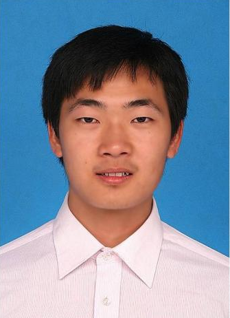 Gan Liu (刘赣)合作论文数School of Pharmaceutical, Sun Yat-sen University8
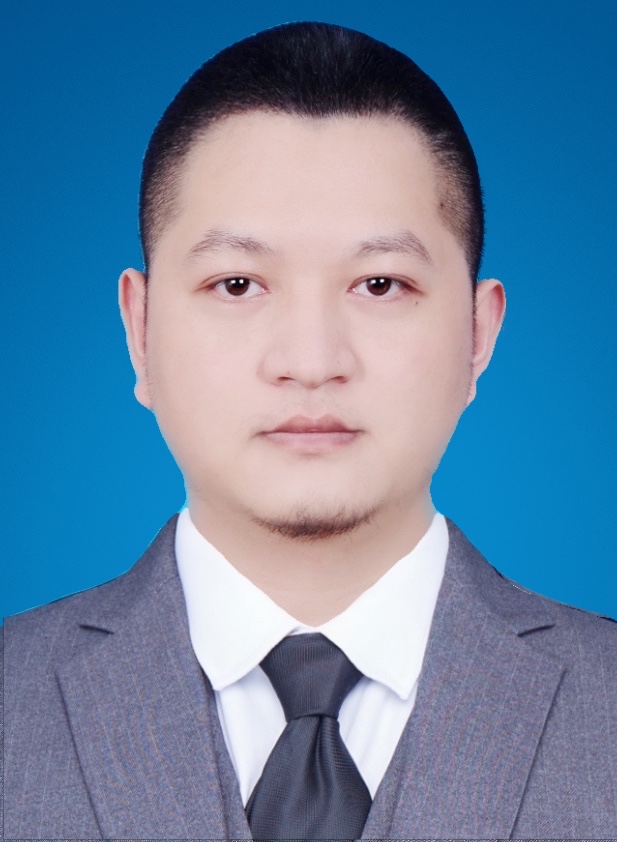 Yuan Li (李源)合作论文数School of Basic Medical Sciences, Southern Medical University8Xianbing Zhu合作论文数Tsinghua Univ, Sch Life Sci, Beijing, Peoples R China7Lili Yan合作论文数7Chang Danfeng合作论文数7
 Yanqi Ye合作论文数Sorrento Therapeutics, Inc.7
 Gianpietro Dotti合作论文数Lineberger Comprehensive Cancer Center, School of Medicine, The University of North Carolina at Chapel Hill7Di Wen合作论文数6Zhangyan Jing合作论文数6Tianyuan Xue合作论文数6
 Guojun Chen合作论文数Department of Biomedical Engineering, Faculty of Medicine and Health Sciences, McGill University6
 Zhaowei Chen (陈兆委)合作论文数College of Chemistry, Fuzhou University;School of Pharmacy, Zhejiang University6
 Wujin Sun合作论文数University of North Carolina at Chapel Hill, North Carolina State University;UNC Eshelman School of Pharmacy, University of North Carolina at Chapel Hill;University of North Carolina at Chapel Hill, University of North Carolina at Chapel Hill6
 Zhongyuan Wang (王中原)合作论文数School Of Life Science And Technology, China Pharmaceutical University6Tianliang Fang合作论文数5Zhongda Lin合作论文数5Baoqi Li合作论文数5ZhenChang Sun (孙振昌)合作论文数5Zhang Lei合作论文数5Fu Xiaorui合作论文数5
 Mingzhi Zhang (张明智)合作论文数He Nan Sui Xian People's Hospital5
 Yu Chang (常宇)合作论文数College of Life Science and Bioengineering, Beijing University of Technology5
 Yi Zheng (郑义)合作论文数清华大学深圳研究生院5
 Sishen Feng (冯思慎)合作论文数Department of Chemical and Biomolecular Engineering, College of Design and Engineering, National University of Singapore5Xusheng Wang (王旭升)合作论文数5
 Jicheng Yu (俞计成)合作论文数College of Pharmaceutical Sciences, Zhejiang University4Xin Li合作论文数4Xiaolong Wu合作论文数4Yanping Wu合作论文数4Yongfeng Gao (高永峰)合作论文数4Li Zhaoming合作论文数4Feifei Nan合作论文数4




