The current tumor-node-metastasis (TNM) staging system is insufficient for predicting the prognosis and chemotherapeutic benefits in stage II-III colorectal cancer (CRC) patients. We aimed to evaluate whether tertiary lymphoid structures (TLS) density and tumor stroma percentage (TSP) allow the prediction of stage II-III CRC survival and chemotherapy benefits. The intratumoral TLS (In-TLS) density, peritumoral TLS (P-TLS) density, and TSP were assessed via whole-slide imaging with hematoxylin and eosin in the training and validation cohorts. The prognostic value of the TLS density and TPS as well as their association with chemotherapy response were assessed. Furthermore, two nomograms for predicting disease-free survival (DFS) and overall survival (OS) were developed and validated. We found that low density P-TLS and high TSP were significantly associated with poor prognosis (P < 0.001). Two nomograms based on depth of invasion, lymph node metastasis, CEA level, P-TLS, and TSP were subsequently developed. The nomograms outperformed the TNM stage in prognosis estimation (C-index: training cohort - DFS, 0.747 vs. 0.649; OS, 0.763 vs. 0.635; validation cohort - DFS, 0.733 vs. 0.641; OS, 0.736 vs. 0.649; P < 0.05 for all). The nomograms could add more net benefit than the TNM stage by decision curve analysis in the two cohorts. Moreover, chemotherapy had no impact on prognosis for patients with low density P-TLS and high TSP in both high-risk stage II disease [DFS, hazard ratios (HR): 0.844 (95
Early detection and optimal treatment could improve the outcomes of patients with colorectal cancer (CRC). No adequate method has been developed to meet these two requirements. Here, we aimed to identify differential circulating tumor DNA (ctDNA) methylation biomarkers associated with CRC, and then establish models for detection, stage stratification and clinical decision-making. A total of 636 participants were included in this prospective study. To identify differential ctDNA methylation biomarkers, we first performed a genome-wide analysis between tumor and adjacent normal tissues using the α-value, which is sensitivity to ctDNA methylation signals. After filtering with PBMC samples, 4965 biomarkers were identified. A panel of 21 biomarkers was selected after shrinkage. A ctDNA methylation-based CRC diagnostic model (cMCD) was constructed. The cMCD yielded a sensitivity of 87.82
Metastatic colorectal cancers (mCRCs) exhibit substantial heterogeneity at the genetic, transcriptomic, histological, and microenvironmental levels, which contributes to therapeutic resistance and variable clinical outcomes. Patient-derived organoids (PDOs) and patient-derived xenografts (PDXs) have emerged as powerful platforms for modeling this complex disease. PDOs faithfully recapitulate tumor architecture, molecular features, and heterogeneity, enabling high-throughput drug screening and personalized treatment response prediction. In addition, PDX models maintain tumor–stroma interactions in vivo and accurately reflect histological and pharmacological phenotypes, supporting studies on treatment response and resistance mechanisms. Recent advances indicate that these models capture intratumoral, intertumoral, and interpatient variability; reveal patterns and mechanisms of drug sensitivity heterogeneity; and can be used to predict chemotherapy efficacy. However, limitations remain for both model types. Innovations such as humanized PDX mouse models and immune‒organoid coculture systems are being developed to overcome these barriers. This review summarizes the latest progress in PDO and PDX applications in research on mCRC heterogeneity, highlights their role in dissecting tumor heterogeneity, and discusses future directions for integrating these models into precision oncology.
This investigation presents an innovative photoelectrochemical (PEC) biosensing platform for the ultrasensitive analysis of the microRNA-141 (miRNA-141) biomarker. The platform utilizes the unique hole-transport characteristics of gallium nitride (GaN) semiconductors and G-quadruplex (G4) structural assemblies. The optimized GaN single-crystal architecture demonstrates superior carrier mobility and photocurrent generation compared to conventional narrow-bandgap photoelectrode materials (e.g., CdS), enabling detection performance that is both stable and reproducible. The guanine-rich DNA secondary structures function as efficient solid-state charge-transfer mediators through it-it stacking interactions, effectively modulating interfacial charge recombination processes. The detection system incorporates a DNA hybridization chain reaction (HCR) mechanism, where target-initiated conformational changes in complementary hairpin probes generate extended nucleic acid nanostructures. Subsequent cation-induced folding (Mg2+/K+ coordination) promotes the formation of G4es, which act as hole traps to facilitate a measurable photocurrent response. This rationally designed biosensor achieves exceptional analytical performance with a linear detection range spanning six orders of magnitude (0.500 fmol/L-10.0 pmol/L) and a sub-femtomolar quantification limit (0.13 fmol/L, S/N = 3). Furthermore, the sensor demonstrates robust performance in complex biological matrices (e.g., human plasma). The synergistic integration of GaN's high photostability and G4-mediated hole trapping endows the sensor with distinct mechanistic advantages over existing literature, including label-free signal transduction, minimized interfacial charge recombination, and enhanced anti-interference capability.
Background: Natural killer (NK) cells are vital for anti-tumor immunity, yet their effector functions are frequently constrained within the tumor microenvironment. Integrin β3 (ITGB3) has been implicated in breast cancer progression and stemness, but whether ITGB3 expression in malignant cells influences NK cell states and contributes to immune evasion remains unclear. Methods: Single-cell RNA sequencing data were utilized to profile the transcriptomic and metabolic divergence of NK cells between ITGB3⁺ and ITGB3⁻ tumor microenvironments. Cell–cell communication and pseudotime trajectory analyses were performed to identify key signaling axes. The SCIPAC framework was employed to map single-cell subsets to the bulk TCGA-BRCA cohort for clinical correlation. The mechanistic findings were validated through in vitro co-culture assays and NKG2A blocking experiments using MDA-MB-231 and BT-474 cell lines. Results: NK cells associated with ITGB3+ tumor microenvironments displayed enhanced cytotoxic and inflammatory transcriptional programs, together with metabolic remodeling and stronger clinical associations with advanced TNM stages. Cell–cell communication analysis revealed intensified predicted interactions between ITGB3+ malignant cells and NK cells, with enrichment of MHC-I-related signaling and HLA-E–KLRC1/KLRC2 interactions. KLRC1 encodes the inhibitory receptor NKG2A, whereas KLRC2 encodes the activating receptor NKG2C. Therefore, these inferred interactions do not by themselves define the net functional direction of HLA-E signaling. Although KLRC2 was transcriptionally enriched in NK cells from the ITGB3+ tumor microenvironment, NKG2A blockade reversed ITGB3-associated suppression of NK-derived IFN-γ secretion, supporting a functional contribution of the inhibitory NKG2A branch. Conclusions: ITGB3 expression in malignant cells is associated with a primed but functionally constrained NK cell state in breast cancer. Increased malignant-cell HLA-E expression and restoration of IFN-γ production following NKG2A blockade support a functional contribution of the inhibitory NKG2A branch, without excluding concurrent activating signaling through NKG2C.
BACKGROUND:Low anterior resection syndrome (LARS) is common after neoadjuvant chemoradiotherapy (nCRT) and sphincter-preserving surgery for rectal cancer and is associated with poor quality of life. However, reliable tools to identify patients at high risk remain limited. This study aims to develop and validate a prediction model for major LARS in patients with rectal cancer after nCRT and sphincter-preserving surgery. PATIENTS AND METHODS:A total of 315 consecutive patients between 2019 and 2021 were retrospectively enrolled (training cohort: 213; independent validation cohort: 102). A distal resection margin collagen score (CSDRM) was derived from multiphoton imaging using least absolute shrinkage and selection operator (LASSO) logistic regression. A prediction nomogram incorporating CSDRM and clinicopathologic factors was developed and evaluated for discrimination, calibration, and clinical utility. RESULTS:The CSDRM was developed on the basis of eight features. Multivariable analysis revealed that the CSDRM (odds ratio [OR] 3.57, 95% confidence interval [CI] 2.56-5.37), tumor distance from the anal verge, and time to stoma closure were independent predictors of major LARS. The CSDRM-integrated nomogram showed good discrimination in the training cohort (area under the receiver operating characteristic curve [AUROC], 0.914, 95% CI 0.863-0.957) and validation cohort (AUROC 0.922, 95% CI 0.851-0.976). Compared with the traditional model, incorporating CSDRM significantly improved discrimination in both the training cohort (AUROC 0.914 versus 0.631; p < 0.001) and validation cohort (AUROC, 0.922 versus 0.619; p < 0.001). CONCLUSIONS:The CSDRM was associated with major LARS after nCRT and sphincter-preserving surgery. The CSDRM-integrated model may support postoperative risk stratification for major LARS in patients with rectal cancer.
RNA modification plays a crucial role in biological processes. This study uses Mendelian randomization to investigate the relationship between RNAm-SNPs and CRC to identify potential protein markers and therapeutic targets. We analyzed RNAm-SNPs and CRC using RMVar and GWAS datasets. eQTL and pQTL analyses were performed to assess associations with gene expression and protein levels. Two-sample MR and summary-data-based MR identified candidate proteins. Single-cell expression analysis, prognostic model construction, protein-protein interaction studies, and druggability evaluations were conducted to identify cell-type enrichment and prioritize targets. Cell experiments further validated key genes in CRC. Six proteins (STX10, TBCA, GRIA4, PPT1, Sperm-associated antigen 2, and IL-21) were linked to CRC risk. These genes are primarily active in fibroblasts and epithelial cells in colon tumor tissue. Three proteins (GRIA4, IL-21, PPT1) are established targets for psychiatric and tumor disorders and may serve as therapeutic targets for CRC. Predictive modeling showed potential for clinical decision-making, and cell experiments confirmed TBCA as protective and GRIA4 as a risk factor in CRC. This study identified protein biomarkers associated with CRC risk, uncovering potential screening biomarkers and therapeutic targets, providing insight into the disease's molecular mechanisms.
Multidrug resistance is a significant barrier in cancer therapy largely due to poorly understood regulatory mechanisms. Here we reveal that certain anticancer drugs can bind to newly synthesized proteins prior to reaching their canonical targets, resulting in various forms of protein damage. This binding disrupts protein functions, particularly those of mitochondrial proteins, resulting in substantial cytotoxicity. The protein damage is further exacerbated by mitochondrial reactive oxygen species generated as a consequence of the initial damage, creating a positive feedback loop. In response, cancer cells rapidly initiate a chain of events, which we term the Protein Damage Response (PDR). This includes damage recognition primarily mediated by protein ubiquitination and subsequent damage clearance via the proteasome system. Notably, patients with advanced, drug-resistant metastatic breast or colon cancers exhibit elevated proteasome activity. In an effort to predict drug resistance, we developed a sensitive kit for detecting proteasome levels, enabling the identification and subtyping of patients with high proteasome activity to support tailored therapeutic strategies. Using a three-dimensional tumor slice culture-based drug sensitivity assay and an investigator-initiated clinical trial, we demonstrate that three clinically approved proteasome inhibitors effectively overcome multidrug resistance in colon and breast cancer patients with elevated proteasome activity.
Surgical decision making for early gastric cancer (EGC) is heavily influenced by its metastasis into the lymph nodes. Currently, the clinicopathological features of EGC cannot be used to accurately distinguish between EGC patients with and without lymph node metastasis. Our retrospective case-matching study included a total of 132 samples from 66 pairs of EGC patients with or without lymph node metastasis and conducted proteomic assays. By comparing the lymph node metastasis group and the nonmetastasis group, we found that two proteins, GABARAPL2 and NAV1, were significantly associated with lymph node metastasis in EGC patients. Our prediction model using protein biomarkers had good prediction accuracy, with an area under the curve (AUC) of 0.87, a sensitivity of 0.78, a specificity of 0.89, and an accuracy of 0.84, which can help distinguish between EGC patients with and without lymph node metastasis and guide the decision-making process for performing tailored surgery.
Glioma is an aggressive brain tumor with a poor prognosis. Establishing an in vitro culture model that closely replicates the cellular composition and microenvironment of the original tumor has been challenging, limiting its clinical applications. Here, we present a novel approach to generate glioma organoids with a microenvironment (GlioME) from patient-derived glioma tissue. These organoids maintain the genetic and epigenetic characteristics of the primary tumor and preserve cell-to-cell interactions within the tumor microenvironment, including resident immune cells. Bulk RNA sequencing, whole exome sequencing, and DNA methylation analysis were used to confirm the molecular similarities between the organoids and primary glioma tissues. Immunofluorescence and flow cytometry were used to assess immune cell viability, comparing GlioME with floating glioma organoids. GlioME exhibited high responsiveness to chemotherapy and targeted therapy, demonstrating its potential for therapeutic screening applications. Notably, GlioME accurately predicted patient response to the recently approved MET inhibitor, vebreltinib. Thus, this organoid model provides a reliable in vitro platform for glioma microenvironment-related research and clinical drug screening.
Colorectal cancer (CRC) represents a significant cause of cancer-related mortality on a global scale. It is a highly heterogeneous cancer, and the response of patients to homogeneous drug therapy varies considerably. Patient-derived tumor organoids (PDTOs) represent an optimal preclinical model for cancer research. A substantial body of evidence from numerous studies has demonstrated that PDTOs can accurately predict a patient’s response to different drug treatments. This article outlines the utilization of PDTOs in the management of CRC across a range of therapeutic contexts, including postoperative adjuvant chemotherapy, palliative chemotherapy, neoadjuvant chemoradiotherapy, targeted therapy, third-line and follow-up treatment, and the treatment of elderly patients. This article delineates the manner in which PDTOs can inform therapeutic decisions at all stages of CRC, thereby assisting clinicians in selecting treatment options and reducing the risk of toxicity and resistance associated with clinical drugs. Moreover, it identifies shortcomings of existing PDTOs, including the absence of consistent criteria for assessing drug sensitivity tests, the lack of vascular and tumor microenvironment models, and the high cost of the technology. In conclusion, despite their inherent limitations, PDTOs offer several advantages, including rapid culture, a high success rate, high consistency, and high throughput, which can be employed as a personalized treatment option for CRC. The use of PDTOs in CRC allows for the prediction of responses to different treatment modalities at various stages of disease progression. This has the potential to reduce adverse drug reactions and the emergence of resistance associated with clinical drugs, facilitate evidence-based clinical decision-making, and guide CRC patients in the selection of personalized medications, thereby advancing the individualized treatment of CRC.
The current tumor‒node‒metastasis (TNM) staging system cannot provide sufficient information for prognosis and chemotherapy benefits in patients with colorectal cancer (CRC). The tumor microenvironment plays a critical role in disease progression and therapeutic response. Here, we developed and validated a multimodal tumor microenvironment signature of CRC (MTMSCRC) using 1314 CRC patients to enhance prognosis and chemotherapy benefit predictions. We found that the MTMSCRC is an independent predictor of prognosis. Furthermore, incorporating the MTMSCRC and clinicopathological characteristics into the integrated nomograms significantly outperformed traditional models and the TNM staging system. Shapley values identified the MTMSCRC as the most important predictor. Moreover, chemotherapy had no impact on prognosis in nomogram-predicted low-risk score patients but was associated with improved prognosis in medium and high-risk score patients. In summary, the MTMSCRC is a valuable prognostic predictor in CRC patients and the integrated nomogram may help identify those who could benefit from chemotherapy.
BackgroundCurrent clinicopathological risk factors lack the precision necessary for accurate prediction of central lymph node metastasis (CLNM) in patients with papillary thyroid cancer (PTC). Structural remodeling of the tumor microenvironment (TME), particularly collagen organization, may play a pivotal role in metastatic dissemination.ObjectiveThe objective of this study was to develop a collagen signature within the TME to predict CLNM in PTC and validate that the new model incorporating it into the assessment alongside clinicopathological risk factors would enhance the predictive accuracy.MethodsIn this retrospective study, we included 350 patients with classic PTC, all of whom underwent thyroidectomy with prophylactic central lymph node dissection. The cases were randomly assigned to a training cohort and a testing cohort with a 6:4 ratio. A total of 142 collagen features in the TME were extracted from second harmonic generation images of tumor specimens. We constructed a collagen signature using a least absolute shrinkage and selection operator (LASSO) regression model. Multivariate logistic regression was used to integrate the signature with clinicopathological variables and construct a nomogram.ResultsThe predictive ability of collagen signature was also validated by AUC of 0.821 in training cohort and AUC of 0.793 in testing cohort. The collagen signature remained an independent predictor after adjustment for tumor size, capsular invasion, and tumor location in the multivariate analysis. Furthermore, the integrated model showed superior predictive performance compared to the clinicopathological model alone (0.842 vs. 0.679, p < 0.001). Decision curve analysis confirmed higher net clinical benefit across a wide range of thresholds.ConclusionsThe collagen signature within the TME represents a promising new biomarker that can effectively predict CLNM in PTC patients, potentially improving clinical decision-making and patient management.
BACKGROUND:Lymph node metastasis is important for the management and surgical procedures of patients with colorectal cancer. Preoperative identification of D3 metastasis could help evaluate the necessity of D3 lymphadenectomy. No adequate noninvasive method has been developed to detect the status of lymph nodes. OBJECTIVE:To establish a circulating tumor DNA methylation-based method to preoperatively identify lymph node metastasis and D3 metastasis in colorectal cancer. DESIGN:This is a prospective and paired diagnostic study. After an analysis of the genome-wide DNA methylation landscape, differential biomarkers were selected. A circulating tumor DNA methylation-based model for identifying lymph node metastasis was constructed with a machine learning algorithm. Its performance was compared with that of traditional methods. A nomogram was constructed by incorporating the circulating tumor DNA methylation-based model and clinicopathological predictors to identify D3 metastasis. In addition, a traditional clinicopathological method was constructed with the same method but without the circulating tumor DNA methylation-based model for identifying D3 metastasis. A comparison of the performance of the 2 models was performed. SETTINGS:A single cancer center in China. PATIENTS:A total of 206 patients with stage I to III colorectal cancer were recruited between June 2022 and September 2023. All patients underwent radical surgical resection and D3 lymphadenectomy without neoadjuvant treatment. MAIN OUTCOME MEASURES:The performance of identifying lymph node metastasis and D3 metastasis. RESULTS:After selecting 27 differential biomarkers, a circulating tumor DNA methylation-based model for identifying lymph node metastasis was constructed, yielding a sensitivity of 82.6% and a specificity of 73.3%. The accuracy was superior to that of CT (77.2% vs 66.5%, p = 0.019). Afterward, a nomogram was constructed by incorporating the circulating tumor DNA methylation-based model and clinicopathological predictors to predict D3 metastasis, which had an accuracy that was also superior to that of the traditional method (82.6% vs 67.4%, p < 0.001). LIMITATION:This study included a small number of patients. CONCLUSIONS:The proposed novel approach based on circulating tumor DNA methylation could accurately identify lymph node metastasis and D3 metastasis in colorectal cancer preoperatively and inform tailored treatment. See Video Abstract . CLINICAL TRIAL REGISTRATION NUMBER:NCT05558436. IDENTIFICACIN PREOPERATORIA DE METSTASIS EN GANGLIOS LINFTICOS EN CNCER COLORRECTAL MEDIANTE FIRMAS DE METILACIN NO INVASIVAS DEL ADN LIBRE EN SANGRE RESULTADOS DE UN ESTUDIO PROSPECTIVO:ANTECEDENTES:La metástasis en los ganglios linfáticos es importante para el tratamiento y los procedimientos quirúrgicos de los pacientes con cáncer colorrectal. La identificación preoperatoria de la metástasis D3 podría ayudar a evaluar la necesidad de una linfadenectomía D3. No se ha desarrollado ningún método no invasivo adecuado para detectar el estado de los ganglios linfáticos.OBJETIVO:Establecer un método basado en la metilación del ADN libre circulante (ctDNA) para identificar preoperatoriamente la metástasis en los ganglios linfáticos y la metástasis D3 en el cáncer colorrectal.DISEÑO:Se trata de un estudio diagnóstico prospectivo y emparejado. Tras un análisis del panorama de la metilación del ADN en todo el genoma, se seleccionaron biomarcadores diferenciales. Se construyó un modelo basado en la metilación del ctDNA para identificar la metástasis en los ganglios linfáticos (cMCL) con un algoritmo de aprendizaje automático. Se comparó su rendimiento con el de los métodos tradicionales. Se construyó un nomograma incorporando el modelo basado en la metilación del ADN libre circulante para identificar la metástasis en los ganglios linfáticos y los predictores clinicopatológicos para identificar la metástasis D3. Además, se construyó un método clinicopatológico tradicional con el mismo método, pero sin el modelo basado en la metilación del ADN libre circulante para identificar la metástasis en los ganglios linfáticos. Se realizó una comparación entre el rendimiento de los dos modelos.ENTORNO:Un único centro oncológico en China.PACIENTES:Se reclutó a un total de 206 pacientes con cáncer colorrectal en estadio I-III entre junio de 2022 y septiembre de 2023. Todos los pacientes se sometieron a una resección quirúrgica radical y a una linfadenectomía D3 sin tratamiento neoadyuvante.PRINCIPALES MEDIDAS DE RESULTADO:El rendimiento de la identificación de la metástasis en los ganglios linfáticos y la metástasis D3.RESULTADOS:Tras seleccionar 27 biomarcadores diferenciales, se construyó el modelo basado en la metilación del ADN libre circulante (ctDNA) para identificar la metástasis en los ganglios linfáticos, que arrojó una sensibilidad del 82,6 % y una especificidad del 73,3 %. La precisión fue superior a la de la TC (77,2 % frente a 66,5 %, p = 0,019). Posteriormente, se construyó un modelo basado en la metilación del ADN libre circulante para identificar la metástasis en los ganglios linfáticos: un nomograma para predecir la metástasis D3. La precisión también fue superior a la del método tradicional (82,6 % frente a 67,4 %, p < 0,001).LIMITACIÓN:Este estudio incluyó un número reducido de pacientes.CONCLUSIONES:El nuevo enfoque propuesto basado en la metilación del ADN libre circulante podría identificar con precisión la metástasis en los ganglios linfáticos y la metástasis D3 en el cáncer colorrectal antes de la operación y servir de base para un tratamiento personalizado. ( AI-generated translation )NÚMERO DE REGISTRO DEL ENSAYO CLÍNICO:NCT05558436.
In clinical practice, lymph node status has an important impact on colon cancer (CC) management and treatment. The role of the tumor microenvironment collagen score and immunoscore in colon cancer lymph node metastasis remains unknown. A total of 249 CC patients who underwent laparoscopic-assisted D3 lymphadenectomy from June 2016 to May 2019 were included. The patients’ clinicopathological data were collected retrospectively. A total of 142 collagen features were extracted by multiphoton imaging and collagen quantification. A collagen score was constructed using a LASSO logistic regression model. Antibodies against CD3 and CD8 were used for immunostaining. The immunoscore was constructed based on the mean densities of CD3 + and CD8 + T cells both in the tumor center and invasion margin on imaging. The lymph node metastasis rate among colon cancer patients was 42.2
Inflammatory bowel disease (IBD) is a life-threatening condition associated with excessive reactive oxygen species (ROS), chronic mucosal inflammation, and gut microbiota dysbiosis. The therapeutic potential of resveratrol (Rv) and mesalazine (Mz) is limited by poor solubility, nonspecific distribution, and low delivery efficiency. To overcome these challenges, we introduce an integrated nanotherapeutic approach where we modify hyaluronic acid (HA) onto the surface of Mz-encapsulated mesoporous Rv-crosslinked polyphosphazene nanobowl (named PHRv@Mz@HA), for multitarget therapy in IBD. Benefiting from the negatively charged surface of HA coating and abundant phenolic hydroxyl groups in PHRv@Mz@HA, it allows for prolonged retention to the gastrointestinal tract and targeted accumulation of the nanomedication to the positively charged inflamed colon regions through electrostatic interactions and bioadhesion. Subsequently, PHRv@Mz@HA specifically binds to CD44-overexpressed inflammatory cells (especially macrophages), thus significantly alleviating oxidative stress and inflammation at IBD lesions. Specifically, mechanistic studies revealed that PHRv@Mz@HA exerted its effects through activating Nrf2/HO-1 signaling pathway for ROS scavenging, while suppressing the inflammatory response by down-regulating TLR4/NF-κB signaling pathway. In the mice models of dextran sulfate sodium- and trinitrobenzenesulfonic acid-induced acute colitis demonstrated that oral administration of PHRv@Mz@HA achieved outperformed therapeutic effects compared with standard drug Mz, as evidenced by elimination of oxidative stress, decreased colonic and systemic inflammation, repaired intestinal barrier, and restored gut microbiota balance. By integrating targeted delivery with bioresponsive release of natural medicine, this work offered a safe and effective intervention for IBD treatment.
OjectivesLow-grade glioma (LGG) is associated with increased mortality owing to recrudescence and the tendency for malignant transformation. Therefore, it is imperative to discover novel prognostic biomarkers as existing traditional prognostic biomarkers of glioma, including clinicopathological features and imaging examinations, are unable to meet the clinical demand for precision medicine. Accordingly, we aimed to evaluate the prognostic value of cyclin D1 (CCND1) expression levels and construct radiomic models to predict these levels in patients with LGGMaterials and MethodsA total of 412 LGG cases from The Cancer Genome Atlas (TCGA) were used for gene-based prognostic analysis. Using magnetic resonance imaging (MRI) images stored in The Cancer Imaging Archive with genomic data from TCGA, 149 cases were selected for radiomics feature extraction and model construction. After feature extraction, the radiomic signature was constructed using logistic regression (LR) and support vector machine (SVM) analyses.ResultsCCND1 was identified as a prognosis-related gene with differential expression in tumor and normal samples and plays a role in regulating both the cell cycle and immune response. Landmark analysis revealed that high-expression levels of CCND1 were beneficial for survival (P < 0.05) in advanced LGG. Four optimal radiomics features were selected to construct radiomics models. The performance of LR and SVM achieved areas under the curve of 0.703 and 0.705, as well as 0.724 and 0.726 in the training and validation sets, respectively.ConclusionElevated levels of CCND1 expression could impact the prognosis of patients with LGG. MRI-based radiomics, especially the AUC values, can serve as a novel tool for predicting CCND1 expression and understanding the correlation between elevated CCND1 expression and prognosis.Availability of Data and MaterialsThe datasets analyzed during the current study are available in the TCGA, TCIA, UCSC XENA and GTEx repository, https://portal.gdc.cancer.gov/, https://www.cancerimagingarchive.net/, https://xenabrowser.net/datapages/, https://www.gtexportal.org/home/.
Accurate prediction of peritoneal recurrence for gastric cancer (GC) is crucial in clinic. The collagen alterations in tumor microenvironment affect the migration and treatment response of cancer cells. Herein, we proposed multitask machine learning-based tumor-associated collagen signatures (TACS), which are composed of quantitative collagen features derived from multiphoton imaging, to simultaneously predict peritoneal recurrence (TACSPR) and disease-free survival (TACSDFS). Among 713 consecutive patients, with 275 in training cohort, 222 patients in internal validation cohort, and 216 patients in external validation cohort, we developed and validated a multitask machine learning model for simultaneously predicting peritoneal recurrence (TACSPR) and disease-free survival (TACSDFS). The accuracy of the model for prediction of peritoneal recurrence and prognosis as well as its association with adjuvant chemotherapy were evaluated. The TACSPR and TACSDFS were independently associated with peritoneal recurrence and disease-free survival in three cohorts, respectively (all P < 0.001). The TACSPR demonstrated a favorable performance for peritoneal recurrence in all three cohorts. In addition, the TACSDFS also showed a satisfactory accuracy for disease-free survival among included patients. For stage II and III diseases, adjuvant chemotherapy improved the survival of patients with low TACSPR and low TACSDFS, or high TACSPR and low TACSDFS, or low TACSPR and high TACSDFS, but had no impact on patients with high TACSPR and high TACSDFS. The multitask machine learning model allows accurate prediction of peritoneal recurrence and survival for GC and could distinguish patients who might benefit from adjuvant chemotherapy.
Importance:The current TNM staging system may not provide adequate information for prognostic purposes and to assess the potential benefits of chemotherapy for patients with stage III colon cancer. Objective:To develop and validate a pathomics signature to estimate prognosis and benefit from chemotherapy using hematoxylin-eosin (H-E)-stained slides. Design, Setting, and Participants:This retrospective prognostic study used data from consecutive patients with histologically confirmed stage III colon cancer at 2 medical centers between January 2012 and December 2015. A total of 114 pathomics features were extracted from digital H-E-stained images from Nanfang Hospital of Southern Medical University, Guangzhou, China, and a pathomics signature was constructed using a least absolute shrinkage and selection operator Cox regression model in the training cohort. The associations of the pathomics signature with disease-free survival (DFS) and overall survival (OS) were evaluated. Patients at the Sixth Affiliated Hospital, Sun Yat-sen University, Guangzhou, China, formed the validation cohort. Data analysis was conducted from September 2022 to March 2023. Main Outcomes and Measures:The prognostic accuracy of the pathomics signature as well as its association with chemotherapy response were evaluated. Results:This study included 785 patients (mean [SD] age, 62.7 [11.1] years; 437 [55.7%] male). A pathomics signature was constructed based on 4 features. Multivariable analysis revealed that the pathomics signature was an independent factor associated with DFS (hazard ratio [HR], 2.46 [95% CI, 2.89-4.13]; P < .001) and OS (HR, 2.78 [95% CI, 2.34-3.31]; P < .001) in the training cohort. Incorporating the pathomics signature into pathomics nomograms resulted in better performance for the estimation of prognosis than the traditional model in a concordance index comparison in the training cohort (DFS: HR, 0.88 [95% CI, 0.86-0.89] vs HR, 0.73 [95% CI, 0.71-0.75]; P < .001; OS: HR, 0.85 [95% CI, 0.84-0.86] vs HR, 0.74 [95% CI, 0.72-0.76]; P < .001) and validation cohort (DFS: HR, 0.83 [95% CI, 0.82-0.85] vs HR, 0.70 [95% CI, 0.67-0.72]; P < .001; OS: HR, 0.80 [95% CI, 0.78-0.82] vs HR, 0.69 [0.67-0.72]; P < .001). Further analysis revealed that patients with a low pathomics signature were more likely to benefit from chemotherapy (eg, combined cohort: DFS: HR, 0.44 [95% CI, 0.28-0.69]; P = .001; OS: HR, 0.43 [95% CI, 0.29-0.64]; P < .001). Conclusions and Relevance:These findings suggest that a pathomics signature could help identify patients most likely to benefit from chemotherapy in stage III colon cancer.
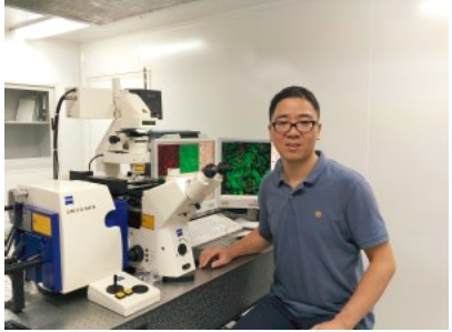 Shuangmu Zhuo (卓双木)合作论文数福建师范大学光电与信息工程学院42
 Dexin Chen合作论文数Southern Med Univ, Nanfang Hosp, Dept Gen Surg, Guangzhou 510515, Guangdong, Peoples R China23Wei Jiang合作论文数22Jianping Lu合作论文数22
 Xiaoyu Dong (董晓瑜)合作论文数20
 Jianxin Chen (陈建新)合作论文数College of Photonic and Electronic Engineering, Fujian Normal University19Jixiang Zheng合作论文数17Botao Yan合作论文数16
 Guoxin Li (李国新)合作论文数School of Clinical Medicine, Tsinghua University;Nanfang Hospital, Southern Medical University16Shuoyu Xu合作论文数Department of General Surgery, Nanfang Hospital, Southern Medical University, Guangzhou, China.13Jiaxin Cheng合作论文数13Guangxing Wang合作论文数11Hongli Ji合作论文数10Zhangyuanzhu Liu合作论文数10Zhiming Li合作论文数10
 Xiufeng Wu (吴秀凤)合作论文数Interdisciplinary Institute for Medical Engineering, Fuzhou University9Huaiming Wang合作论文数8Mingyuan Feng合作论文数8Meiting Fu合作论文数Southern Med Univ, Clin Med Coll 1, Guangzhou 510515, Guangdong, Peoples R China8Xiumin Liu合作论文数8Weisheng Chen合作论文数8Shijie Wang合作论文数8WeiFeng Zhu (朱伟峰)合作论文数8Yaowen Hu合作论文数7Yuting Tang合作论文数7Qi Sun合作论文数7Yan-long CHEN (陈彦龙)合作论文数7
 Hui Wang (王慧)合作论文数Zhongshan School of Medicine, Sun Yat-sen University7
 Gang Chen (陈刚)合作论文数Fujian Cancer Hospital;Interdisciplinary Institute for Medical Engineering, Fuzhou University7Deng Hai-Jun合作论文数7
 Shusen Xie (谢树森)合作论文数Fujian Normal University7Zexi Lin合作论文数6Yandong Zhao合作论文数6Ting Wang合作论文数6Jie Tan合作论文数6
 Chuxia Deng (鄧初夏)合作论文数Faculty of Health Sciences, University of Macau;University of Western Australia;Nanchong Medical College;Cancer Hospital, Beijing University;Luzhou Medical College;Wuhan University;Institute of Biotechnology6
 Mingang Ying (应敏刚)合作论文数福建省肿瘤医院6Zexin Chen合作论文数5Wei Wu合作论文数5
 Yanan Wang (王亚楠)合作论文数Beijing Children’s Hospital, Capital Medical University5Fanni Li合作论文数5
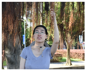 Nenrong Liu (刘嫩容)合作论文数福建师范大学物理与能源学院5Wenju Liu合作论文数Southern Med Univ, Nanfang Hosp, Dept Gen Surg, Guangzhou 510515, Guangdong, Peoples R China4Liyan Lin合作论文数4Weisong Xue (薛巍松)合作论文数4Dongfang Li合作论文数4Yuan-Hong Gao合作论文数4DaJia Lin (林达佳)合作论文数4
 Hao Liu合作论文数Department of General Surgery, Nanfang Hospital, Southern Medical University4




