BACKGROUND:This prospective observational study evaluated three-dimensional soft tissue contour changes over a 90-day healing period following bilateral premolar extractions, with and without alveolar ridge preservation (ARP), using intraoral scanning and 3D digital analysis. METHODS:Eighteen orthodontic patients requiring bilateral premolar extractions were enrolled. In each patient, the extraction socket with relatively lower buccal bone height was assigned to alveolar ridge preservation (ARP), while the contralateral site was left to heal spontaneously. Intraoral scans were obtained immediately before extraction and at 30 days and 90 days post-extraction. Linear horizontal soft tissue changes were measured at 2 mm (H2) and 4 mm (H4) below the gingival margin, while vertical changes were assessed at the central aspect of the extraction socket. Surface displacement was analyzed using root mean square (RMS) distance, and volumetric changes were quantified using region-of-interest segmentation. RESULTS:Significant time-dependent changes in soft tissue contours were observed (p < .001), with greater horizontal reduction at H2 than H4, and more pronounced buccal changes (p < .05). At day 90, mean horizontal reduction at H2 buccal was 1.84 ± 0.69 mm (ARP) and 1.72 ± 0.68 mm (control); corresponding H2 lingual reductions were 1.34 ± 0.89 mm and 1.16 ± 0.29 mm. Vertical reduction peaked at 30 days with partial recovery by day 90. RMS-based displacement was greater buccally. Volumetric reduction was approximately 17% in both groups. No statistically significant differences were found between groups. CONCLUSIONS:Intraoral scanning combined with digital superimposition proved effective for evaluating soft tissue changes after extraction. While ARP remains a valuable approach for alveolar bone preservation, its additional impact on soft tissue contour was limited in sites with favorable baseline conditions. TRIAL REGISTRATION:Clinical Research Information Service (CRIS), KCT0010923. Registered 19 August 2025. Retrospectively registered.
PURPOSE This study aimed to investigate whether new-onset periodontitis or apical periodontitis in the adjacent teeth affects osseointegrated dental implants in a beagle dog model. METHODS One control group and 2 experimental groups (periodontitis and apical periodontitis groups) were defined based on the presence of experimental periodontitis or apical periodontitis, with 1 beagle dog randomly assigned to each group. The mandibular second and fourth premolars on both sides of the 3 beagles were extracted. Eight weeks after extraction, 4 bone-level implant fixtures, 2 on both sides of each mandible, were placed in each beagle. Six weeks after implant surgery, healing abutments were connected. After sufficient osseointegration, plaque control was performed in the control group, while periodontitis and apical periodontitis were induced in the experimental groups. The beagles were euthanized for histological analyses 20 weeks after induction of experimental periodontitis. Statistical analyses were performed using the Kruskal-Wallis test with the Bonferroni correction to compare the 3 groups. RESULTS The implants in the control and apical periodontitis groups were well-maintained, while those in the periodontitis group showed clinical signs of inflammation with bone resorption. The bone-to-implant contact (BIC) and bone area values in the periodontitis group were lower than those in the other groups. The distance between the implant shoulder and the first BIC was significantly greater in the periodontitis group than in the control group (P<0.05). CONCLUSIONS The presence of periodontitis in adjacent teeth can pose a risk to dental implants, potentially resulting in peri-implantitis. However, this was not observed for apical periodontitis. Within the limitations of this study, periodontal care is necessary to reduce the impact of periodontitis in adjacent teeth on osseointegrated implants.
PURPOSE:This study aimed to evaluate the long-term cumulative survival rate (CSR) of dental implants with micro-threads in the neck over a 10-year follow-up period and to examine the factors influencing the survival rate of dental implants.METHODS:This retrospective study was based on radiographic and dental records. In total, 151 patients received 490 Oneplant® dental implants with an implant neck micro-thread design during 2006-2010 in the Department of Periodontology of Seoul National University Dental Hospital. Implant survival was evaluated using Kaplan-Meier analysis. Cox proportional hazard regression analysis was used to identify the factors influencing implant failure.RESULTS:Ten out of 490 implants (2.04%) failed due to fixture fracture. The CSR of the implants was 97.9%, and no significant difference was observed in the CSR between external- and internal-implant types (98.2% and 97.6%, respectively, P=0.670). In Cox regression analysis, 2-stage surgery significantly increased the risk of implant failure (hazard ratio: 4.769, P=0.039). There were no significant differences in influencing factors, including sex, age, implant diameter, length, fixture type, location, surgical procedure, bone grafting, and restoration type.CONCLUSIONS:Within the limitations of this retrospective study, the micro-thread design of the implant neck was found to be favorable for implant survival, with stable clinical outcomes.
PURPOSE:The aim of this study was to retrospectively evaluate the survival and failure rates of RESTORE® implants over a follow-up period of 10-15 years at a university dental hospital and to investigate the factors affecting the survival rate of these dental implants. METHODS:A total of 247 RESTORE® dental implants with a resorbable blast media (RBM) surface inserted in 86 patients between March 2006 and April 2011 at the Department of Periodontology of Seoul National University Dental Hospital were included. Patients with follow-up periods of less than 10 years were excluded, and data analysis was conducted based on dental records and radiographs. RESULTS:Over a 10- to 15-year period, the cumulative survival rate of the implants was 92.5%. Seventeen implants (6.88%) were explanted due to implant fracture (n=10, 4.05%), peri-implantitis (n=6, 2.43%), and screw fracture (n=1, 0.4%). The results of univariate regression analysis using a Cox proportional hazards model demonstrated that implants placed in male patients (hazard ratio [HR], 4.542; 95% confidence interval [CI], 1.305-15.807; P=0.017) and implants that supported removable prostheses (HR, 15.498; 95% CI, 3.105-77.357; P=0.001) showed statistically significant associations with implant failure. CONCLUSIONS:Within the limitations of this retrospective study, the RESTORE® dental implant with an RBM surface has a favorable survival rate with stable clinical outcomes.
In an aging society, quality of life improvement is emerging as an important issue, and as implants are accepted as the core of oral rehabilitation treatment, competition for leadership in developing related technologies is intensifying. In this trend, unlike what is evident in the literature, the patent landscape shows the status of industrial-based technology development. A database analysis of a total of 32,237 dental implant patents shows improvements in technology, diverse geographical characteristics, and new advances toward technological convergence in this field. Technologically, dental implant technology has shown a tendency to develop from conventional implant materials and surface treatment technologies to new material technologies making use of substances such as pure zirconium and tantalum or software technologies related to diagnosis and prognosis. Regionally, dental implant technology, which was developed mainly in Europe and the Unites States in the past, is growing explosively in East Asian countries accompanied by the recent growth of the Asian market. In summary, dental implant technology seems to be developing while trying to converge with various technological areas based on the local market environment. Therefore, it is necessary to develop a new dental implant material technology that is highly applicable to the development of hybrid information/communication technology and is suitable for a new manufacturing method. Our study may provide important information to help basic and translational researchers and their financial supporters set their research directions in advancing the development of dental implants.
Embryonic stem cells have been a popular research topic in regenerative medicine owing to their pluripotency and applicability. However, due to the difficulty in harvesting them and their low yield efficiency, advanced cell reprogramming technology has been introduced as an alternative. Dental stem cells have entered the spotlight due to their regenerative potential and their ability to be obtained from biological waste generated after dental treatment. Cell reprogramming, a process of reverting mature somatic cells into stem cells, and transdifferentiation, a direct conversion between different cell types without induction of a pluripotent state, have helped overcome the shortcomings of stem cells and raised interest in their regenerative potential. Furthermore, the potential of these cells to return to their original cell types due to their epigenetic memory has reinforced the need to control the epigenetic background for successful management of cellular differentiation. Herein, we discuss all available sources of dental stem cells, the procedures used to obtain these cells, and their ability to differentiate into the desired cells. We also introduce the concepts of cell reprogramming and transdifferentiation in terms of genetics and epigenetics, including DNA methylation, histone modification, and non-coding RNA. Finally, we discuss a novel therapeutic avenue for using dental-derived cells as stem cells, and explain cell reprogramming and transdifferentiation, which are used in regenerative medicine and tissue engineering.
This study aimed to histologically and histomorphometrically evaluate osseointegration following simultaneous implant placement and maxillary sinus augmentation. Three retrospective human cases are described in which implants were placed at the maxillary sinus site augmented with deproteinized bovine bone mineral (DBBM) and later retrieved due to implant fracture after 5 to 8 years of occlusal loading. The removed implants with bone were processed for histologic evaluation, and bone-to-implant contact (BIC), bone area (BA), and mirror-image bone area (MIBA) were measured. Mature lamella bone was mainly observed, and some unabsorbed grafted bone particles remained in all cases. The measured values of BIC, BA, and MIBA in the three consecutive threads with the highest values were 86.0% to 91.2%, 65.8% to 91.9%, and 73.0% to 90.4%, respectively, and there were no signs of inflammation. Within the limits of this study, these cases demonstrate successful bone formation after maxillary sinus bone augmentation with DBBM and simultaneous implant placement.
Purpose: This study aimed to identif y the trend of horizontal sof t tissue contour changes in extraction sockets using data obtained by intraoral scanners, and to investigate the effect of ridge preservation. Materials and Methods: In nine patients, 18 teeth were extracted, including both premolars, and ridge preservation with deproteinized bovine bone mineral and an absorbable collagen membrane was performed on only one side, and a linear change in width was observed. Intraoral scanning was performed immediately before extraction and at 30- and 90 days af ter extraction. Superimposition and measurements of the three-dimensional scan images were performed. Results: In the experimental group, a decrease in the horizontal width of 0.845 mm buccally and 0.307 mm lingually at 4 mm below the baseline was observed 90 days af ter tooth extraction, and in the control group, an average decrease of 0.73 mm and 0.21 mm was observed, respectively. The vertical decrease in gingival height measured 90 days af ter tooth extraction was 0.8 mm in the experimental group and 0.46 mm in the control group. Conclusion: The horizontal ridge contour during the healing process of healthy mandibular premolar tooth extraction showed a tendency to decrease over time, and a greater change was observed as the height of the measurement point was closer to the reference gingival margin. The effects of ridge preservation were not evident.
PURPOSE:This study aimed to evaluate the effectiveness of the partial split-flap technique with a K-incision on vertical guided bone regeneration (vGBR) and to retrospectively analyze the clinical and radiographic outcomes of dental implantation using this approach.METHODS:In total, 78 patients who received 104 dental implants with vGBR, categorized as (1) pre-GBR and post-implantation and (2) simultaneous GBR and implantation, were enrolled. Data analysis was based on periapical radiographs, clinical photos, and dental records. The 2-sample t-test was used to compare the 2 surgical procedures.RESULTS:The baseline vertical bone level, augmented bone height (ABH), and treatment duration were significantly higher in the pre-GBR procedure group. The survival rates of the implants were 96.1% and 94.8% in implant- and patient-based analyses, respectively. In Cox regression analysis, high rates of implant failure were found in the presence of ABH of ≥4 mm, smoking, and diabetes.CONCLUSIONS:Within the limitations of this retrospective study, the partial split-flap technique using a K-incision for vGBR showed stable clinical outcomes and favorable dental implant survival.
Background The Encyclopedia of DNA Elements (ENCODE) project has advanced our knowledge of the functional elements in the genome and epigenome. The aim of this article was to provide the comprehension about current research trends from ENCODE project and establish the link between epigenetics and periodontal diseases based on epigenome studies and seek the future direction. Main body Global epigenome research projects have emphasized the importance of epigenetic research for understanding human health and disease, and current international consortia show an improved interest in the importance of oral health with systemic health. The epigenetic studies in dental field have been mainly conducted in periodontology and have focused on DNA methylation analysis. Advances in sequencing technology have broadened the target for epigenetic studies from specific genes to genome-wide analyses. Conclusions In line with global research trends, further extended and advanced epigenetic studies would provide crucial information for the realization of comprehensive dental medicine and expand the scope of ongoing large-scale research projects.
Objectives We previously suggested an ovariectomy (OVX)-induced osteoporotic rat model showing an impaired alveolar bone defect healing. This study aimed to evaluate and compare the effects of recombinant human bone morphogenetic protein-2 (rhBMP-2) and recombinant human platelet-derived growth factor-BB (rhPDGF-BB) on alveolar bone defect healing in OVX-induced osteoporotic rats. Materials and Methods A total of forty-one female rats were divided into four groups: a collagen group ( n =10), a PDGF-BB group ( n =11), a BMP-2 group ( n =10), and a control group ( n =10). Four months after OVX, alveolar bone drill-hole defects were created and grafted with collagen gel, rhPDGF-BB/collagen gel, or rhBMP-2/collagen gel. The defects in the control group were not grafted with any material. Defect healing was evaluated by histological, histomorphometric, and microcomputed tomographic (micro-CT) analyses at 2 and 4 weeks. Results According to the micro-CT analysis, the BMP-2 group exhibited the greatest bone volume fraction among all groups, while the PDGF-BB group did not show significant differences compared with the collagen group. The histomorphometric analysis showed a significantly larger amount of new bone area in the BMP-2 group than in the control and collagen groups at 4 weeks; however, the PDGF-BB group did not reach significant superiority compared with the other groups. Conclusions Alveolar bone regeneration was significantly enhanced by the local use of rhBMP-2/collagen gel compared with the use of rhPDGF-BB/collagen gel in OVX-induced osteoporotic rats. Clinical Relevance A treatment modality using rhBMP-2 may be a promising approach to promote alveolar bone regeneration in patients suffering from postmenopausal osteoporosis.
Purpose: Direct intraoral scanning and superimposing methods have recently been applied to measure the dimensions of periodontal tissues. The aim of this study was to analyze various correlations between labial gingival thickness and underlying alveolar bone thickness, as well as clinical parameters among 3 tooth types (central incisors, lateral incisors, and canines) using a digital method. Methods: In 20 periodontally healthy subjects, cone-beam computed tomography images and intraoral scanned files were obtained. Measurements of labial alveolar bone and gingival thickness at the central incisors, lateral incisors, and canines were performed at points 0-5 mm from the alveolar crest on the superimposed images. Clinical parameters including the crown width/crown length ratio, keratinized gingival width, gingival scallop, and transparency of the periodontal probe through the gingival sulcus were examined. Results: Gingival thickness at the alveolar crest level was positively correlated with the thickness of the alveolar bone plate (P<0.05). The central incisors revealed a strong correlation between labial alveolar bone thickness at 1 and 2 mm, respectively, inferior to the alveolar crest and the thickness of the gingiva at the alveolar crest line (G0), whereas G0 and labial bone thickness at every level were positively correlated in the lateral incisors and canines. No significant correlations were found between clinical parameters and hard or soft tissue thickness. Conclusions: Gingival thickness at the alveolar crest level revealed a positive correlation with labial alveolar bone thickness, although this correlation at identical depth levels was not significant. Gingival thickness, at or under the alveolar crest level, was not associated with the clinical parameters of the gingival features, such as the crown form, gingival scallop, or keratinized gingival width.
Background Although the presence of prosthetic restorations has been associated with plaque accumulation, gingivitis, and periodontitis, there is a lack of large epidemiological investigations providing credible evidence on the association of prosthetic crowns with periodontitis. The purpose of the study was to analyze the association between the number of prosthetic crowns and the presence of periodontitis. Methods This study was based on the Seventh Korea National Health and Nutrition Examination Survey (2016-2018). A total of 12,689 participants over the age of 19 years were surveyed. Multivariate logistic regression analyses were used to identify the association between the number of prosthetic crowns and periodontitis after adjusting for potential confounders, including demographic variables, socio-economic characteristics, oral health-related variables, and oral and systemic clinical variables. Results The odds ratio of periodontitis showed statistically significant differences in the anterior and posterior regions, and the prevalence of periodontal disease increased as the number of crown prostheses increased. Participants with 6–10 and 11 prosthetic crowns had 1.24 and 1.28 times higher prevalence of periodontitis, respectively, than patients with no prosthetic crown. Conclusions The results of this study show that the number of prosthetic crowns present in adults is related to the prevalence of periodontitis.
Purpose: In this study, we aimed to evaluate the clinical validity of the modified tunneling technique using minimal soft tissue harvesting and volume-stable collagen matrix in the anterior mandible. Methods: In total, 27 anterior mandibular teeth and palatal donor sites in 17 patients with >= 1 mm of gingival recession (GR) were analyzed before and after root coverage. For the recipient sites, vertical vestibular incisions were made in the interdental area and a subperiosteal tunnel was created with an elevator. After both sides of the marginal gingiva were tied to one another, a prepared connective tissue graft and volume-stable collagen matrix were inserted through the vestibular vertical incision and were fixed with resorbable suture material. The root coverage results of the recipient site were measured at baseline (T0), 3 weeks (T3), 12 weeks (T12), and the latest visit (Tl). For palatal donor sites, a free gingival graft from a pre-decided area avoiding the main trunk of the greater palatine artery was harvested using a prefabricated surgical template at a depth of 2 mm after de-epithelization using a rotating bur. In each patient, the clinical and volumetric changes at the donor sites between T0 and T3 were measured. Results: During an average follow-up of 14.5 months, teeth with denuded root lengths of 1-3 mm (n=12), 3-6 mm (n=11), and >6 mm (n=2) achieved root coverage of 97.01%+/- 7.65%, 86.70%+/- 5.66%, and 82.53%+/- 1.39%, respectively. Miller classification I (n=12), II (n=10), and III (n=3) teeth showed mean coverage rates of 97.01%+/- 7.65%, 86.91%+/- 5.90%, and 83.19%+/- 1.62%, respectively. At the donor sites, an average defect depth of 1.41 mm (70.5%) recovered in 3 weeks, and the wounds were epithelized completely in all cases. Conclusions: The modified tunneling technique in this study is a promising treatment modality for overcoming GR in the anterior mandible.
The oral microbiome is an important part of the human microbiome. The oral cavity has the second largest microbiota after the intestines, and its open structure creates a special environment. With the development of technology such as next-generation sequencing and bioinformatics, extensive in-depth microbiome studies have become possible. They can also be applied in the clinical field in terms of diagnosis and treatment. Many microbiome studies have been performed on oral and systemic diseases, showing a close association between the two. Understanding the oral microbiome and host interaction is expected to provide future directions to explore the functional and metabolic changes in diseases, and to uncover the molecular mechanisms for drug development and treatment that facilitate personalized medicine. The aim of this review was to provide comprehension regarding research trends in oral microbiome studies and establish the link between oral microbiomes and systemic diseases based on the latest technique of genome-wide analysis.
BACKGROUND AND PURPOSE Significant research and development (R&D) has been conducted to make the best dental implants while developing various patent applications and registrations. In this study, we evaluated the current status of patents on dental implants and identified the future direction of R&D progress. MATERIALS AND METHODS A total of 29 711 patents related to dental implants were reviewed. These were published between 1909 and 2020 and retrieved from the Derwent Innovation patent database. The patents were grouped into three categories depending on the implant components: fixture, abutment, and artificial teeth. RESULTS The category with most patents was "abutment," and the most cited patent was "screw-type dental implant anchor." Global patenting trends over the past 20 years showed that both applicants and applications increased in the early 2010s; however, these have since been on the decline. Currently, the United States holds the largest number of patents, and Nobel Biocare Holding AG is the top assignee. Technic maturation prediction analysis showed that the current dental implant technology is in the "decline stage." CONCLUSION Trend analysis of the dental implant patent indicates the main contributors of development are profit-oriented companies. Recent reduction in the number of new patent applications suggests the technology is in the mature declining stage. The emergence of new materials or technologies that may close the gap in clinical unmet needs would reverse the trend.
It is widely accepted that sandblasted/large-grit/acid-etched (SLA) surfaces of titanium (Ti) have a higher osteogenic potential than machined ones. However, most studies focused on differential gene expression without elucidating the underlying mechanism for this difference. The aim of this study was to evaluate how the surface roughness of dental Ti implants affects their osteogenic potential. Mouse preosteoblast MC3T3-E1 cells were seeded on machined and SLA Ti discs. The cellular activities of the discs were analyzed using confocal laser scanning microscopy, proliferation assays, and real-time polymerase chain reaction (PCR). DNA methylation was evaluated using a methylation-specific PCR. The cell morphology was slightly different between the two types of surfaces. While cellular proliferation was slightly greater on the machined surfaces, the osteogenic response of the SLA surfaces was superior, and they showed increased alkaline phosphatase (Alp) activity and higher bone marker gene expression levels (Type I collagen, Alp, and osteocalcin). The degree of DNA methylation on the Alp gene was lower on the SLA surfaces than on the machined surfaces. DNA methyltransferase inhibitor stimulated the Alp gene expression on the machined surfaces, similar to the SLA surfaces. The superior osteogenic potential of the SLA surfaces can be attributed to a different epigenetic landscape, specifically, the DNA methylation of Alp genes. This finding offers novel insights into epigenetics to supplement genetics and raises the possibility of using epidrugs as potential therapeutic targets to enhance osteogenesis on implant surfaces.
Periodontal disease is a major public health issue, and various periodontal therapies have been performed to regenerate periodontal tissues. The periodontium is a complex structure composed of specialized tissues that support the teeth, and most periodontal surgeries are invasive procedures, including a resection of the gingiva or the alveolar bone. The periodontal wound healing process is slightly different from cutaneous wound healing and is similar to fetal healing, being almost scar-free. The aim of this review article is to provide an overview of periodontal wound healing and discuss various surgical and pharmaceutical approaches to achieve stable wound healing and improve the treatment outcomes. In addition, detrimental and limiting factors that induce a compromised prognosis are discussed, along with the perspective and future direction for successful periodontal tissue regeneration.
Although the presence of prosthetic restorations has been associated with plaque accumulation, gingivitis, and periodontitis, there is a lack of large epidemiological investigations providing evidence on the association of prosthetic crowns with periodontitis. This study aimed to analyze the association between the number of prosthetic crowns and the presence of periodontitis. This study was based on the Seventh Korea National Health and Nutrition Examination Survey (2016–2018). A total of 12,689 participants over the age of 19 years were surveyed. Multivariate logistic regression analyses were used to identify the association between the number of prosthetic crowns and periodontitis after adjusting for potential confounders, including demographic variables, socio-economic characteristics, oral health-related variables, and oral and systemic clinical variables. The odds ratio of periodontitis showed statistically significant differences in the anterior and posterior regions, and the prevalence of periodontal disease increased as the number of crown prostheses increased. Participants with 6–10 and 11 prosthetic crowns had 1.24 and 1.28 times higher prevalence of periodontitis, respectively, than patients with no prosthetic crown. The results of this study show that the number of prosthetic crowns present in adults is related to the prevalence of periodontitis.
 Yong-Moo Lee合作论文数School of Dentistry, Seoul National University130
 In-Chul Rhyu合作论文数125Chong-Pyoung Chung合作论文数113Yang-Jo Seol合作论文数68
 Tae Il Kim合作论文数Department of Public Administration, College of Political Science & Economics, Korea University66Soo Boo Han合作论文数59
 Ki-Tae Koo合作论文数44Kyoung-Hwa Kim合作论文数Intellectual Biointerface Engineering Center (IBEC),, Korea Science and Engineering Foundation (KOSEF),43
 Yoon Jeong Park合作论文数College of Pharmacy, Ewha Womans University32S H Choi合作论文数30
 Young-Dan Cho合作论文数27CP Chung合作论文数21Kim Sungtae合作论文数20
 Seung Jin Lee合作论文数Department of Pharmacy, College of Pharmacy, Ewha Womans University17IC Rhyu合作论文数15
 Hyun-Mo Ryoo合作论文数Department of Molecular Genetics, School of Dentistry, Seoul National University15Soo-Boo Han合作论文数14Seung-Yun Shin合作论文数Samsung Medical Center, Sungkyunkwan University School of Medicine14YM Lee合作论文数13SB Han合作论文数10Su-Hwan Kim合作论文数10
 Seong-Joo Heo合作论文数10Yun-Jeong Kim合作论文数9Jue Yeon Lee合作论文数8
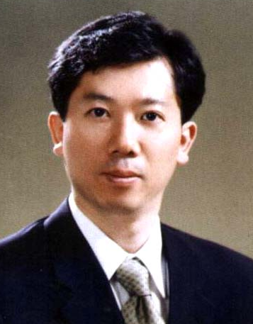 Jun Hyeog Jang合作论文数Depart.of Biochemistry, College of Medicine, Inha University8Seung-Min Yang合作论文数7Seung-Beom Kye合作论文数7YJ Seol合作论文数6
 Sang-Hoon Rhee合作论文数Seoul National University6Park Jun-Beom合作论文数6Jae-Eun Chung合作论文数5Seung-Jin Lee合作论文数5
 Ulf ME Wikesjo合作论文数Loma Linda University5Kim Hyun-Ju合作论文数5Kihwan Bae合作论文数5
 Kyung Mi Woo合作论文数School of Dentistry, Seoul National University;Dental Research Institute, Seoul National University5Sang-Cheol Lee合作论文数4chul woo lee合作论文数4byungdo hahm合作论文数4Da-Jeong Namgung合作论文数4JY Lee合作论文数4
 Woo Jin Kim合作论文数4
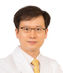 Park Ji-Man合作论文数Department of Prosthodontics &College of Dentistry, Seoul National University;College of Dentistry, Seoul National University4Rhyu I C合作论文数4
 Kim Pil-Jong合作论文数School of Dentistry, Seoul National University4Chong-Hyun Han合作论文数4
 Jeong-Hwa Baek合作论文数School of Dentistry, Seoul National University4byungkun yang合作论文数3




