Objective: Clinical cardiac CT multiphase reconstructions generally provide acceptable image quality in end-diastole (ED) or end-systole (ES) phases, but in other phases may exhibit motion artifacts, especially in the right coronary artery (RCA). This limits ground-truth availability in 4D cardiac CT imaging research. We aim to construct a 4D cardiac CT dataset that is generally suitable to serve as pseudo ground truth. Methods: We propose Coronary Mask Guided Registration (CMGR) to produce a motion-preserved, artifact-reduced, and continuous-time 4D cardiac CT sequence from the clinical multiphase reconstruction of each patient. For artifact reduction, CMGR uses the ED or ES phase as the reference phase and warps the reference volume with deformation fields to produce the sequence. For motion preservation, CMGR registers the reference phase to each non-reference phase of the multiphase reconstruction. To capture the motion of both the RCA and other cardiac structures in each registration, CMGR regularizes RCA masks and incorporates them into image-domain registration. Time-continuity is achieved by interpolating the deformation fields for non-reference phases to arbitrary times. Results: CMGR outperformed representative image-domain registration methods in capturing RCA motion and providing reasonable RCA shape, and showed competitive performance in capturing whole-heart motion. Additionally, CMGR reduced motion artifacts from clinical multiphase reconstructions, and intermediate CMGR frames generally provided plausible transitions between discrete cardiac phases. Conclusion: CMGR provides an effective approach for constructing continuous-time 4D cardiac CT datasets. Significance: The dataset can be used in system design simulations and in reconstruction algorithm development, thereby facilitating advances in cardiac CT imaging.
X-ray scatter has been a serious concern in computed tomography (CT), leading to image artifacts and distortion of CT values. The linear Boltzmann transport equation (LBTE) is recognized as a fast and accurate approach for scatter estimation. However, for multi-spectral CT, it is cumbersome to compute multiple scattering components for different spectra separately when applying LBTE-based scatter correction. In this work, we propose a Matrixed-Spectrum Decomposition accelerated LBTE solver (MSD-LBTE) that can be used to compute X-ray scatter distributions from CT acquisitions at two or more different spectra simultaneously, in a unified framework with no sacrifice in accuracy and significant reduction in computation compared with conventional solvers in theory. First, a matrixed-spectrum solver of the LBTE is obtained by introducing an additional label dimension to expand the phase space. Then, we propose a “spectrum basis” for the LBTE and a principle of selection of basis using the QR decomposition, along with the above solver to construct the MSD-LBTE. Based on MSD-LBTE, a unified scatter correction method can be established for multi-spectral CT.We validate the effectiveness and accuracy of our method by comparing it with the Monte Carlo method. We also evaluate the scatter correction performance using two different phantoms for kV-switching dual-energy spectral CT, and using an elliptical phantom in a numerical simulation for kV-modulation enabled CT scans, validating that our proposed method can significantly reduce the computational cost at multiple spectra and effectively reduce scatter artifact in reconstructed CT images.
Crystallographic texture strongly influences the macroscopic properties of materials, making its precise characterization important for materials research. Conventional monochromatic X-ray diffraction is constrained by low X-ray flux in laboratory systems and inaccessibility of synchrotron-based instruments for industrial use. Polychromatic X-ray diffraction is promising, but complex diffraction patterns limit its use in texture analysis. Here we show a laboratory-based methodology for rapid and quantitative texture characterization of strongly fiber-textured materials using polychromatic X-ray diffraction without monochromators or energy-resolving detectors. We find that X-ray spectrum and texture information are inherently decoupled within the diffraction patterns of such symmetric systems, which simplifies the analysis. Using high-performance fiber-textured magnets as a representative benchmark, we demonstrate that this methodology reduces measurement time and enables spatially resolved analysis. This methodology provides an efficient method for texture characterization of fiber-textured magnets and has potential applications to other materials with similar texture symmetry. Crystallographic texture governs material performance, but conventional X-ray diffraction is often slow and difficult to access. Here, the authors introduce a lab-based polychromatic XRD method that enables fast, quantitative and spatially resolved texture analysis of strongly fiber-textured materials.
This work aims to achieve both high current emission density and high emission current of carbon nanotube (CNT) cathodes for high-power X-ray generation applications. High-purity small-diameter CNT materials were obtained, and a novel “five-state” electrophoretic deposition method was proposed to fabricate CNT cathodes. For an emission area of 10 mm × 0.45 mm, a high and stable cathode emission current of 350 mA was achieved, corresponding to an emission current density of 7.8 A/cm2. An X-ray dose rate of 39.49 mGy/s@50 cm was measured under a tube potential of 120 kV, cathode current of 100 mA, and pulse width of 10 ms. The focal spot size of the X-ray source, measured using a slit camera, was 0.98 mm (width) × 1.05 mm (length) at 15% max intensity, and the pulse width range was 100 µs–100 ms. Through continuous testing at 200 mA emission current, 100 µs pulse width, and 0.3% duty cycle for 400 h, the CNT cathode is estimated to exhibit a lifetime of approximately 5085 h, demonstrating stable and reliable durability. This study, for the first time, simultaneously realizes multi-A/cm2-level emission current density, hundreds-of-milliampere emission current, and hundreds-of-millisecond operating pulse width for CNT cathodes.
This study conducted an in-depth investigation on the forward radiation field characteristics of a multi-beam X-ray source. The study revealed distribution characteristics at different distances, beam number, spacing, and tube voltage. Based on multi-beam X-ray source with 45 linearly arranged focal spots, the dose field distribution was analyzed through Monte Carlo simulations and validated by measurement data. A radiation measurement platform was established, and measurement data verified the accuracy of the Monte Carlo simulation results. The research results show that under an anode voltage of 160 kV and an anode current of 15 mA, the forward radiation field of the 45 focal spot multi-beam X-ray source decreases from 62.2 mGy/s at the center to 22.3 mGy/s at the edges at a distance of 20 cm from the focal spot distribution line, and to 6 mGy/s at 80 cm forward distance. Unlike the peaked radiation pattern of a single-beam X-ray tube, this configuration exhibits a gentle-slope distribution and an end truncation effect, referred to as a quasi-line-source characteristic. Further simulations demonstrated that the near-field region in front of the focal spot distribution line shows an alternating peak–valley pattern, which disappears in the far-field. For a 160 kV multi-beam X-ray source, the positions of these peaks and valleys correspond closely to the focal spot spacing and shift closer as the tube voltage increases. The findings of this work provide useful references for the radiation field characterization, imaging dose evaluation, physical system design, and shielding design of multi-beam X-ray imaging systems.
Digital chest tomosynthesis (DCT) has been clinically validated to offer significant advantages in diagnostic efficiency for pulmonary diseases and radiation dose reduction. Emerging stationary DCT (sDCT) systems can further shorten acquisition time and eliminate motion artifacts caused by X-ray source movement and patient respiration. This work focuses on the development of a multi-beam X-ray source for mobile sDCT systems by specification definition, source design, and experimental validation. The developed X-ray tube integrates 63 focal spots arranged linearly over a length of 816 mm. X-rays are emitted through seven segmented windows, achieving an angular span of 36° at a source image distance (SID) of 120 cm, with full coverage of a detector area of 35.6 cm × 43.2 cm. The tube operates at a maximum anode voltage of 140 kV, maximum anode current of 20 mA, and 24 mAs per scan, with a focal spot size of IEC 0.6. The developed multi-beam X-ray source achieves multiple key performance breakthroughs and provides an alternative source architecture for future sDCT implementation, with the potential to facilitate further system performance optimization and engineering development.
Multi-source stationary computed tomography (MSS-CT) offers significant advantages in medical and industrial applications due to its gantry-less scan architecture and/or capability of simultaneous multi-source emission. However, the lack of anti-scatter grid deployment in MSS-CT results in severe forward and/or cross scatter contamination, presenting a critical challenge that necessitates an accurate and efficient scatter correction. In this work, ComptoNet, an innovative end-to-end deep learning framework for scatter estimation in MSS-CT, is proposed, which integrates Compton-scattering physics with deep learning techniques to address the challenges of scatter estimation effectively. Central to ComptoNet is the Compton-map, a novel concept that captures the distribution of scatter signals outside the scan field of view, primarily consisting of large-angle Compton scatter. In ComptoNet, a reference Compton-map and/or spare detector data are used to guide the physics-driven deep estimation of scatter from simultaneous emissions by multiple sources. Additionally, a frequency attention module is employed for enhancing the low-frequency smoothness. Such a multi-source deep scatter estimation framework decouples the cross and forward scatter. It reduces network complexity and ensures a consistent low-frequency signature with different photon numbers of simulations, as evidenced by mean absolute percentage errors (MAPEs) that are less than 1.26%. Conducted by using data generated from Monte Carlo simulations with various phantoms, experiments demonstrate the effectiveness of ComptoNet, with significant improvements in scatter estimation accuracy (a MAPE of 0.84%). After scatter correction, nearly artifact-free CT images are obtained, further validating the capability of our proposed ComptoNet in mitigating scatter-induced errors.
Multi-segment static computed tomography (MS-staticCT) is a generalized and efficient configuration of static CT systems, achieving high temporal resolution imaging by sequentially firing x-ray sources, instead of rotation. However, it contains numerous geometric parameters. Due to the dense arrangement of both the x-ray sources and detectors within their respective configurations, there are some coupled illumination relationships where some x-ray sources simultaneously illuminate multiple detectors. To address these calibration challenges, we propose a geometric calibration method based on ordered subsets. We categorize two types of ordered subsets of sources and detectors: source subsets and detector subsets. Each source subset includes a group of sources that illuminate the same detectors, along with the illuminated detectors. Similarly, each detector subset includes a group of detectors illuminated by the same sources, along with the sources that illuminate them. The calibration of the sources in source subsets and the detectors in detector subsets is performed alternately until convergence, ensuring that the calibrated geometry to accurately describe all the illumination relationships. These calibration steps are detailed in a workflow. During each step, the estimations for different ordered subsets are independent and parallelizable to significantly improving computational efficiency. A calibration phantom is involved in our method. During the calibration, we iteratively estimate the parameters by minimizing the average re-projection error (aRPE) of the balls in the calibration phantom. We evaluated the proposed method by simulation and actual experiments. The aRPE was reduced to 0.0087 mm and the reconstructed images were clear without obvious misalignment in simulation. Compared to estimating all parameters together, our method improved computational efficiency by a factor of 2.20. The targeted spatial resolution (2.5 lp·mm-1) of an actual MS-staticCT system was obtained. These results verified the efficiency and accuracy of the proposed method.
Reconstructing CT images from incomplete projection data remains challenging due to the ill-posed nature of the problem. Diffusion bridge models have recently shown promise in restoring clean images from their corresponding Filtered Back Projection (FBP) reconstructions, but incorporating data consistency into these models remains largely underexplored. Incorporating data consistency can improve reconstruction fidelity by aligning the reconstructed image with the observed projection data, and can enhance detail recovery by integrating structural information contained in the projections. In this work, we propose the Projection Embedded Diffusion Bridge (PEDB). PEDB introduces a novel reverse stochastic differential equation (SDE) to sample from the distribution of clean images conditioned on both the FBP reconstruction and the incomplete projection data. By explicitly conditioning on the projection data in sampling the clean images, PEDB naturally incorporates data consistency. We embed the projection data into the score function of the reverse SDE. Under certain assumptions, we derive a tractable expression for the posterior score. In addition, we introduce a free parameter to control the level of stochasticity in the reverse process. We also design a discretization scheme for the reverse SDE to mitigate discretization error. Extensive experiments demonstrate that PEDB achieves strong performance in CT reconstruction from three types of incomplete data, including sparse-view, limited-angle, and truncated projections. For each of these types, PEDB outperforms evaluated state-of-the-art diffusion bridge models across standard, noisy, and domain-shift evaluations.
Diffusion-based models have demonstrated remarkable effectiveness in image restoration tasks; however, their iterative denoising process, which starts from Gaussian noise, often leads to slow inference speeds. The Image-to-Image Schrödinger Bridge (I2SB) offers a promising alternative by initializing the generative process from corrupted images while leveraging training techniques from score-based diffusion models. In this paper, we introduce the Implicit Image-to-Image Schrödinger Bridge (I3SB) to further accelerate the generative process of I2SB. I3SB restructures the generative process into a non-Markovian framework by incorporating the initial corrupted image at each generative step, effectively preserving and utilizing its information. To enable direct use of pretrained I2SB models without additional training, we ensure consistency in marginal distributions. Extensive experiments across many image corruptions—including noise, low resolution, JPEG compression, and sparse sampling—and multiple image modalities—such as natural, human face, and medical images— demonstrate the acceleration benefits of I3SB. Compared to I2SB, I3SB achieves the same perceptual quality with fewer generative steps, while maintaining or improving fidelity to the ground truth.
Limited-view computed tomography (CT) presents significant potential for reducing radiation exposure and expediting the scanning process. While deep learning (DL) methods have exhibited promising results in mitigating streaking artifacts caused by a reduced number of projection views, their generalization remains challenging. In this work, we proposed a DL-driven alternative Bayesian reconstruction method (DLBayesian) that efficiently integrates data-driven priors and data consistency constraints. DLBayesian comprises three stages: group-level embedding, significance evaluation, and individual-level consistency adaptation. Firstly, DL network parameters are optimized to learn how to eliminate the general limited-view artifacts on a large-scale paired dataset. Then, we introduced a significance score to quantitatively evaluate the contribution of parameters in DL models as a guide for the subsequent individual-level adaptation. Finally, in the Bayesian adaptation stage, an alternative Bayesian reconstruction further optimizes the DL network parameters precisely according to the projection data of the target case. We validated DLBayesian with sparse-view (90 views) projections from a circular trajectory CT and a special data missing case from a multi-segment linear trajectory CT. The results underscore DLBayesian's superior generalization capabilities across variations in patients, anatomic structures, and data distribution, as well as excelling in contextual structure recovery compared to networks solely trained via supervised loss. Real experiments on a dead rat demonstrate its capability in practical CT scans.
With the increasing demand for intraoral dental health assessment, it has become important to obtain more detailed and informative visual data. To obtain high-resolution dental imaging, this study conducts research on advanced X-ray imaging technologies. This study proposes adopting multi-beam X-ray source for achieving high-resolution 3D intraoral imaging. Key parameters such as focal spot size, detector pixel size, system magnification and line-pair resolution are systematically analyzed. A theoretical framework is established to derive the resolution formula for X-ray imaging, identifying key factors that affect imaging resolution. An experimental platform is constructed to conduct performance testing on the multi beam X-ray tube. The results show that X-ray tube is worked on an anode voltage of 70 kV and a current of 7 mA. The measured focal spot sizes ranged from 0.43 mm to 0.52 mm, with an average of 0.47 mm and a standard deviation of 0.028. Line-pair resolution tests confirmed that all focal spots achieved the target resolution of 16 lp/mm. Systematic analysis of the X-ray imaging system demonstrates that, by employing a multi-beam X-ray tube and a detector with 18.5 μm pixels and a frame rate of 20 fps under a magnification factor of 1.04, the system achieved high-resolution imaging with resolution of 31.3 μm (16 lp/mm) for each focal spot, enabling the acquisition of seven projection angles within 1 s. The experimental results conclusively validate the clinical potential of multi-beam X-ray source in detecting various dental pathologies, demonstrating its reliability and suitability for high-resolution intraoral imaging applications.
In this work, we investigate the feature of projection sampling and analytical reconstruction algorithms for a Static CT with sources and detectors distributed in a Multi-Segment manner (MS-StaticCT). MS-StaticCT is a generalized configuration of previous static linear CT systems offering enhanced design flexibility and utilization efficiency in both X-ray source and detector components. By analyzing the imaging geometry of single-segment source and detector pairs, we delved into the Radon space properties of MS-StaticCT and proposed a data sufficiency condition for system design. To explore the impact of the unique sampling characteristics of MS-StaticCT on reconstruction quality, we derived analytical algorithms under two popular pipelines filtered-backprojection (MS-FBP) and differentiated backprojection filtration (MS-DBF), and assessed their performance. Due to the non-uniform sampling and singular points between segments, the global filtration process of MS-FBP requires local rebinning. The local nature of differentiation enables convenient filtration without rebinning. Besides, to address insufficient data caused by optical obstruction by sources and detectors, we incorporated multiple imaging planes and designed a generalized weighting function that efficiently utilizes conjugate projections. Simulation studies on numerical phantoms and clinical CT data demonstrate the feasibility of MS-StaticCT and the proposed reconstruction algorithms. The results highlighted MS-DBF's superiority in accuracy and spatial resolution for multi-segment geometries without compromising noise performance compared to MS-FBP whose performance depends on the number of detector segments involved for each focal spot. Our study provides a comprehensive understanding of the essential data structure and basic reconstruction tailored for systems characterized by linear source trajectories and detectors.
Trace detection and X-ray security inspection technology respectively serve as the "nose" and "eyes" of security applications, detecting explosives through "smell" and "vision" perspectives. Following decades of innovation and deployment, they are currently playing critical roles in the integrated aviation security solution. This study explores the prospective fusion of these two technologies for aviation security. Starting with basic technical principles and comparisons, considering security, efficiency, and passenger experience, the study examines industrial trends and discusses the bottlenecks of existing solutions based on standalone security equipment. Subsequently it elaborates the novel concept of fusing trace detection and X-ray computed tomography (X-CT) in security inspection. According to qualitative analysis and discrete event simulation, the study predicts the effectiveness of such fusion. The study further discusses the challenges faced in the development of an automatic trace detection system, elaborates on its concrete innovations and different methods of fusion, and envisions the trends of fusing multiple security detection technologies. According to industry reality, this study analyzes the domestic and international application prospects of this technology in aviation security from a rational and optimistic perspective, aiming to visualize a blueprint for technology developers and end users of security equipment.
Objective. While deep learning (DL) methods have exhibited promising results in mitigating streaking artifacts caused by limited-view computed tomography (CT), their generalization to practical applications remains challenging. To address this challenge, we aim to develop a novel approach that integrates DL priors with targeted-case data consistency for improved artifact suppression and robust reconstruction. Approach. We propose an alternative penalized weighted least squares reconstruction framework by strategic optimization of a DL model (PWLS-SOM). This framework combines data-driven DL priors with data consistency constraints in a three-stage process: (1) Group-level embedding: DL network parameters are optimized on a large-scale paired dataset to learn general artifact elimination. (2) Significance evaluation: A novel significance score quantifies the contribution of DL model parameters, guiding the subsequent strategic adaptation. (3) Individual-level consistency adaptation: PWLS-driven strategic optimization further adapts DL parameters for target-specific projection data. Main results. Experiments were conducted on sparse-view (90 views) circular trajectory CT data and a multi-segment linear trajectory CT scan with a mixed data missing problem. PWLS-SOM reconstruction demonstrated superior generalization across variations in patients, anatomical structures, and data distributions. It outperformed supervised DL methods in recovering contextual structures and adapting to practical CT scenarios. The method was validated with real experiments on a dead rat, showcasing its applicability to real-world CT scans. Significance. PWLS-SOM reconstruction advances the field of limited-view CT reconstruction by uniting DL priors with PWLS adaptation. This approach facilitates robust and personalized imaging. The introduction of the significance score provides an efficient metric to evaluate generalization and guide the strategic optimization of DL parameters, enhancing adaptability across diverse data and practical imaging conditions.
In this work, we proposed the Projection Embedded Schrödinger Bridge (PESB) for CT sparse view reconstruction. PESB constructs Schrödinger Bridges between the distribution of Filtered Back-Projection (FBP) reconstructed images and the distribution of clean images conditioned on measured projections. By embedding projections into the marginal conditions, data consistency is inherently incorporated into the generative process. Experimental results validate the effectiveness of PESB, demonstrating its superior performance in CT sparse view reconstruction compared to several diffusion-based models.
Multi-source stationary computed tomography (MSS-CT) offers significant advantages in medical and industrial applications due to its gantryless scan architecture and capability of simultaneous multi-source emission. However, the lack of anti-scatter grid deployment in MSS-CT leads to severe forward and cross scatter contamination, necessitating accurate and efficient scatter correction. In this work, we propose ComptoNet, an innovative decoupled deep learning framework that integrates Compton-scattering physics with deep learning for scatter estimation in MSS-CT. The core innovation lies in the Compton-map, a representation of large-angle Compton scatter signals outside the scan field of view. ComptoNet employs a dual-network architecture: a conditional encoder-decoder network guided by reference Compton-maps and spare detector data for cross scatter estimation, and a frequency U-Net with attention mechanisms for forward scatter correction. Experiments on Monte Carlo-simulated data demonstrate ComptoNet's superior performance, achieving a mean absolute percentage error of 0.84% on scatter estimation. After correction, CT images show nearly artifact-free quality for all test phantoms, validating ComptoNet's robustness in mitigating scatter-induced errors across diverse photon counts and phantoms comparing with other methods.
Objective. Low-dose interior tomography integrates low-dose CT (LDCT) with region-of-interest (ROI) imaging which finds wide application in radiation dose reduction and high-resolution imaging. However, the combined effects of noise and data truncation pose great challenges for accurate tomographic reconstruction. This study aims to develop a novel reconstruction framework that achieves high-quality ROI reconstruction and efficient extension of recoverable region to provide innovative solutions to address coupled ill-posed problems. Approach. We conducted a comprehensive analysis of projection data composition and angular sampling patterns in low-dose interior tomography. Based on this analysis, we proposed two novel deep learning-based reconstruction pipelines: (1) deep projection extraction-based reconstruction (DPER) that focuses on ROI reconstruction by disentangling and extracting noise and background projection contributions using a dual-domain deep neural network; and (2) DPER with progressive extension (DPER-Pro) that enhances DPER by a progressive 'coarse-to-fine' strategy for missing data compensation, enabling simultaneous ROI reconstruction and extension of recoverable regions. The proposed methods were rigorously evaluated through extensive experiments on simulated torso datasets and real CT scans of a torso phantom. Main results. The experimental results demonstrated that DPER effectively handles the coupled ill-posed problem and achieves high-quality ROI reconstructions by accurately extracting noise and background projections. DPER-Pro extends the recoverable region while preserving ROI image quality by leveraging disentangled projection components and angular sampling patterns. Both methods outperform competing approaches in reconstructing reliable structures, enhancing generalization, and mitigating noise and truncation artifacts. Significance. This work presents a novel decoupled deep learning framework for low-dose interior tomography that provides a robust and effective solution to the challenges posed by noise and truncated projections. The proposed methods significantly improve ROI reconstruction quality while efficiently recovering structural information in exterior regions, offering a promising pathway for advancing low-dose ROI imaging across a wide range of applications.
Compared to single-energy computed tomography (CT), dual- or multiple-energy cone-beam CT (CBCT) has potential of offering better image quality and material differentiation capability. However, an accurate and fast scatter estimation is highly demanded, as the X-ray scattering influences imaging quality, resulting in inaccurate material decomposition and image artifacts. The linear Boltzmann transport equation (LBTE) is considered to be a fast and accuracy approach for scatter estimation. In this work, we introduce a new label dimension in LBTE (LBTE-L) and developed a unified and highly efficient scatter estimation method, which can calculate scatter signals at multiple different spectra in a single computation. We validate its effectiveness and accuracy by comparing it with the Monte Carlo Method and by applying scatter correction on the actual data measured in a spectral CBCT tabletop system.
Sparse-view computed tomography (CT) has great potential in reducing radiation dose and accelerating the scan process. Although deep learning (DL) methods have exhibited promising results in mitigating streaking artifacts caused by very few projections, their generalization remains a challenge. In this work, we proposed a DL-driven alternative Bayesian reconstruction method that efficiently integrates data-driven priors and the data consistency constraints. This methodology involves two stages: universal embedding and consistency adaptation respectively. In the embedding stage, we optimize DL parameters to learn and eliminate the general sparse-view artifacts on a large-scale paired dataset. In the subsequent consistency adaptation stage, an alternative Bayesian reconstruction further optimizes the DL parameters according to individual projection data. Our proposed technique is validated within both image-domain and dual-domain DL frameworks leveraging simulated sparse-view (90 views) projections. The results underscore the superior generalization and context structure recovery of our approach compared to networks solely trained via supervised loss.
 Li Zhang (张丽)合作论文数Department of Engineering Physics, Tsinghua University239
 Liang Li (李亮)合作论文数Department of Engineering Physics, Tsinghua University135
 Yuxiang Xing (邢宇翔)合作论文数130
 Kejun Kang (康克军)合作论文数Department of Engineering Physics, Tsinghua University126Ziran Zhao (赵自然)合作论文数120
 Yongshun Xiao (肖永顺)合作论文数Department of Engineering Physics, Tsinghua University69
 Yinong Liu (刘以农)合作论文数Department of Engineering Physics, Tsinghua University41
 Hewei Gao (高河伟)合作论文数Department of Engineering Physics, Tsinghua University40
 Yingxin Wang (王迎新)合作论文数Department of Engineering Physics, Tsinghua University37
 Jianping Cheng (程建平)合作论文数Department of Engineering Physics, Tsinghua University;Beijing Normal University35Zhifeng Huang (黄志峰)合作论文数24
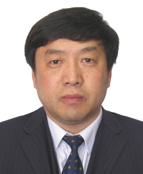 Yuanjing Li (李元景)合作论文数Department of Engineering Physics, Tsinghua University23WenHuan Gao (高文焕)合作论文数20wanlong wu (吴万龙)合作论文数20Zhentian Wang (王振天)合作论文数19xilei luo (罗希雷)合作论文数17
 Ge Wang (王革)合作论文数Biomedical Imaging Center, Rensselaer Polytechnic Institute17
 Xin Jin (金欣)合作论文数Tsinghua Shenzhen International Graduate School17l i yuanjing合作论文数16haifeng hu合作论文数16Ming Chang合作论文数16
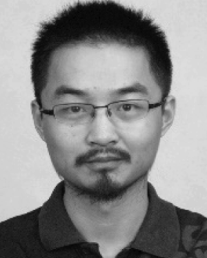 Siyuan Zhang合作论文数15bin sang (桑斌)合作论文数14Fan Guo合作论文数13Lingbo Qiao (乔灵博)合作论文数Nuctech Company Limited13
 Moran Wang (王沫然)合作论文数Institute of Engineering Thermophysics, Department of Engineering Mechanics, School of Aerospace Engineering, Tsinghua University;Laboratory of Multiscale Interfacial Transport, Department of Engineering Mechanics, School of Aerospace Engineering, Tsinghua University;Center for Nano and Micro Mechanics, Tsinghua University13
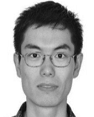 Wei Jia合作论文数12Qitian Miao (苗齐田)合作论文数12
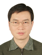 Xuewu Wang (王学武)合作论文数Department of Engineering Physics, Tsinghua University12Yuanyuan Liu (刘圆圆)合作论文数11Dufan Wu合作论文数11
 Yingkang Jin (金颖康)合作论文数Nuctech Company Limited11shangmin sun合作论文数10
 Xinbin Li合作论文数10zhimin zheng (郑志敏)合作论文数10shuo cao合作论文数10
 Jianmin Li (李荐民)合作论文数Department of Engineering Physics, Tsinghua University10
 Duan Xinhui合作论文数10
 Zahed Siddique合作论文数School of Aerospace and Mechanical Engineering, University of Oklahoma10Ronglan Xu (徐荣栏)合作论文数10Yidi Yao合作论文数9Wenyuan Bi合作论文数9
 Ruizhe Li合作论文数9Zhiyang Yao合作论文数9Jia Hao合作论文数9Yigang Yang (杨祎罡)合作论文数9
 Zongjun Shen (沈宗俊)合作论文数Nuctech Company Limited9Qingping Huang (黄清萍)合作论文数Nuctech Company Limited9




