3D bioprinting has revolutionized wound management by addressing the clinical challenges posed by chronic wounds and acute skin defects. While traditional skin substitutes serve critical roles in wound healing, current research focuses on optimizing bioprinting techniques, bioink formulations, and functional biological dressings to enhance tissue regeneration. The field is rapidly advancing through integration with nanotechnology, organoid technology, and microfluidics, while emerging approaches such as AI-assisted, in situ, and 4D bioprinting offer new therapeutic dimensions. This technological evolution necessitates continued innovation to overcome the existing limitations in clinical translation.
Successful re-epithelialization requires keratinocytes to undergo partial epithelial-mesenchymal transition (pEMT), yet endogenous mechanisms maintaining homeostatic quiescence remain poorly understood. Here, we identified family with sequence similarity 19 member A5 (FAM19A5) as a novel negative regulator of cutaneous wound healing. FAM19A5 was predominantly expressed in keratinocytes and significantly downregulated during wound repair. Exogenous FAM19A5 administration dose-dependently delayed wound closure and impaired re-epithelialization by suppressing pEMT, as evidenced by increased E-cadherin and decreased vimentin expression. In vitro, FAM19A5 inhibited keratinocyte migration without affecting proliferation. Mechanistically, FAM19A5 knockdown enhanced peroxisome proliferator-activated receptor delta (PPARD) nuclear translocation by 2.3-fold and upregulated downstream targets matrix metallopeptidase 1, angiopoietin-like 4, and Snail. PPARD antagonist GSK3787 substantially attenuated FAM19A5 knockdown-induced migration, whereas PPARD agonist GW501516 recapitulated key features of FAM19A5 deficiency. These findings suggest the FAM19A5-PPARD-Snail axis as a proposed regulatory mechanism that suppresses keratinocyte migration and maintains the epithelial phenotype, offering potential therapeutic targets for managing impaired wound healing.
Vulvovaginal rejuvenation procedures are increasingly popular. Hyaluronic acid (HA) filler injection, a significant representative of non-surgical method, can cause a rare but serious complication: vascular embolism. Mild cases may present as skin necrosis and ulceration, while severe cases can be life-threatening. The possible complications caused by vulvovaginal injection of hyaluronic acid were systematically reviewed. A 47-year-old Asian woman underwent a vulvovaginal rejuvenation procedure with HA fillers for labia majora, labia minora, and vaginal rejuvenation. She experienced a vascular occlusion event characterized by severe pain and necrotic skin ulceration. 7 days after the HA injection, she received superselective intra-arterial thrombolysis. Necrotic wounds were treated with CGF via topical application and local injection. Three cases of vascular complications have been reported previously. This paper describes the fourth. After immediate intra-arterial thrombolytic therapy (IATT), angiography showed improved blood supply in the right internal pudendal artery. The patient’s severe pain was relieved, and the necrosis was under control. The patient achieved complete wound healing within 5 days following CGF treatment and was subsequently discharged from the hospital. Serious vascular embolic events can be caused by hyaluronic acid injection for vulvovaginal rejuvenation, which can be treated with superselective IATT. For wound repair resulting from compromised skin perfusion caused by HA injection, CGF therapy offers a viable alternative. This journal requires that authors assign a level of evidence to each article. For a full description of these Evidence-Based Medicine ratings, please refer to the Table of Contents or the online Instructions to Authors www.springer.com/00266.
BACKGROUND:Skin T cells are essential for maintaining the skin's immune barrier and promoting early wound healing. Exosomes from adipose-derived stem cells (ADSCs-exo) can accelerate wound healing and reduce inflammation, but their impact on skin T-cell inflammation is unclear. OBJECTIVES:This study aims to explore ADSCs-exo's regulatory effects on skin T cells and wound inflammation. METHODS:ADSCs-exo were isolated by differential ultracentrifugation. An in vitro inflammation model using the human skin T-cell line HuT 78 was established to analyze the effects of ADSCs-exo on T-cell activation markers, inflammatory cytokines, and PI3K/Akt signaling. Apoptosis in HuT 78 cells was assessed with Calcein-AM/PI staining. A full-thickness skin injury model in C57 mice was used to evaluate ADSCs-exo's impact on dendritic epidermal T cells (DETCs) and inflammatory cytokine expression. RESULTS:Phorbol 12-myristate 13-acetate (PMA) enhanced interleukin (IL)-2, IL-17A, tumor necrosis factor alpha, and interferon gamma mRNA expression elevated T cell activation marker CD25 and reduced Akt/PI3K phosphorylation in HuT 78 cells, while inducing apoptosis. Although ADSCs-exo alone showed no CD25 modulation, their co-administration with PMA attenuated CD25 expression, inhibited IL-2 and IL-17A, and enhanced Akt/PI3K phosphorylation compared with PMA alone. Furthermore, ADSCs-exo can reverse the proapoptotic effect of PMA. In vivo, DETCs comprised 1% of mouse epidermal cells and increased at the wound margins postinjury. ADSCs-exo reduced both DETC recruitment and IL-17A levels during early wound healing. CONCLUSIONS:ADSCs-exo inhibited PMA-induced skin T-cell activation and inflammatory cytokine expression. Although acute trauma increased DETC expression at the wound site, ADSCs-exo inhibited early DETC and IL-17A expression, preventing excessive inflammation.
The global prevalence of alopecia areata (AA) is estimated to be around 2%. However, the global disease burden of AA and current research hotspots in this field have received limited attention. The disability adjusted life years (DALYs), incidence rates, and prevalence rates of AA from 1990 to 2019 were collected from the Global burden of disease (GBD) 2019 database and analyzed using R software to elucidate the temporal trend in disease burden associated with AA. VOSviewer software was employed to cluster keywords within the field of AA for identification of research hotspots. On a global scale, areas with low socio-demographic index (SDI) exhibited the highest increase in DALYs, incidence rates, and prevalence rates for AA from 1990 to 2019, while regions with high SDI observed the most substantial decrease. However, it is noteworthy that high SDI regions continued to bear the highest burden of AA. AA imposes a higher disease burden on women than men within the same age bracket. Young individuals (aged between 25 and 39 years) experience a greater disease burden compared to other age cohorts. Bibliometric analysis reveals that recent research focus in the field of AA primarily revolves around clinical trials and evaluating various treatment modalities such as Janus kinases (JAK) inhibitors and platelet rich plasma. The disease burden of AA may still be on the rise worldwide. This study further validates the gender- and region-specific impacts of AA and its associated burden, offering valuable guidance for prevention strategies and resource allocation. JAK inhibitors and platelet rich plasma are currently favored by researchers, and further high-quality studies are required to assess their long-term efficacy and safety more comprehensively.
BACKGROUND:Although intravascular interventional thrombolysis therapy is beneficial for patients suffering from impaired vision caused by hyaluronic acid (HA) embolism, there is no medical evidence-base for the efficacy of this treatment. OBJECTIVES:The aim of this study was to show that intra-arterial thrombolysis therapy (IATT) can effectively improve visual function after impairment caused by facial injection of HA within a certain time window, and to propose a clinically relevant model of retinal ischemia and reperfusion in nonhuman primates. METHODS:Ophthalmic artery (OA) embolization in rhesus monkeys was induced by injecting HA into the OA, and reperfusion was achieved by IATT with hyaluronidase immediately and at 1, 4, and 24 hours after embolization. Digital subtraction angiography and fundus fluorescein angiography were used to evaluate blood flow before and after retinal reperfusion. Retinal structure and function before and after reperfusion were evaluated by electroretinography, hematoxylin and eosin staining, and transmission electron microscopy. scRNA-seq and bioinformatics analyses were used to detect retinal changes. RESULTS:Angiography confirmed complete ophthalmic arterial embolization after intra-arterial HA injection, and reperfusion after IATT with hyaluronidase. Electroretinography indicated retinal dysfunction following ischemia and recanalization, and it was found that recanalization of the OA at 1, 4, or even 24 hours could improve the visual function of the embolized eye, but some impairment of visual function was nevertheless observed. Histological damage in the retinal cells occurred postembolization. Single-cell sequencing indicated that rhodopsin cytokine expression decreased with longer embolization times. CONCLUSIONS:The monkey model presented in this study closely simulated retinal blood flow during OA ischemia and reperfusion in clinical practice and could help elucidate the molecular mechanisms underlying the visual impairment caused by retinal tissue cell ischemia.
Background: Alopecia is a global dermatological challenge. Adipose-derived stem cells (ADSC) show therapeutic potential, but their mechanisms in promoting hair regrowth, particularly under oxidative stress conditions, remain unclear..Objective: To investigate ADSC's role in promoting hair regrowth by mitigating oxidative stress.Methods: Using H₂O₂-stressed HaCaT cells, ADSC's protective effects were evaluated via conditioned medium (CM) and co-culture. Assessments included cell viability, colony formation, ROS, MDA, antioxidant enzymes, and 8-OHdG. Nrf2 activation was analyzed by immunofluorescence and Western blot. A mouse radiation injury model validated findings.Results: Non-pretreated ADSC offered limited oxidative protection to HaCaT cells. Conversely, H₂O₂-pretreated ADSC significantly enhanced HaCaT viability and proliferation in both CM and co-culture systems. This involved paracrine activation of the Nrf2 pathway in HaCaT cells, boosting antioxidant enzymes, accelerating ROS clearance, and reducing lipid peroxidation. These effects were reversible with Nrf2 inhibition. In vivo, CM from H₂O₂-stimulated ADSC promoted hair regrowth in irradiated mice, outperforming CM from non-pretreated ADSC by activating Nrf2 and reducing tissue oxidative damage.Conclusions: Oxidative stress potentiates the protective capacity of ADSC against oxidative via Nrf2-dependent paracrine mechanisms, offering a promising strategy for alopecia treatment.
BackgroundHypertrophic scar (HS) is a fibroproliferative disorder resulting from abnormal healing of skin tissue after injury. Although various therapies are currently employed in clinical to treat HSs, there is no widely accepted standard therapy. Micro-plasma radiofrequency (MPR) and autologous chyle fat grafting are emerging treatments for this condition, and they have demonstrated promising therapeutic outcomes in clinical applications. The aim of this study is to investigate the effectiveness and safety of combining MPR with autologous chyle fat grafting for the treatment of HSs.MethodsWe performed a retrospective study on patients diagnosed with HS in a single center between January 2020 and December 2023. According to the treatments, patients were divided into three groups, with 6 months follow-up. The single therapy group received MPR alone for two times. The combined therapy Group 1 first received the MPR treatment followed by the combined treatment. The combined therapy Group 2 first received the combined treatment and then received the MPR treatment. The effectiveness of treatment was evaluated using the Vancouver Scar Scale (VSS) and the Patient Scar Assessment Scale (PSAS). The Visual Analog Scale (VAS) was used to assess the patients' pain on the day of treatment and 1 day after treatment. Adverse events and complications were recorded to assess the safety of treatment.ResultsA total of 73 patients diagnosed with HS were enrolled in this study, including 35 patients in the single therapy group, 18 patients in the combined therapy Group 1, and 20 patients in the combined therapy Group 2. After the treatments were completed, all three groups exhibited significant effectiveness. The two combined therapy groups scored lower after treatments in the VSS, which includes height, vascularity, pliability, and total scores, as well as in the PSAS, which includes color, stiffness, thickness, and total scores, compared to the single therapy group, with a statistically significant difference. Regarding pain response to treatment, there was no statistical difference in VAS among the three groups. No statistical difference in the overall incidence of adverse events was observed among the three groups, and no severe complications were recorded.ConclusionsThis study revealed the combination of MPR and autologous chyle fat grafting showed superior effectiveness compared to MPR alone in treating HSs, without any observed increase in overall adverse event frequency. For patients diagnosed with HS, this combination therapy stands as a promising and effective clinical intervention.
Hypertrophic scar (HTS) is a prevalent chronic inflammatory skin disorder characterized by abnormal proliferation and extracellular matrix deposition. N-Myc downstream regulated gene 2 (Ndrg2) is a cell stress response gene related to cell proliferation, differentiation and various fibrotic diseases. However, the role of Ndrg2 in HTS is unknown and warrants further investigation. In this study, we confirmed that the expression of Ndrg2 was increased in HTS of human and a bleomycin-induced fibrosis mouse model. We then used Ndrg2 knockout mice and found Ndrg2 deletion could significantly reduce the synthesis of collagen and alleviate skin fibrosis. In addition, the proliferation and migration of Ndrg2-interfered HTS-derived fibroblasts decreased and those of Ndrg2-overexpressed normal skin-derived fibroblasts increased. Further, by western blot analysis, we verified that the expression of phosphorylated-PI3K, PI3K, phosphorylated-AKT and AKT were all increased after Ndrg2 overexpressed in normal skin-derived fibroblasts. Moreover, PI3K inhibitor (LY294002) administration significantly rescued the effect of Ndrg2 overexpression on skin fibrosis. In summary, our results demonstrated that Ndrg2 could promote HTS fibrosis by mediating PI3K/AKT signaling pathway. Our data suggest that Ndrg2 may be a promising therapeutic target for HTS.
Mesenchymal stem cell (MSC) -derived exosomes, especially adipose-derived mesenchymal stem cell exosomes (ADSC-Exos), have emerged as a promising alternative for skin damage repair with anti-inflammatory, angiogenic and cell proliferation effects while overcoming some of the limitations of MSC. However, the mechanism by which ADSC-Exos regulates inflammatory cells during wound healing remains unclear. This study investigated how ADSC-Exos regulate macrophages to promote wound healing. ADSC-Exos were isolated using ultracentrifugation, with subsequent quantification of exosomes particle number. To investigate their role in wound healing, the effects of ADSC-Exos on inflammation, angiogenesis, collagen deposition and macrophage polarization were evaluated through immunohistochemical staining, immunofluorescence and western blotting. Changes in gene expression associated with ADSC-Exos-induced macrophage polarization were analyzed using qPCR. RNA sequencing was performed to identify differentially expressed genes affected by ADSC-Exos. The critical role of IL-33 in the wound healing process was further confirmed using Il33−/− mice. Additionally, co-culture experiments were conducted to explore the effects of IL-33 on keratinocyte proliferation, collagen deposition and epithelialization. ADSC-Exos inhibited the expression of TNF-α and IL-6, induced M2 macrophage polarization, promoted collagen deposition and angiogenesis, and accelerated wound healing. RNA sequencing identified IL-33 as a key mediator in this process. In Il33−/− mice, impaired wound healing and decreased M2 macrophage polarization were observed. The co-culture experiments showed that IL-33 enhanced keratinocyte function through activation of the Wnt/β-catenin signaling pathway. These findings highlight the therapeutic potential of ADSC-Exos in wound healing by modulating IL-33. ADSC-Exos promote wound healing by regulating macrophage polarization and enhancing IL-33 release which drives keratinocyte proliferation, collagen deposition and epithelialization via the Wnt/β-catenin signaling pathway. These findings provide a mechanistic basis for the therapeutic potential of ADSC-Exos in tissue repair and regeneration.
Disrupted wound healing poses significant challenges to both patients and healthcare systems. Human umbilical cord-derived mesenchymal stem cells (hUC-MSCs), isolated from Wharton's Jelly, offer numerous advantages over MSCs from other tissue sources. These cells have emerged as a promising therapeutic modality for accelerating wound healing and improving healing quality. However, direct injection of these cells into wounds often results in low cell viability, transient retention, and overall limited efficacy, thereby restricting their clinical application. In this study, we evaluated the use of a naturally derived hydrogel scaffold derived from human decellularized adipose matrix (hDAM) as a delivery vehicle for hUC-MSCs. The hDAM hydrogel demonstrated no cytotoxicity or hemolysis and was capable of in vivo degradation. It exhibited excellent biocompatibility with hUC-MSCs, supporting their adhesion, survival, proliferation and could facilitate hUC-MSCs transportation. Furthermore, the hydrogel enhanced the angiogenic paracrine activity of hUC-MSCs, particularly increasing the secretion of EGF compared to cells cultured on plastic plates. In a porcine model of excisional full-thickness skin wounds, wounds treated with the hDAM hydrogel delivered hUC-MSCs exhibited accelerated wound healing and increased neovascularization. Our findings suggest that the hDAM hydrogel is an ideal bioactive scaffold for the clinical-grade hUC-MSCs delivery, enhancing their regenerative potential and accelerating wound healing.
Autologous fat grafting (AFG) is widely used to treat progressive hemifacial atrophy (PHA), but accurate measurement of tissue defects in PHA patients remains a challenge due to varied facial defects and complex anatomy. This retrospective study monitored patients undergoing AFG over 12–16 months. Using 3D computed tomography (CT), the midsagittal plane of the face was scanned, mirror-image analysis (MIA) was carried out, and soft tissue volume loss was predicted across facial regions. The patients were evaluated at four intervals (preoperatively and at 3, 6, and 12 months postoperatively) by calculating the asymmetry index (AI), asymmetry rate (AR), preoperative soft tissue volume deficiency (Preop STVD), volume deficiency rate (VDR), and percentage volume maintenance (PVM). Patient characteristics, satisfaction, and outcomes were also analyzed. Fifteen patients (mean age 22) underwent an average of 1.9 AFG surgeries. Preoperative soft tissue defect volume was measured at 101.96 ± 82.52 cm3, with an average fat injection volume of 93.38 ± 73.71 cm3. Tissue defect rates ranged from 1.43 to 10.16 www.springer.com/00266 .
BACKGROUND:Postinjection alopecia is a rare but significant complication of facial fillers, likely caused by compromised microcirculation and follicular damage. Effective treatments are needed, but regenerative therapies specifically for this etiology lack systematic investigation. OBJECTIVE:This study evaluated the efficacy and safety of combined autologous chylated fat (ACF) and concentrated growth factors (CGF) for treating acute alopecia following facial filler injection. METHODS:In this single-center, retrospective case series, nine female patients with acute alopecia (2-4 weeks post-HA filler) were treated between June 2021 and June 2024. Treatment involved three monthly subcutaneous injections of a 1:1 ACF and liquid CGF mixture (0.1 mL/cm2). Outcomes were assessed at 6 months posttreatment initiation, including hair density, diameter, terminal/vellus ratio, anagen/telogen ratio, Global Aesthetic Improvement Scale (GAIS), and adverse events. RESULTS:All nine patients completed the 6-month follow-up. Significant improvements (p < 0.001) were observed at 6 months compared to baseline: hair density increased from 32.7 ± 13.4 to 92.4 ± 23.4 hairs/cm2, hair diameter from 52.3 ± 15.2 to 75.8 ± 21.7 μm, terminal/vellus ratio from 0.31 ± 0.11 to 1.27 ± 0.35, and anagen proportion from 18.4% ± 6.5% to 63.7% ± 14.9%. GAIS scores indicated high satisfaction. Treatment was well-tolerated, with only one patient reporting transient erythema. CONCLUSION:Combined ACF and CGF represent a promising and effective treatment for post-filler alopecia, offering a novel therapeutic option for early intervention and improved esthetic outcomes.
Level of Evidence IV This journal requires that authors assign a level of evidence to each article. For a full description of these Evidence-Based Medicine ratings, please refer to the Table of Contents or the online Instructions to Authors www.springer.com/00266 .
To investigate the efficacy of micro-plasma radio-frequency (MPRF) in enhancing the healing of infected skin ulcers. This study involved ten patients diagnosed with infected skin ulcers between May and December 2023. Treatments were administered using MPRF every three days until the ulcers healed completely. We assessed microbial infection status, wound healing progression, healing duration, and changes in patient-reported symptoms such as pain and itching. We also monitored for adverse effects, including erythema and blisters, and evaluated scar formation post-healing. Cultures from the wounds of all ten patients became negative after 2-8 MPRF treatments. After 7–15 treatments, over 90
The incidence of skin and soft tissue nontuberculous mycobacteria infection (SSTNI) is increasing. But it may be challenging to diagnose and treat SSTNI. To summarize course, clinical features, diagnosis and treatment of SSTNI and discuss the association between SSTNI and procedures. A retrospective case series study was conducted among patients diagnosed with SSTNI after surgical procedures. Medical history was collected, disease course and characteristics were analyzed. 49 patients were collected. Among them, 39 (80 www.springer.com/00266 .
The management of chronic wounds has presented a significant dilemma, which is evident not only in clinical treatment but also in the substantial burden it places on medical resources. The global COVID-19 pandemic in 2020 is likely to further exacerbate this trend. Therefore, it is imperative to delve deeper into the impact of chronic wound on disease burden across different regions and populations. In this study, we focused on decubitus ulcers (DU) as representative chronic wounds and utilized data from the Global Burden of Disease (GBD) 2019 database ( http://ghdx.healthdata.org/gbd-results-tool ) pertaining to age, gender, region, year and socio-demographic index (SDI) group. Disability-adjusted life years (DALYs) and mortality were utilized as indicators to assess the burden of DU. The analysis and visualization were performed using R software (version 4.2.3). A decrease in the global ASRs of DALYs and mortality for DU was observed across most regions between 1990 and 2019. The reduction in burden was particularly significant in regions characterized by a high SDI, while regions with a high-middle SDI experienced an increase. The burden of DU increased with age for both males and females, with males generally experiencing a higher burden compared to females. Strengthening population-based data on the prevalence of DU and implementing dynamic monitoring at the public health level will enable policymakers to develop evidence-based strategies for efficient allocation of healthcare resources.
Arterial embolism is a rare complication caused by hyaluronic acid (HA) injection. However, it is one of the most serious complications. Once it happens, the complication would have a great and long-term impact on patients. Intra-arterial recanalization has been reported for recovering the visual acuity in patients with visual loss caused by hyaluronic acid. There is little report about the benefits of superselective intra-arterial recanalization therapy for skin wounds caused by hyaluronic acid vascular embolization. Eight patients who had received the superselective intra-arterial recanalization therapy were retrospectively reviewed. Hyaluronidase was injected into the facial artery by superselective intra-arterial recanalization therapy, followed by symptomatic treatment. The facial artery recanalization was successfully performed and no interventional procedure-related adverse events happened. Arterial embolization accompanies by the interruption or reduction of blood supply, followed by ochrodermia, pain, numbness, swelling, yellowish white secreta and even necrosis on skin wound area. Early detection of skin blood supply disorders and early recovery of blood supply are very critical to treat facial artery embolization caused by HA. After superselective intra-arterial recanalization therapy, the blood supply to facial skin was restored and skin wounds recovered in all patients. Only 1 patient was left with small and superficial scars. Superselective intra-arterial recanalization therapy is an effective and safe method that can alleviate skin wounds caused by HA vascular embolization. This journal requires that authors assign a level of evidence to each article. For a full description of these Evidence-Based Medicine ratings, please refer to the Table of Contents or the online Instructions to Authors www.springer.com/00266 .
Background: Hypertrophic scar (HTS) is a prevalent chronic inflammatory skin disorder characterized by abnormal proliferation and extracellular matrix deposition and the precise mechanisms underlying HTS remain elusive. This study aimed to identify and validate potential immune-related genes associated with hypertrophic scar formation. Methods: Skin samples from normal (n = 12) and hypertrophic scar tissues (n = 12) were subjected to RNA-seq analysis. Differentially expressed genes (DEGs) and significant modular genes in Weighted gene Co-expression Network Analysis (WGCNA) were identified. Subsequently, functional enrichment analysis was performed on the intersecting genes. Additionally, eight immune-related genes were matched from the ImmPort database. Validation of NRG1 and CRLF1 was carried out using an external cohort (GSE136906). Furthermore, the association between these two genes and immune cells was assessed by Spearman correlation analysis. Finally, RNA was extracted from normal and hypertrophic scar samples, and RT-qPCR, Immunohistochemistry staining and Western Blot were employed to validate the expression of characteristic genes. Results: A total of 940 DEGs were identified between HTS and normal samples, and 288 key module genes were uncovered via WGCNA. Enrichment analysis in key module revealed involvement in many immune-related pathways, such as Th17 cell differentiation, antigen processing and presentation and B cell receptor signaling pathway. The eight immune-related genes (IFI30, NR2F2, NRG1, ESM1, NFATC2, CRLF1, COLEC12 and IL6) were identified by matching from the ImmPort database. Notably, we observed that activated mast cell positively correlated with CRLF1 expression, while CD8 T cells exhibited a positive correlation with NRG1. The expression of NRG1 and CRLF1 was further validated in clinical samples. Conclusion: In this study, two key immune-related genes (CRLF1 and NRG1) were identified as characteristic genes associated with HTS. These findings provide valuable insights into the immune-related mechanisms underlying hypertrophic scar formation.
 Guiwen Zhou合作论文数49LinYing Lai (赖琳英)合作论文数34
 Jiake Chai (柴家科)合作论文数Chinese People's Liberation Arm General Hospital33Xiao Xu合作论文数29Qiang Fu合作论文数27
 Huifeng Song (宋慧锋)合作论文数Fourth Medical Center of PLA General Hospital25
 Zihao Lin (林子豪)合作论文数Department of Plastic and Reconstructive Surgery,Changzheng Hospital,Second Military Medical University of PLA,Shanghai ,China23Hongfan Ding合作论文数22BaoJu Chen (陈宝驹)合作论文数19
 Yanqiu Wu (吴焱秋)合作论文数Chinese PLA General Hospital17MingHuo Xu (许明火)合作论文数16YaoZhong Zhao (赵耀忠)合作论文数14
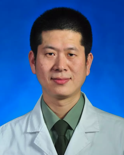 Liming Liang (梁黎明)合作论文数Chinese People's Liberation Army General Hospital13Can Zheng合作论文数12Shiyi Li合作论文数12XiaoHai Zhu (朱晓海)合作论文数12Tongtong Yan合作论文数11Kui Ma合作论文数11Qian Wu合作论文数10Xuer Sun合作论文数10Sa Jing (京萨)合作论文数10Ruiqi Bai (白瑞琪)合作论文数9Huijuan Fu合作论文数9Boya Yu合作论文数9JunNan Chen (陈俊男)合作论文数9YouTao Yu (于友涛)合作论文数8
 Zhiyong Sheng (盛志勇)合作论文数Chinese PLA General Hospital8Fanting Meng (孟凡婷)合作论文数7Yun-chao ZHOU (周云超)合作论文数7XiangBin Yuan (袁相斌)合作论文数7GuoDong Teng (滕国栋)合作论文数7
 Xiaobing Fu (付小兵)合作论文数Chinese PLA General Hospital;School of Medicine, Nankai University7Jinxiu Yang合作论文数7
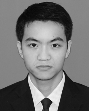 Yi Yang合作论文数6
 Ningfei Liu (刘宁飞)合作论文数Shanghai Ninth People's Hospital, Shanghai Jiao Tong University School of Medicine6XU Xiao (徐潇)合作论文数5Xin-yue BAI (白馨月)合作论文数5Lixia Zhang合作论文数5TongZhu Sun (孙同柱)合作论文数5JianMing Wu (吴建明)合作论文数5
 Songlin Yang (杨松林)合作论文数Shanghai Sixth People's Hospital Affiliated to Shanghai Jiao Tong University5Peixuan Zhang合作论文数4QingLian He (何清濂)合作论文数4XinJi Yang (杨新吉)合作论文数4Hong Wu (吴宏)合作论文数4Kang Chen (陈康)合作论文数4




