Cerebral infarction is the second leading cause of death in the world,and has become the most serious cause of disability and death in China.Acute ischemic stroke(AIS)is the most common type of cerebral infarction,accounting for about 80%of all cerebral infarctions.Hemorrhagic transformation(HT)is one of the natural regression processes in pa-tients with AIS,and is the most serious complication after treatments[such as intravenous thrombolysis(IVT)or endovascu-lar thrombectomy(EVT)],which brings a heavy burden for patients and their families and even the all society.Accurate pre-diction and evaluation are of important clinical significance.In recent years,imaging research has focused on the value of CT and MRI in evaluating HT.The diagnostic value of CT plain scan is limited.Before AIS treatment,an early diagnostic score≤7 points in the Alberta Cerebral Infarction Plan is associated with the occurrence of HT(P=0.033),and high-density middle cerebral artery sign is an independent risk factor for the occurrence of HT(OR=10.334).For AIS patients treated with thrombectomy within 2-7 days,dual energy CT scanning at 24 hours after therapy had a high efficacy for prediction of HT occurrence,with a sensitivity of 82.5%and specificity of 100%.CT angiography suggests that patients with high thrombus burden had a higher probability of developing HT(OR=1.28).The incidence of HT in AIS patients with good collateral circulation is low.CT perfusion imaging parameters,including surface permeability,cerebral blood volume,Tmax,etc.,have good predictive value for predicting HT occurence.In MRI plain scan,high signal on FLAIR can predict the occurrence of HT.The volume of high signal areas on MRI diffusion-weighted imaging can predict HT,with area under the ROC curve of 0.78.Brush like sign,and micro bleeding lesions on sensitivity weighted MRI indicate the occurrence of HT.Enhanced T1 weighted imaging of MRI shows a significant correlation between brain parenchymal enhancement and HT oc-currence(P<0.05).Perfusion weighted imaging of MRI shows a decrease in cerebral blood flow(CBV)in the infarcted area may predict HT occurence.In addition,the CT and MRI image post-processing system RAPID has improved the evaluation efficiency for HT occurrence.In the future,personalized imaging detection methods and processes should set up based on the hardware and local medical conditions of each emergency center for HT management.
Background A non-invasive tool for tumor regression grade (TRG) evaluation is urgently needed for gastric cancer (GC) treated with neoadjuvant chemotherapy (NAC). Purpose To develop and validate a radiomics signature (RS) to evaluate TRG for locally advanced GC after NAC and assess its prognostic value. Material and Methods A total of 103 patients with GC treated with NAC were retrospectively recruited from April 2018 to December 2019 and were randomly allocated into a training cohort (n = 69) and a validation cohort (n = 34). Delineation was performed on both mixed and iodine-uptake images based on dual-energy computed tomography (DECT). A total of 4094 radiomics features were extracted from the pre-NAC, post-NAC, and delta feature sets. Spearman correlation and the least absolute shrinkage and selection operator were used for dimensionality reduction. Multivariable logistic regression was used for TRG evaluation and generated the optimal RS. Kaplan–Meier survival analysis with the log-rank test was implemented in an independent cohort of 40 patients to validate the prognostic value of the optimal RS. Results Three, five, and six radiomics features were finally selected for the pre-NAC, post-NAC, and delta feature sets. The delta model demonstrated the best performance in assessing TRG in both the training and the validation cohorts (AUCs=0.91 and 0.76, respectively; P>0.1). The optimal RS from the delta model showed a significant capability to predict survival in the independent cohort ( P<0.05). Conclusion Delta radiomics based on DECT images serves as a potential biomarker for TRG evaluation and shows prognostic value for patients with GC treated with NAC.
Objectives: To prolong the survival, the value of a computed tomography-based radiomic score (RS) in stratifying survival and guiding personalized chemotherapy strategies in far-advanced gastric cancer (FGC) was investigated. Materials and Methods: This retrospective multicenter study enrolled 283 FGC patients (cT4a/bNxM0-1) from three centers. Patients from one center were randomly divided into the training (n = 166) and internal validation (n = 83) cohorts, whereas the external validation cohort (n = 34) consisted of patients from the two other centers. The RS was calculated for each patient to predict progression-free survival (PFS). Features from the primary tumor and main metastasis (peritoneum, liver, and lymph node) were integrated in the training cohort and then validated for its ability to stratify PFS and overall survival (OS) in the validation cohort. The association between the RS and efficacy of neoadjuvant intraperitoneal and systemic (NIPS) therapy was also explored. Results: The RS demonstrated a favorable prognostic ability to predict PFS in all cohorts (training: C-index 0.83, 95% confidence interval [CI]: 0.788-0.872; internal validation: C-index 0.75, 95% CI: 0.682-0.818; external validation: C-index 0.76, 95% CI: 0.669-0.851; all p < 0.05), as well as an excellent ability to stratify the PFS and OS in both the whole population and metastatic subgroups (p < 0.05). Patients with a low score were more likely to undergo surgery after perioperative chemotherapy ( p < 0.05). Furthermore, only high-scoring patients with peritoneal metastasis benefited from NIPS. Conclusion: The RS may be an effective risk stratifier for the outcomes of FGC patients and may be used to select patients who can benefit from NIPS therapy.
Objective:To investigate the impact of the deep learning reconstruction algorithm TrueFidelity TM for Gemstone Spectral Imaging (TF-GSI) and the adaptive statistical iterative reconstruction algorithm (ASiR-V, hereinafter referred to as ASiR-V) based on phantom and animal models on the image quality of dual-energy CT images. Methods:GE Revolution Apex CT was used to scan the ACR 464 phantom and a mouse model of gastric cancer with lymph node metastasis ( n=16). TF-GSI and ASiR-V were separately used to reconstruct middle and high-grade images (TF-GSI-M, TF-GSI-H, ASiR-V-50%, and ASiR-V-100%) on the phantom and mouse based on virtual monoenergetic images at 70 keV. The task transfer function (TTF) of bone and acrylic, image noise power spectrum (NPS), and detectability index (d′) of the phantom images were evaluated. One-way ANOVA analysis was used to compare the image noise, signal-to-noise ratio (SNR) and contrast-to-noise ratio (CNR) for brain and liver on images of mice. The consistency of the two reconstruction-algorithm images (TF-GSI-H and ASiR-V100%) in the detection of small lesions by two radiologists (A and B) was evaluated using kappa test. Results:In terms of the phantom, the TF-GSI-H group had the best performance in TTF, NPS, and d′. Compared to ASiR-V-100%, the TTF50% of bone and acrylic in the TF-GSI-H group increased by 2.4% and 8.9%, respectively; the NPS peak decreased by 54.1%, compared to ASiR-V-100%; the d′ of bone and acrylic in the TF-GSI-H group relative to ASiR-V-100% increased by 52.7% and 59.5%, respectively. The TF-GSI group had reduced image noise compared to the ASiR-V group, and both SNR and CNR of the two tissues increased, but the differences between the groups were not statistically significant (all P>0.05). The two reconstruction-algorithm images showed good consistency in image evaluation by the two radiologists (A, Kappa=0.875, P<0.001; B, Kappa=0.625, P=0.012). In terms of the detection of micro-metastases in mice, the TF-GSI group outperformed the ASiR-V group (average accuracy: 83.5% vs 71.9%; average sensitivity: 77.8% vs 61.2%; average specificity: 85.7% vs 85.7%). Conclusion:Compared with iterative reconstruction algorithm, the DLIR algorithm showed improved spatial resolution, reduced image noise, and enabled detectability of micro-lesion for images from dual-energy CT.
Objectives To evaluate robustness of dual-energy CT (DECT) radiomics features of virtual unenhanced (VUE) image and virtual monoenergetic image (VMI) among different imaging platforms. Methods A phantom with sixteen clinical-relevant densities was scanned on ten DECT platforms with comparable scan parameters. Ninety-four radiomic features were extracted via Pyradiomics from VUE images and VMIs at energy level of 70 keV (VMI 70keV ). Test–retest repeatability was assessed by Bland–Altman analysis. Inter-platform reproducibility of VUE images and VMI 70keV was evaluated by coefficient of variation (CV) and quartile coefficient of dispersion (QCD) among platforms, and by intraclass correlation coefficient (ICC) and concordance correlation coefficient (CCC) between platform pairs. The correlation between variability of CT number radiomics reproducibility was estimated. Results 92.02% and 92.87% of features were repeatable between scan–rescans for VUE images and VMI 70keV , respectively. Among platforms, 11.30% and 28.39% features of VUE images, and 15.16% and 28.99% features of VMI 70keV were with CV < 10% and QCD < 10%. The average percentages of radiomics features with ICC > 0.90 and CCC > 0.90 between platform pairs were 10.00% and 9.86% in VUE images and 11.23% and 11.23% in VMI 70keV . The CT number inter-platform reproducibility using CV and QCD showed negative correlations with percentage of the first-order radiomics features with CV < 10% and QCD < 10%, in both VUE images and VMI 70keV ( r 2 0.3870–0.6178, all p < 0.001). Conclusions The majority of DECT radiomics features were non-reproducible. The differences in CT number were considered as an indicator of inter-platform DECT radiomics variation. Critical relevance statement: The majority of radiomics features extracted from the VUE images and the VMI70keV were non-reproducible among platforms, while synchronizing energy levels of VMI to reduce the CT number value variability may be a potential way to mitigate radiomics instability. Graphical Abstract
Hemorrhagic transformation (HT) is one of the common complications in patients with acute ischemic stroke (AIS). This study aims to investigate the value of different thresholds of Tmax generated from perfusion-weighted MR imaging (PWI) and the apparent diffusion coefficient (ADC) value in the prediction of HT in AIS. A total of 156 AIS patients were enrolled in this study, with 55 patients in the HT group and 101 patients in non-HT group. The clinical baseline data and multi-parametric MRI findings were compared between HT and non-HT groups to identify indicators related to HT. The optimal parameters for predicting HT and the corresponding cutoff values were obtained using the receiver operating characteristic curve analysis of the volumes of ADC < 620 × 10−6 mm2/s and Tmax > 6 s, 8 s, and 10 s. The results showed that the volumes of ADC < 620 × 10−6 mm2/s and Tmax > 6 s, 8 s, and 10 s in the HT group were all significantly larger than that in the non-HT group and were all independent risk factors for HT. Early measurement of the volume of Tmax > 10 s had the highest value, with a cutoff lesion volume of 10.5 mL.
目的:探讨基于CT图像的纹理分析对CT引导下经皮肺穿刺出血并发症的预测价值.方法:回顾性分析130例行CT引导下经皮肺穿刺活检患者的术前平扫图像,术区有大于或等于2级肺出血者为有出血组,0级或1级肺出血者为无/少量出血组.首先随机选取100例作为训练组,采用MaZda软件,分别手动勾画出平扫肺窗图像上预穿刺路径周边的肺野作为感兴趣区(ROI),分别通过Fisher系数、分类错误概率联合平均相关系数(POE+ACC)、交互信息(MI)法筛选出区分出血组及无/少量出血组最具有价值的纹理特征,然后分别采用原始数据分析(RDA)、主要成分分析(PCA)、线性分类分析(LDA)和非线性分类分析(NDA)四种特征分类统计方法进行判断,结果以错判率形式表示;最后再根据得到的最优纹理参数及特征分类方法分别对另外30例图像加以验证.结果:以穿刺路径周边的肺野作为ROI时,最低错判率为11.00%(11/100),该结果出现在特征选择方法采用POE+ACC或MI,特征分类统计方法采用NDA时,以此结果进行验证的错判率分别为13.33%(4/30)和16.67%(5/30),两者差异无统计学意义.结论:分析预穿刺路径周边肺野的纹理特征有助于预测CT引导下肺穿刺并发出血的风险,为选择合适的穿刺路径以减少肺出血并发症提供依据.
目的:比较MRI不同序列组合评估子宫内膜癌宫颈间质浸润及术前分期的准确性.方法:选取128例经病理证实的子宫内膜癌患者的术前MRI,包括:T2WI、T1WI、DWI及DCE-MRI序列,不同序列组合对子宫内膜癌宫颈间质浸润、术前分期进行评估,与术后病理结果对照,计算子宫内膜癌宫颈间质浸润的灵敏度、特异度、阳性预测值、阴性预测值和准确度,并采用配对卡方检验比较不同序列组合对宫颈间质浸润及术前分期评估的准确度.结果:常规T2WI+T1WI诊断子宫内膜癌宫颈间质浸润的准确度为88.3%,DCE+T2WI为97.7%,DWI+T2WI为95.3%,DCE+T2WI诊断宫颈间质浸润的准确度要高于DWI+T2WI和T2WI+T1WI,其中DCE+T2WI与T2WI+T1WI之间差异具有统计学意义.T2WI+T1WI判断子宫内膜癌术前分期准确度为78.1%,DCE+T2WI为91.4%,DWI+T2WI为93.8%,DWI+T2WI与DCE+T2WI评估子宫内膜癌分期的准确度要明显高于T2WI+T1WI,差异具有统计学意义.结论:DWI+T2WI与DCE+T2WI评估子宫内膜癌宫颈间质浸润及术前分期具有相当的准确度,诊断效能均优于T2WI+T1WI.
目的:探究能谱CT成像定量参数在非实性肺结节空气支气管征中的价值.方法:回顾性分析我院2019年7月—2021年3月收治的264例经手术切除并被证实为肺腺癌的非实性肺结节(270个)患者,其中82个病灶出现空气支气管征,再根据其支气管走行进行分类.最后分别记录其结节最大径、浸润程度、动静脉期的能谱参数,并进行统计学分析.采用ROC曲线分析各参数预测空气支气管征分型的诊断效能.结果:82个出现空气支气管征的病灶中,27个病灶内支气管管腔走行正常(正常型),55个支气管管腔扩张、走行扭曲或闭塞(异常型).肺腺癌的浸润程度在正常型与异常型空气支气管征之间存在显著差异(P<0.05).动脉期碘含量(IC)值、动脉期及静脉期标准化碘含量(NIC)值、结节最大径在2组空气支气管征分型中出现显著差异(P<0.05).动脉期IC值、动脉期及静脉期NIC值在2组空气支气管征中均具有良好的诊断效能,ROC曲线下面积分别为0.735、0.728和0.679.结论:能谱CT成像参数在不同空气支气管征分型结节中具有显著差异性;异常空气支气管征还可以提示病灶的浸润性,更有助于诊断非实性肺结节.
目的:探讨CT对肝硬化患者食管静脉曲张(EV)诊断和严重程度评估的应用价值.方法:回顾性选择本院收治的慢性乙型病毒性肝炎肝硬化患者138例,以胃镜诊断为标准,评价CT对EV的诊断价值,分析EV的CT分级和胃镜分级的一致性.通过CT测量食管静脉直径、门静脉直径、脾静脉直径、肝脏体积、脾脏体积和肝脏体积/脾脏体积,分析上述指标对EV的诊断价值及与EV分级的相关性.结果:CT对EV的诊断灵敏度为96.4%,特异度为82.1%,阳性预测值为95.5%,阴性预测值为85.2%,准确度为93.5%.EV的CT分级和胃镜分级具有较强的一致性(Kappa值=0.638,P<0.001).不同EV分级患者的CT定量指标差异具有统计学意义(P<0.05),随着EV分级增加,食管静脉直径、门静脉直径、脾静脉直径和脾脏体积有逐渐升高的趋势,而肝脏体积和肝脏体积/脾脏体积有逐渐降低的趋势.食管静脉直径、门静脉直径、脾静脉直径、肝脏体积、脾脏体积和肝脏体积/脾脏体积均对EV有较好的诊断价值(曲线下面积分别为0.915、0.771、0.884、0.748、0.870、0.866,均P<0.001).当上述指标各取值3.015 mm、14.010 mm、8.600 mm、3103.955 cm3、905.660 cm3、2.820时,敏感度分别为100.0%、92.9%、96.4%、64.3%、89.3%、92.9%,特异度分别为73.6%、53.6%、74.5%、80.9%、79.1%、67.3%.EV分级与食管静脉直径、门静脉直径、脾静脉直径、脾脏体积呈正相关(r分别为0.794,0.566,0.744,0.719,均P<0.001),与肝脏体积、肝脏体积/脾脏体积呈负相关(r分别为-0.528,-0.770,均P<0.001).结论:CT成像可用于肝硬化患者EV诊断和严重程度评估,可作为胃镜的替代检查手段.
目的:观察分析正常人群中副中结肠动脉(AMCA)的发生率及分布规律.方法:采用回顾性研究方法,收集2021年1月至6月在上海瑞金医院北部院区同时进行上下腹增强CT检查的正常人群331例,发现存在AMCA者97例.将检查的静脉期原始图像进行多平面重建,记录AMCA起源、伴行静脉及与胰腺毗邻关系,并测量AMCA起始点直径,与胰腺最近点的距离.结果:AMCA发生率为29.3%(97/331),66.0%起源于肠系膜上动脉(64/97)、17.5%起源于肠系膜下动脉(17/97)、16.5%起源于胰十二指肠下动脉(16/97).AMCA直径为(2.0±0.24)mm,距离胰腺最近点为(2.1±1.4)mm.AMCA均可见伴行静脉,72.2%AMCA的伴行静脉汇入肠系膜下静脉(70/97),20.6%汇入肠系膜上静脉(20/97),6.2%汇入脾静脉(6/97),1%汇入空肠静脉(1/97).结论:多层螺旋CT增强扫描能清晰显示AMCA起源、伴行静脉与胰腺毗邻关系,能为临床术前提供准确的血管评估.
目的 探索基于扩散峰度成像(DKI)扫描参数的推算序列代替常规扩散加权成像(DWI)序列扫描的应用价值.方法 回顾性分析2019年11月至2020年10月瑞金医院确诊的直肠癌患者62例,其手术前2周内均行MR扫描,将DKI图像经后处理推算出b=1000 s/mm2扩散图像,两名医师分别对推算序列与常规DWI序列的b=1000s/mm2的图像进行比较.结果 DKI推算序列的主观图像质量、信噪比(SNR)、对比噪声比(CNR)及信号强度比(SIR)均优于常规DWI序列(P <0.050).两者术前T分期的准确率分别为91.94% (57/62)、74.19%(46/62),差异有统计学意义(P <0.050);N分期的准确率分别为70.97% (44/62)、66.13% (41/62),差异无统计学意义(P >0.050).结论 DKI推算序列具有代替常规DWI序列、减少扫描时间、优化扫描流程及T分期诊断效能的潜在应用价值.
目的:探讨常规MRI纹理分析在高原地区脑囊虫病和其他病因引起的颅内囊性病变的鉴别诊断价值.方法:纳入30例脑囊虫病(脑囊虫病组)和20例其他病因(对照组)引起的颅内囊性变患者的病例资料,所有患者均接受常规MRI检查进行诊断,包括T1WI、T2WI、液体衰减反转恢复(FLAIR)序列及T1WI增强.利用3D-slicer软件手动勾画囊性变囊腔的感兴趣区(ROI),并提取相应的影像组学纹理参数数据,包括第10和第90百分位数、四分位距、最小值、最大值、平均值、中位数、偏度、峰度、方差、均匀性、值域、均方根、能量和熵.使用独立样本t检验或秩和检验比较不同来源囊性变的MRI图像纹理参数的差异,并绘制受试者工作特征(ROC)曲线,评价其鉴别诊断囊性变病理来源的效能.结果:FLAIR的纹理参数对鉴别诊断价值最大,其脑囊虫组的熵、四分位距、最大值、值域、方差均显著大于对照组,而均匀性显著小于对照组(P<0.05),其中方差的ROC曲线下面积(AUC)最大,为0.845 5,其灵敏度为90.91%,特异度为80%.增强T1WI纹理参数中,脑囊虫病组的熵和四分位距显著大于对照组,而偏度和均匀性显著小于对照组(P<0.05),其中均匀性的AUC最大,为0.875,其灵敏度为100%,特异度为83.33%.T2WI纹理参数中,脑囊虫病组的峰度和均匀性都显著小于对照组(P<0.05),其中峰度的AUC最大,为0.84,其灵敏度为70%,特异度为90%.而2组的T1WI的纹理参数中未见显著差异.结论:高原藏区不同来源颅内囊性变的常规MRI纹理参数间存在显著差异,纹理分析能为脑囊虫病所致颅内囊性变的鉴别诊断提供可靠、量化的客观依据.
目的:探讨胰腺神经内分泌肿瘤(pNET)的CT征象对其侵袭性行为的预测价值.方法:回顾性分析经手术病理确诊、临床及CT资料完整、术前均行胰腺CT平扫及增强检查的120例pNET,分析病灶的部位、形态、包膜、有无囊变和钙化、有无胰管扩张、肿瘤的强化方式,测量肿瘤最大径、最短径以及实性部分平扫、动脉期及门脉期密度,并计算动脉期强化差值、门脉期强化差值.根据手术病理结果将肿瘤分为侵袭组和无侵袭组,比较两组间CT征象的差异.使用绘制受试者操作特征(ROC)曲线评价肿瘤最大径、最短径、动脉期差值和门脉期差值对pNET的侵袭性行为的预测价值.结果:侵袭组和无侵袭组pNET在发病部位、胰管扩张、囊变、钙化及强化方式无差异,在形态、完整包膜上有差异.两组在最大径、最短径、动脉期强化差值、门脉期强化差值定量特征有差异,其ROC曲线下面积为0.693、0.69、0.73和0.64,具有判别效能.对6个差异有统计学意义的影像特征通过多变量Logistic回归分析,结果显示动脉期强化差值为判断pNET侵袭性行为的独立预测因素,动脉期强化差值最佳临界点为90.1HU(灵敏度0.714,特异性0.656,阳性预测值64.5%,阴性预测值72.4%,准确率68.3%).结论:胰腺神经内分泌肿瘤体积大、形态不规则、包膜不完整或无包膜,肿瘤实性成分动脉期、门脉期强化差值低等征象提示肿瘤具有侵袭性行为,其中动脉期强化差值为pNET侵袭性行为的独立预测因素.
ObjectiveThe aim of the study was to evaluate the computed diffusion-weighted images (DWI) in image quality and diagnostic performance of rectal cancer by comparing with the acquired DWI.MethodsA total of 103 consecutive patients with primary rectal cancer were enrolled in this study. All patients underwent two DWI sequences, namely, conventional acquisition with b = 0 and 1,000 s/mm2 (aDWIb1,000) and another with b = 0 and 700 s/mm2 on a 3.0T MR scanner (MAGNETOM Prisma; Siemens Healthcare, Germany). The images (b = 0 and 700 s/mm2) were used to compute the diffusion images with b value of 1,000 s/mm2 (cDWIb1,000). Qualitative and quantitative analysis of both computed and acquired DWI images was performed, namely, signal-to-noise ratio (SNR), contrast-to-noise ratio (CNR), and signal intensity ratio (SIR), and also diagnostic staging performance. Interclass correlation coefficients, weighted κ coefficient, Friedman test, Wilcoxon paired test, and McNemar or Fisher test were used for repeatability and comparison assessment.ResultsCompared with the aDWIb1,000 images, the cDWIb1,000 ones exhibited significant higher scores of subjective image quality (all P <0.050). SNR, SIR, and CNR of the cDWIb1,000 images were superior to those of the aDWIb1,000 ones (P <0.001). The overall diagnostic accuracy of computed images was higher than that of the aDWIb1,000 images in T stage (P <0.001), with markedly better sensitivity and specificity in distinguishing T1–2 tumors from the T3–4 ones (P <0.050).ConclusioncDWIb1,000 images from lower b values might be a useful alternative option and comparable to the acquired DWI, providing better image quality and diagnostic performance in preoperative rectal cancer staging.
目的:本研究应用能谱CT扫描成像,研究胰腺导管腺癌的能谱定量多参数,探讨胰腺导管腺癌CT能谱成像的参数特点及潜在应用价值.方法:回顾性分析经病理证实为胰腺导管腺癌,并均行胰腺能谱CT动脉晚期、门静脉期增强扫描的61例患者.记录胰腺癌病灶和胰腺实质的单能量CT值、有效原子序数(Effective-Z)、碘(水)基值、水(碘)基值.采用Wilcoxon符号秩和检验(非正态分布)或配对t检验(正态分布)比较胰腺癌和胰腺实质分别在动脉晚期和门静脉期间相应上述CT能谱成像参数的差异,并绘制相应能谱曲线.结果:动脉晚期胰腺导管腺癌单能量CT值明显低于周围正常胰腺,胰腺导管腺癌门静脉期单能量CT值高于动脉期,低于周围正常胰腺,差异在低能量段相当显著;胰腺导管腺癌与胰腺实质动静脉期能量段越低差异越明显,能量段越高,差异越小;胰腺导管腺癌与胰腺实质动静脉期的单能量CT值、配对基物质碘(水)基值、Effective-Z所对应均化浓度值有显著性差异,具有统计学意义,水(碘)值差异无统计学意义.结论:胰腺导管腺癌CT能谱成像的能谱参数具有一定特征.动静脉期胰腺癌单能量CT值、碘(水)基值、有效原子序数均低于相应胰腺实质,动脉晚期低于门静脉期,能量段越低差异越明显.综合应用多能量段和能谱多参数成像有助于提高能谱CT密度分辨力,具有为小胰腺癌早诊断提供影像信息的潜在价值.
2020年新冠肺炎疫情催生人们的生产生活方式迅速变革,同时,远程影像会诊在国家政策指导下获得有力推动.但不同的运营模式产生的效益不同,如何提升运营效率、质量及降低成本,形成良性循环是远程影像会诊获得长足发展的关键.本文探讨"互联网+"时代远程影像会诊运营模式和新技术的发展,并提出相应的建议,以期为我国远程医疗的良性发展提供借鉴与参考.
目的:利用CT诊断技术对胸腺上皮性肿瘤作出明确临床分期,帮助临床对患者行术前手术评估及为术后治疗提供参考.方法:经病理证实的31例胸腺上皮肿瘤均行CT平扫及增强检查,对病变的形态、密度、强化方式、周围结构有无侵犯及种植情况进行回顾性分析,结合Masaoka-Koga临床分期评估术中所见与CT分期的一致性.结果:在31例胸腺上皮肿瘤手术病理分期中,Ⅰ期14例,Ⅱ期4例,Ⅲ期4例,Ⅳ期9例,其术前CT分期与手术病理分期高度一致.其中肿瘤形状、轮廓、大小、肿瘤内部密度、强化方式、钙化、有或没有相邻结构(纵隔胸膜、肺、心包、纵隔血管)的侵犯与Masaoka-Koga分期均相关.结论:利用CT诊断可以对胸腺上皮性肿瘤的临床分期作出准确评估,并对临床治疗包括手术方案制订提供一定的帮助.
Objective: We intend to evaluate the diagnostic efficacy of dual-energy CT radio-mics model based on Iodine Map (IM) in the application of preoperative re-staging of serosal invasion in locally advanced gastric cancer (LAGC) after neoadjuvant chemotherapy (NAC) treatment. Methods: A retrospective study was conducted on 155 patients with LAGC who were treated with standard NAC before operation (including 110 cases in training group and 45 cases in testing group). Two radiologists analyzed all the CT images and carried out the classification. After the semi-automatic drawing of region of interest volume (VOI), we extracted 1226 imaging features from each lesion based respectively on IM and 120kVp images. We adopted Spearman related analysis, Least Absolute Shrinkage and Selection Operator (LASSO) to punish Logistic regression in order to acquire important feature by getting rid of unstable and redundant features. Through multi-factor Logistic regression analysis, we established two prediction models (120kVp and IM-120kVp) based on the features selected respectively by 120kVp and 120kVp combined with IM. Results: Two radio-mics models both showed great prediction accuracy and efficiency in training and testing groups (IM-120kVp: AUC: training group, 0.953, testing group, 0.879; 120kVp: AUC: training group, 0.940, testing group, 0.831). The diagnostic accuracy of both models in the testing group (IM-120kVp: 84.4%, 120kVp: 80.0%) were higher than manual classification (68.9%). The diagnostic efficacy of IM-120kVp model was better than manual classification both in training (P<0.001) and testing groups (P=0.034). Conclusion: The radio-mics model based on dual-energy CT shows convincing diagnostic efficacy in differentiating serosal invasion in preoperative re-staging for LAGC patients after NAC treatment.
目的:研究评价能谱CT成像的功能参数在缺血性心脏病患者心功能评估方面的临床应用价值.方法:收集我院40例行心功能检查的缺血性心脏病病人,患者均行宝石能谱CT高分辨率扫描模式的回顾性心电门控冠脉CTA扫描和延迟1 min双能量能谱成像(GSI)模式扫描.在一周内行MRI心脏电影成像.在GSI浏览器能谱分析软件上获得能谱CT 40~140keV单光子能量图像及碘基物质密度图像,分别测得缺血心肌、正常心肌能谱曲线和碘浓度(IC).心功能软件自动计算出左室舒张末期容积、收缩末期容积和射血分数.将所得结果和MRI评估结果做对照分析.结果:能谱CT缺血心肌、正常心肌能谱曲线斜率(K值)之间存在显著差异,正常心肌能谱曲线斜率>缺血心肌能谱曲线斜率;缺血心肌与正常心肌之间碘浓度存在显著差异,缺血心肌碘浓度显著低于正常心肌碘浓度,能谱CT缺血心肌对应MRI电影图像均有相应节段室壁运动度减弱;能谱CT计算出的舒张末期容积、收缩末期容积和射血分数与MRI电影图像计算出的数值有较高的相关性和一致性.结论:能谱CT结合心功能分析软件能够定量评估缺血性心脏病患者的心肌供血情况以及心功能情况.
 Kemin Chen (陈克敏)合作论文数Ruijin Hospital, Shanghai Jiao Tong University School of Medicine102
 Fuhua Yan (严福华)合作论文数Zhoushan Branch, Shanghai Ruijin Hospital50
 Qi Song (宋琦)合作论文数Ruijin Hospital, Shanghai Jiao Tong University School of Medicine33
 Huan Zhang (张欢)合作论文数Ruijin Hospital, Shanghai Jiao Tong University School of Medicine30
 LianJun Du (杜联军)合作论文数29HuaWei Ling (凌华威)合作论文数27DING Bei (丁蓓)合作论文数26Lifang Pang合作论文数18Huanhuan Liu合作论文数14JingCi Xu (徐敬慈)合作论文数14WenJie Yang (杨文洁)合作论文数14Jingwen Tan合作论文数11Shi Cen合作论文数10
 Wenjie Yang (杨文洁)合作论文数School of Medicine, Shanghai Jiao Tong University10Wen-ting SU (苏文婷)合作论文数9YuLian Bo (卜玉莲)合作论文数9WenQiang Fang (方文强)合作论文数9
 Xiaozhu Lin (林晓珠)合作论文数Ruijin Hospital, School of Medicine, Shanghai Jiao Tong University9Min Rao (饶敏)合作论文数8Bo Liu合作论文数8KanMin Yao (姚侃敏)合作论文数7DaMing Wu (吴达明)合作论文数7Baisong Wang合作论文数6Jianying Li合作论文数6
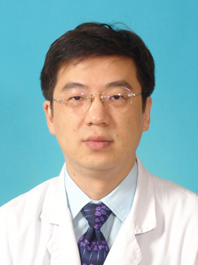 Weiwu Yao (姚伟武)合作论文数Tong Ren Hospital, Shanghai Jiao Tong University School of Medicine6
 Lan Wang (王兰)合作论文数Shanghai Center for Systems Biomedicine, Shanghai Jiao Tong University6Yingqian Ge合作论文数5Lan ZHU (朱兰)合作论文数5Yongjing Guan合作论文数5Lan Zhu (朱岚)合作论文数5FuRong Sun (孙芙蓉)合作论文数5Lei Shi (石磊)合作论文数5
 Yong Chen合作论文数Case Comprehensive Cancer Center, Case Western Reserve University;Department of Radiology, School of Medicine, Case Western Reserve University5Weihuan Fang (房炜桓)合作论文数5
 Xiaoyi Ding (丁晓毅)合作论文数Zhoushan Branch, Shanghai Ruijin Hospital5
 Hao Jiang (江浩)合作论文数Peking University People's Hospital5
 Yihan Xia合作论文数4Lingyun Wang合作论文数4Zhihan Xu合作论文数4Xiao-wei ZHAO (赵晓薇)合作论文数4CHEN Xiaoyong (陈小勇)合作论文数4Zhen-fang WU (吴振芳)合作论文数4XiaoYuan Gao (杲霄源)合作论文数4XiaoYan Chen (陈晓炎)合作论文数4HaiPeng Dong (董海鹏)合作论文数4ShaoGuang Huang (黄绍光)合作论文数4GuoXiang He (何国祥)合作论文数4




