NSI-566 is a primary allogeneic human neural stem cell line derived from a single fetal CNS. Here we present 7 years of follow-up data from a double-blind, randomized, sham-surgery controlled Phase 2a clinical study of NSI-566 intracerebral transplantation in chronic ischemic stroke patients with stable hemiparesis for 6-24 months. Method: A total of 23 patients were randomized into 2 groups where one received 7.2x10 7 NSI-566 cells in about 45 cell deposits around the 3D perimeter of MRI-defined stroke cavity in a single surgery (n=12) and the other received sham surgery (n=11) who were offered NSI-566 transplantation 12 months later. Changes in their upper and lower motor functions were assessed by Fugl-Meyer Motor Score (FMMS, normal=100) by independent blinded stroke rehabilitation specialists. Final follow-up of all available patients was carried out at 48-84 months after the NSI-566 transplantation. Results/Conclusions: Altogether 19 patients received a single dose of NSI-566 of whom 18 completed at least 12 months of follow-up. The degree of motor improvement by FMMS highly correlated with the degree of stroke cavity filling by new tissue from the 566 transplantation. Eight 566 patients showed no filling of the cavity in MRI and no significant change in FMMMS scores for the entire duration. Seven patients had the new tissue primarily within the cavity itself with little merging/integrating with the brain tissues surrounding the cavity. These patients gradually gained partial motor function with clinically meaningful 14-23 points of change from baseline several years after the transplantation. The slow motor gain presumably corresponds to the period for the new tissue from transplantation to integrate with the host brain circuitry. The remaining three patients showed almost complete filling of their cavity with new tissue by 12 months and gained 25-51 FMMS points from baseline of motor improvement that reached 76-87% of normal motor function in combined lower and upper limb movements. The upper extremity function improved up to 36 points and reached up to 89% of normal. The lower extremity function improved up to 8 points and reached up to 82% of normal. The motor improvement of patients who received NSI-566 (n=18) were statistically significant by 2-way ANOVA analysis (p<0.001) against the blinded Sham (n=11). The maximum motor improvement gained remained stable at their final follow-up visit, 74-84 months since the cell transplantation.
Cell localization plays a crucial role in pathology image analysis and is traditionally accomplished through density map regression. However, the use of broad Gaussian kernels in generating ground truth density maps often leads to susceptibility to background noise, resulting in density loss. Narrowing the Gaussian kernel could alleviate this issue, but current methods struggle with density maps generated using narrow kernels. To address this challenge, the diffusion model presents a viable solution by modeling complex distributions and maintaining stability during density map training. In this work, we explore the use of the diffusion model to recover density maps from fully Gaussian noise and present the first Diffusion model for Cell localization. Additionally, we design an Attention map Guidance mechanism that enables the diffusion model to generate higher-quality samples with a moderate guidance scale. Given the noise present at intermediate steps of the diffusion process, we also incorporate a regression branch to estimate cell counts during training. We conduct extensive experiments on several public datasets to validate the effectiveness of the proposed method. The experimental results demonstrate that the proposed method achieves notable improvements in localization and counting performance across multiple datasets.
BACKGROUND:Silent corticotroph adenomas (SCAs) are an aggressive pituitary neuroendocrine tumor (PitNET) subtype lacking effective medical therapies and showing a high rate of recurrence. The molecular mechanisms driving their proliferation remain poorly understood. Although metabolic reprogramming is a hallmark of cancer, neither lipid metabolism nor its regulatory pathways have been systematically investigated in SCAs. METHODS:We performed integrative analyses of bulk and single-cell RNA sequencing datasets from SCAs, functioning corticotroph adenomas (FCAs), and normal pituitary tissue to characterize GAL expression and associated signaling pathways. Mechanistic studies employed GAL gain- and loss-of-function models, RNA sequencing, luciferase reporter assays, lipidomics, and pharmacological inhibition. Regulation of GAL by MYCN was assessed through promoter transactivation assays. The therapeutic efficacy of SREBP1 inhibition (fatostatin) was evaluated both in vitro and in vivo. RESULTS:We identified a marked downregulation of galanin (GAL) in SCAs and demonstrated that GAL loss promotes tumor cell proliferation. GAL deficiency activated the PI3K-Akt-mTOR signaling cascade, resulting in increased activity of SREBP1, a key transcriptional regulator of lipogenesis. Enhanced fatty-acid synthesis provided metabolic support for SCA growth. We further uncovered a linear regulatory axis in which MYCN directly upregulates GAL; however, MYCN downregulation in SCAs suppresses GAL expression, thereby enabling SREBP1-driven lipogenesis. Pharmacological inhibition of SREBP1 with fatostatin reduced lipogenesis and significantly suppressed SCA growth in vitro and in vivo. CONCLUSIONS:Our findings reveal a previously unrecognized MYCN-GAL-SREBP1 lipogenic axis that drives SCA proliferation. SREBP1-dependent lipogenesis represents a promising and druggable therapeutic vulnerability for the treatment of SCAs.
BackgroundParkinson’s disease (PD) is a progressive neurodegeneration disease characterized by dopaminergic (DA) neuron loss, with chronic neuroinflammation. Subthalamic nucleus deep brain stimulation (STN-DBS) is clinically effective for the relief of parkinsonism motor symptoms. Here, we used a homemade device to investigate the effect of STN-DBS on neuroprotection and chronic neuroinflammation in a unilateral 6-hydroxydopamine (6-OHDA)-induced PD rat model.MethodMale Sprague–Dawley rats received 6-OHDA injections into the striatum, followed by ipsilateral STN electrode implantation and high-frequency stimulation. Motor function was assessed by open-field and apomorphine-induced rotation tests. DA neuron survival, glial phenotype changes, and nuclear factor (NF)-κB pathway activity in the nigrostriatal system were evaluated using Western blotting, immunofluorescence, and RT-qPCR.ResultsSTN-DBS improved motor deficits and decreased the loss of tyrosine hydroxylase-positive neurons. It promoted astrocytes presented a neuroprotective phenotype and increased expression of brain-derived neurotrophic factor, and microglia mainly presented an anti-inflammatory M2 phenotype instead of a pro-inflammatory M1 phenotype. These effects may be associated with IκB-α stabilization, the suppression of NF-κB hyperactivation, and the consequent reduction in the release of downstream pro-inflammatory cytokines.ConclusionOur findings highlight that our homemade device for STN-DBS is capable of inhibiting NF-κB, modulating glial phenotypes, mitigating neuroinflammation, and ultimately ameliorating parkinsonism deficits.
Rationale and Objectives This study aims to establish a nomogram predictive model capable of identifying high-grade tumors among IDH-mutant astrocytomas exhibiting positive T2-FLAIR mismatch sign (T2FM) before surgery. Materials and Methods We collected T2FM-positive IDH-mutant astrocytomas from three distinct centers. Using data from one center as the training set (154 cases) to establish predictive model. Data from the remaining two centers served as external validation set (29 cases) to evaluate the model’s performance. The assessment of the predictive model included the receiver operating characteristic (ROC) curve, calibration curve, decision curve analysis, area under the curve (AUC), sensitivity, and specificity. Results In the clinical features, we identified female sex (p=0.02) and the presence of enhancement within the tumor in contrast-enhanced T1-weighted imaging (p<0.001) as independent predictors of high-grade T2FM-positive IDH-mutant astrocytomas. We also found that radiomics features, such as LeastAxisLength, could aid in distinguishing WHO grades. Based on clinical and radiomics features, we developed an combined predictive model, which demonstrated superior performance compared to models relying solely on clinical or radiomics features. The combined predictive model achieved AUC of 0.819 and 0.858, sensitivity of 0.615 and 0.500, and specificity of 0.941 and 1.000, in the training and validation sets, respectively. Conclusion We developed a predictive model based on gender, contrast enhancement, and radiomics features to predict high-grade tumors among T2FM-positive IDH-mutant astrocytomas with high specificity but low sensitivity.
Background: Among primary intracranial neoplasms in adults, glioblastoma multiforme stands out for both its prevalence and its exceptionally invasive character. Uric acid-related genes (UARGs) may enhance tumor cell invasiveness and drug resistance by promoting oxidative stress responses. This study aimed to elucidate uric acid-driven mechanisms in glioblastoma, focusing on risk stratification and therapeutic vulnerability. Methods: Transcriptomic profiles of GBM were retrieved from TCGA and GEO repositories, followed by performing differentially expressed analysis, univariate Cox and LASSO regression, in order to screen prognostic UARGs and construct a risk model. Then, prognostic analyses were expanded by performing immune microenvironment analysis, drug sensitivity analysis, tumor mutation analysis, independent prognostic analysis, and nomogram construction. Additionally, dataset GSE162631 was interrogated to pinpoint pivotal cell subsets and to map intercellular communication as well as pseudo-time analysis. Results: A risk model incorporating six prognostic UARGs (TIMP1, PLAUR, CTSB, KLF10, RARRES2, and PTPRN) was constructed and identified as a favorable prognostic signature. Resting dendritic cells and drugs (including acetalax and trametinib) were found to be associated with GBM patients' risk stratification. Low-risk patients showed relatively higher mutation rates of PTEN and TP53. A nomogram was developed based on RARRES2 and PTPRN, which exhibited favorable predictive performance for GBM prognosis. Furthermore, scRNA-seq profiling identified dendritic cells (DCs), macrophages, and T cells as key populations in the tumor microenvironment. Intercellular communication inference indicated relatively strong DCs-macrophage crosstalk, and pseudo-time analysis linked prognostic UARG expression to the differentiation trajectory of critical cell subsets. Conclusions: This study identified uric acid-related genes as potential independent indicators of clinical outcomes in glioblastoma progression. A novel prognostic UARG-associated signature was developed and validated, which showed potential in predicting GBM patient outcomes.
Deep brain stimulation (DBS) is a reversible and adjustable neurostimulation technique, and has been established as a significant therapeutic modality in functional neurosurgery although its mechanisms are not yet fully understood. The primary indications for DBS include movement disorders such as Parkinson's disease (PD) and dystonia, essential tremor (ET), and Huntington's disease (HD). In recent years, its application has expanded to encompass the treatment of various other neurological and psychiatric conditions, including epilepsy, depression, obsessive-compulsive disorder (OCD), Alzheimer's disease (AD), and Tourette syndrome (TS), although the indications for DBS currently approved by the Food and Drug Administration (FDA) are PD, dystonia, ET, OCD, and epilepsy. In this review, we summarize the prevailing hypotheses regarding the mechanisms through which DBS ameliorates neurological and psychiatric disorders. We also describe the advancements in DBS for treating PD, dystonia, epilepsy, and other brain disorders. Furthermore, we outline the limitations associated with DBS treatment to date. Through this comprehensive analysis, we aim to enhance the understanding of DBS applications in neurological and psychiatric diseases and suggest potential avenues for technological advancement.
Closed head injury (CHI) provokes a prominent neuroinflammation that may lead to long-term health consequences. Microglia plays pivotal and complex roles in neuroinflammation-mediated neuronal insult and repair following CHI. We previously reported that induced neural stem cells (iNSCs) can block the effects of CXCL12/CXCR4 signaling on NF-κB activation in activated microglia by CXCR4 overexpression. Here we aim to uncover the mechanism of CXCR4 upregulation in iNSCs. We performed bioinformatic analysis to detect the differentially expressed genes in iNSCs after co-cultured with LPS-activated microglia. Subsequently, we predicted the target genes and performed gain- and loss-of-functional studies, dualluciferase reporter, RNA immunoprecipitation, biotin-coupled miRNA pulldown, fluorescence in situ hybridization and cell transplantation assays to further elucidate the mechanism underlying the immunoregulatory effects of iNSCs. Student’s t-test and one-way analysis of variance (ANOVA) with Tukey’s post hoc test were used to determine statistical significance. Our results indicated that Malat1 could act as a sponge of miR-139-5p to modulate the expression of CXCR4 that exerted significant influence on the immunoregulatory effects of iNSCs on the secretion of CXCL12, TNF-α and IGF-1 by activated microglia. Furthermore, Malat1 inhibition blocked the immunoregulatory effects of iNSC grafts on microglial activation as well as neuroinflammation in the injured cortices of CHI mice. Interestingly, NF-κB activation in iNSCs augmented the immunoregulatory effects of iNSCs on microglial activation by activating the axis of Malat1/miR-139-5p/Cxcr4. Notably, we found that TNF-α secreted by activated microglia could bind to TNFR1 at the surface of iNSCs to trigger NF-κB activation in iNSCs. In short, our findings reveal a novel role of Malat1 in the immunomodulatory effects of iNSCs on microglial activation, suggesting that transplanted iNSCs may self-perceive the changes of the activated state of microglia and thus make prudential regulation of the neuroinflammation following CHI.
PURPOSE: Glioma is a highly malignant primary neoplasm of the central nervous system. Temozolomide (TMZ) is the first-line chemotherapeutic drug for glioma, commonly employed in conjunction with radiotherapy to improve overall patient outcomes, but the resistance significantly limits its efficacy. Identifying regulatory molecular targets that influence glioma progression and TMZ sensitivity is critical improve treatment outcomes. Even though SIRT7 has been implicated in tumorigenesis, the regulatory mechanisms of SIRT7 and the role of its upstream microRNAs in glioma progression remain unclear. METHODS: We constructed SIRT7 knockdown and overexpression in glioma cells lines, and detected the tumor phenotypes. Moreover, both in - vitro and in - vivo experiments were carried out to assess the influence of SIRT7 expression levels on the treatment effectiveness of temozolomide (TMZ) in glioma. RESULTS: Our research indicated that SIRT7 is significantly over - expressed in tumor specimens obtained from glioma patients. This over - expression is associated with the tumor stage and a unfavorable prognosis. In addition, reducing the expression of SIRT7 can effectively impede the advancement of glioma cells. It has been confirmed that SIRT7 serves as a downstream target of miR-148a-3p. When there is an upregulation of miR-148a-3p, it suppresses the proliferation of glioma cells. Moreover, it causes tumor cells to become arrested in the G1 phase and stimulates cell death via apoptosis. Interestingly, SIRT7 knockdown enhanced TMZ-induced cytotoxicity in vitro and potentiated TMZ antitumor effects in glioblastoma xenografts. CONCLUSIONS: The aforementioned results suggested the miR-148a-3p/SIRT7 axis drives glioma progression and modulates TMZ sensitivity, and targeting this axis may represent a promising therapeutic approach to overcome TMZ resistance in glioma.
During inkjet bioprinting, cells are subjected to direct shear stress as they pass through the nozzles, causing reversible deformation of the cell membranes and potentially triggering subcellular changes, such as activation of molecular pathways, leading to beneficial outcomes. I n this study, neural progenitor N E-4C cells were printed through 30 mu m thermal inkjet nozzles. Compared to manually pipetted cells (control group), bioprinted cells (inkjet group) exhibited several distinct changes, such as reduced cell proliferation during the first four days after bioprinting, increased tolerance to high-concentration retinoic acid, and significantly elevated expression of the early neuronal marker class III beta-tubulin, indicating enhanced neuronal differentiation. Furthermore, RNA sequencing and enrichment analysis further revealed upregulation of cell metabolism pathways in the bioprinted group. Collectively, these findings suggest that inkjet bioprinting may be a promising strategy to accelerate neural tissue formation, warranting further studies.
Subarachnoid hemorrhage (SAH) induces multifaceted brain injuries, with white matter injury (WMI) exhibiting dual pathological features resembling traumatic brain injury and cerebral ischemia. Inflammatory responses triggered by SAH lead to extensive axonal and myelin disintegration in white matter, resulting in severe neurological dysfunction. Myelin regeneration post-injury primarily relies on promoting the differentiation of oligodendrocyte precursor cells (OPCs) into mature oligodendrocytes (OLs). We observed significant alterations in myelin basic protein (MBP) levels in human SAH brain tissues, paralleled by reduced MBP expression in rat brains post-SAH. SAH rats exhibited marked neurological deficits compared to sham group, alongside disrupted myelin integrity. Dynamic changes in OPC and OL populations were identified post-SAH. Miconazole (MCZ), an antifungal medication approved by Food and Drug Administration (FDA), has previously demonstrated neurorestorative properties. Using the mitogen-activated protein kinase (MAPK) pathway inhibitor GSK1120212, it was observed that the MAPK pathway could be effectively reverted, thereby counteracting the effects induced by MCZ. Our findings reveal that MCZ restores MBP expression, enhances OPC-to-OL differentiation, and accelerates myelin regeneration by activating MAPK signaling pathway. This provides a novel therapeutic strategy for mitigating neurological deficits in SAH patients.
The recurrence of glioma after treatment has remained an intractable problem for many years. Recently, numerous studies have explored the pivotal role of the mouse double minute 2 (MDM2)/p53 pathway in cancer treatment. Lysine phosphate phosphohistidine inorganic pyrophosphate phosphatase (LHPP), a newly discovered tumor suppressor, has been confirmed in numerous studies on tumors, but its role in glioma remains poorly understood. Expression matrices in The Cancer Genome Atlas (TCGA) and Chinese Glioma Genome Atlas (CGGA) databases were analyzed using gene set enrichment analysis (GSEA), revealing significant alterations in the p53 pathway among glioma patients with high LHPP expression. The overexpression of LHPP in glioma cells resulted in a reduction in cell proliferation, migration, and invasive ability, as well as an increase in apoptosis and alterations to the cell cycle. The present study has identified a novel inhibitory mechanism of LHPP against glioma, both in vivo and in vitro. The results demonstrate that LHPP exerts anti-glioma effects via the MDM2/p53 pathway. These findings may offer a new perspective for the treatment of glioma in the clinic.
During inkjet bioprinting, cells are subjected to shear stress directly while passing through the nozzles, which would cause reversible deformation on cell membranes. Moreover, other subcellular-level changes such as activation of gene pathways might also occur during inkjet bioprinting, leading to beneficial results. In this study, neural progenitor NE-4C cells were printed through 30μm thermal inkjet nozzles. Compared with manually pipetted cells (control group), a series of changes have occured on inkjet-bioprinted cells (inkjet group): Cell proliferation was down-regulated during the initial 4 days after bioprinting. Meanwhile, inkjet group exhibited stronger tolerance to high-concentration retinoic acid (RA). Most importantly, the expression level of early neuron marker tuj-1 was significantly higher in inkjet group, indicating the promotion of neuronal differentiation efficiency. Furthermore, RNA sequence and enrichment analysis was performed which had shown that cell-metabolism pathways were upregulated in inkjet group. These beneficial effects collectively suggested that inkjet bioprinting might be a promising strategy to accelerate neural tissue formation while further studies are performed.
People celebrate the 100th anniversary of the discovery of human EEG from various perspectives and envision its promising future. In 1948, 20 years after the discovery of human EEG, the first EEG equipment was introduced in China. A long and slow period of development followed. The number of EEG papers from China, about 20 a year, represented about 1/80 of the global total by the 70th anniversary of EEG in 1994. After a steady rise, Chinese EEG research reached about 1/3 of the global total by the 100th anniversary of EEG in 2024. Academic organizations related to EEG were established during this process, along with the widespread use of EEG in thousands of clinical hospitals and closely collaborating with international colleagues. Chinese and global EEG research are being bridged in this article to benefit mankind in the future by jointly creating more advancements in EEG technology.
Motor function recovery after complete spinal cord injury remained as a challenge in medical field, while one of the key approaches is promoting the local microenvironments. In this research, we performed a conjugated therapy by transplantation of neural stem cell (NSC) scaffolds and umbilical cord mesenchymal stem cell derived exosomes (ucMSC-exos) for the treatment of complete transactional spinal cord injury (SCI). We first demonstrated the anti-inflammatory effects of ucMSC-exos in vitro and found that ucMSC-exos could regulate microglia polarization from M1 to M2, an anti-inflammatory phenotype. Besides, ucMSC-exos also promoted NSC proliferation and neural differentiation during in vitro culturing. On the other hand, core-shell hydrogel microfibers were used as transplantation scaffolds for both small and large SCI defects. The core-shell microfibers could carry large amounts of NSCs in the core portion and the shell portion is highly permeable for nutrient and metabolite transportation. In in vivo experiments, we found that conjugated transplantation of ucMSC-exos and NSC microfibers could decreased inflammatory cytokines at lesion sites, gave rise to more neurons and promoted angiogenesis, thus comprehensively improved the local microenvironment while compared with transplantation of NSC scaffolds only. These beneficial results were in accordance with those in vitro experiments and further led to better locomotor function recovery. In summary, this research has demonstrated that that conjugated transplantation of ucMSC-exos and NSC microfibers could make a potential tool for complete SCI repair.
Ischemic stroke is a leading cause of disability and mortality, with neuroinflammation playing a key role in post-stroke injury. The molecular mechanisms remain incompletely defined. This study explored the functions of IRF7 and its downstream target ISG15 in stroke-associated neuroinflammation and prognosis of ischemic stroke. Bioinformatic analysis of transcriptomic datasets from microglia and ischemic brain tissues identified both molecules as hub genes. Their expression was validated in a mouse transient middle cerebral artery occlusion (tMCAO) model and in microglial cultures exposed to oxygen-glucose deprivation/reoxygenation (OGD/R). The effects of these molecules were assessed using siRNA knockdown, conditioned media assays with SY5Y neuronal cells, in vivo overexpression of ISG15, histological and functional assessments. Both IRF7 and ISG15 were significantly upregulated after stroke. IRF7 knockdown reduced ISG15 expression, whereas ISG15 knockdown did not affect IRF7, suggesting a unidirectional regulatory relationship. Conditioned media from microglia treated with siIRF7 or siISG15 increased SY5Y cell mortality, with a stronger effect in the siISG15 group, highlighting neuroprotective role of ISG15. ISG15 knockdown also enhanced microglial migration. Conversely, microglial ISG15 overexpression in vivo promoted a shift toward reduced neuroinflammation, improved neuronal survival, and enhanced functional recovery. Mechanistically, ISG15 stabilized NLRP3 protein but more strongly decreased its mRNA stability through accelerated degradation. These findings demonstrate that the activation of IRF7/ISG15 axis in microglia inhibits NLRP3 Expression and improve the prognosis of ischemia/reperfusion in mice, with ISG15 exerting stronger neuroprotective effects. Targeting microglial ISG15 may offer a promising therapeutic strategy for ischemic stroke. MAIN POINTS ### Competing Interest Statement The authors have declared no competing interest. * List of abbreviations : OGD/R : oxygen-glucose deprivation/reoxygenation BBB : blood-brain barrier NF-κB : nuclear factor-κB TLRs : Toll-like receptors IRFs : interferon regulatory factors LPS : lipopolysaccharide CpG-ODN : CpG oligodeoxynucleotides tMCAO : transient middle cerebral artery occlusion CCA : right common carotid artery ECA : external carotid artery ICA : internal carotid artery MCA : middle cerebral artery TTC : triphenyltetrazolium chloride PFA : paraformaldehyde PMSF : phenylmethylsulfonyl fluoride TBST : Tris-buffered saline with Tween 20 NCM : normal culture medium PS : Penicillin-Streptomycin PPI : Protein-protein interaction ISG15⁻/⁻ : ISG15 knockout HGF : hepatocyte growth factor DMEM : Dulbecco’s Modified Eagle’s Medium FBS : fetal bovine serum DEGs : Differentially expressed genes GO : Gene Ontology KEGG : Kyoto Encyclopedia of Genes and Genomes GSEA : Gene set enrichment analysis SD : standard deviation pIRF7 : phosphorylated IRF7 Il-1βmat : active form of Il-1β GSDMDmat : active form of GSDMD GSDMDpre : precursor of GSDMD NC : negative control CHX : cycloheximide TBI : traumatic brain injury siISG15 : siRNA targeting ISG15 siIRF7 : siRNA targeting IRF7 Sichuan Provincial Science and Technology Program Project, 2023NSFSC0643, 2023YFS0050, 2024YFHZ0009
OBJECTIVE:This study aimed to develop an advanced method for preoperative planning and surgical guidance using open-source artificial intelligence (AI)-assisted rapid 3D color multimodal image fusion (MIF) and augmented reality (AR) in extracerebral tumor surgical procedures. METHODS:In this prospective trial of 130 patients with extracerebral tumors, the authors implemented a novel workflow combining FastSurfer (AI-based brain parcellation), Raidionics-Slicer (deep learning tumor segmentation), and Sina AR projection. Comparative analysis between AI-assisted 3D-color MIF (group A) and manual-3D-monochrome MIF (group B) was conducted, evaluating surgical parameters (operative time, blood loss, resection completeness), clinical outcomes (complications, hospital stay, modified Rankin Scale [mRS] scores), and technical performance metrics (processing time, Dice similarity coefficient [DSC], 95% Hausdorff distance [HD]). RESULTS:The AI-3D-color MIF system achieved superior technical performance with brain segmentation in 1.21 ± 0.13 minutes (vs 4.51 ± 0.15 minutes for manual segmentation), demonstrating exceptional accuracy (DSC 0.978 ± 0.012 vs 0.932 ± 0.029; 95% HD 1.51 ± 0.23 mm vs 3.52 ± 0.35 mm). Clinically, group A demonstrated significant advantages with shorter operative duration, reduced intraoperative blood loss, higher rate of gross-total resection, lower complication incidence, and better postoperative mRS scores (all p < 0.05). CONCLUSIONS:The integration of open-source AI tools (FastSurfer/Raidionics) with AR visualization creates an efficient 3D-color MIF workflow that enhances anatomical understanding through color-coded functional mapping and vascular relationship visualization. This system significantly improves surgical precision while reducing perioperative risks, representing a cost-effective solution for advanced neurosurgical planning in resource-constrained settings.
Background: Approximately 8.94%-44.44% of nonenhancing adult-type diffuse gliomas are identified as glioblastomas. Our purpose is to develop a nomogram that can predict glioblastomas from nonenhancing adult-type diffuse gliomas. Methods: Nonenhancing adult-type diffuse gliomas were collected from Beijing Tiantan Hospital and TCIA public database. Univariate and multivariate logistic regression were performed to screen features on the training set. The features with P < .05 in multivariate logistic regression were used to establish the prediction model. The testing and validation sets were used to test the model. Results: A total of 557 and 67 nonenhancing adult-type diffuse gliomas were collected from Beijing Tiantan Hospital and TCIA, respectively. The T2-FLAIR mismatch sign exhibited 100% specificity but low sensitivity (<30%) in ruling out glioblastoma. Age, tumor location, rADC((kurtosis)), and rADC((median)) were identified as independent predictors and employed for developing the prediction model. The AUC of the model was 0.901, 0.861, and 0.945 in the training, testing, and validation set, respectively. The best cutoff value of nomoscore was 138.5, which achieved sensitivity of 0.935, 0.714, and 0.895, specificity of 0.777, 0.782, and 0.8775 in the training, testing, and validation sets, respectively. Survival analysis shown that patients with nomoscore above 138.5 had significantly poorer survival time than those with scores below 138.5. Conclusions: Positive T2-FLAIR mismatch sign can effectively rule out glioblastoma in nonenhancing adult-type diffuse gliomas with high specificity. Nonenhancing adult-type diffuse gliomas with nomoscore above 138.5 are highly suspicious for glioblastoma or nonglioblastoma with a poor prognosis.
 Xiaodan Jiang (姜晓丹)合作论文数Zhujiang Hospital of Southern Medical University265
 Yiquan Ke (柯以铨)合作论文数南方医科大学珠江医院神经外科138
 Yingqian Cai (蔡颖谦)合作论文数120MouXuan Du (杜谋选)合作论文数88Yu-Xi Zou合作论文数88Shizhong Zhang合作论文数83LiHua Chen (陈立华)合作论文数60
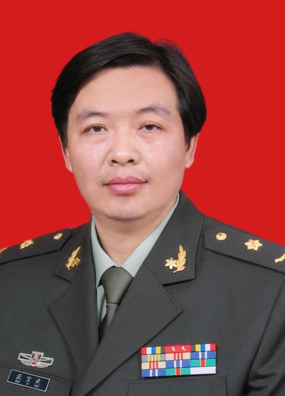 Yiwu Dai (戴宜武)合作论文数The General Hospital of the People's Liberation Army (PLAGH)60TieLin Li (李铁林)合作论文数47
 Chuanzhi Duan (段传志)合作论文数ZhuJiang Hospital of Southern Medical University47
 ZhiJun Yang (杨志军)合作论文数Chinese PLA General Hospital44
 Hongdian Zhang (张洪钿)合作论文数Chinese PLA General Hospital42Hongtian Zhang合作论文数41ZhenZhou Chen (陈镇洲)合作论文数40Wenjin Chen (陈文锦)合作论文数38Wangming Zhang (张旺明)合作论文数36ZhiLin Yang (杨志林)合作论文数36Gao Mou合作论文数34YanWu Guo (郭燕舞)合作论文数33QiuJing Wang (汪求精)合作论文数32YunJun Li (李运军)合作论文数32Hao Zhao (赵浩)合作论文数Chinese Pla Medical School, Chinese Pla General Hospital32ChunSen Shen (沈春森)合作论文数30Qiang Xu (徐强)合作论文数30WenDe Li (李文德)合作论文数29
 Qun Wei (魏群)合作论文数Chinese PLA General Hospital29Zhu Jian-Wei合作论文数28
 Qinghua Wang (王清华)合作论文数南方医科大学珠江医院27Zhaotao Wang合作论文数26Yizhao Chen合作论文数26
 Xiangyu Wang (王向宇)合作论文数The First Affiliated Hospital of Jinan University26
 JiangHong He (何江弘)合作论文数Chinese PLA General Hospital25Zhiliang Liu (刘智良)合作论文数25Kai Sun (孙恺)合作论文数24GuLan Zhou (周谷兰)合作论文数23ChengYi Luo (罗成义)合作论文数23Chunyang Liang (梁春阳)合作论文数21YongChun Luo (罗永春)合作论文数20Yigang Yu (郁毅刚)合作论文数School of Medicine, Xiamen University;Nanchang University;Gannan Medical University20Ping Peng (彭苹)合作论文数19JinBao Gao (高进宝)合作论文数19Bin Yu (于斌)合作论文数19Dang Yuan-Yuan合作论文数19Ling-Sha Qin合作论文数19m y zhang合作论文数17Li-Hua Chen合作论文数17Qin Dong合作论文数17Jia Weiqiang合作论文数17
 Yi Yang (杨艺)合作论文数陆军总医院17




