There is a growing urgency to develop effective strategies for studying and targeting glioblastoma including, which are crucial for tumor initiation and resistance to therapy. Human-immunodeficiency-virus-type-1-based viral particles have emerged as valuable tools in neurology, aiding in the exploration of neural development and tumorigenesis, with promising clinical applications on the horizon. We targeted proteolipid plasmolipin, found on glial cell surfaces, acting as an entry receptor for the McERV endogenous murine retrovirus, with replication-defective vectors carrying either a fluorescent marker or the suicide gene TK.007, pseudotyped with the McERV envelope protein. We evaluated these vectors’ efficiency in delivering genes to glioblastoma and neuronal cells both in vitro and in vivo, using intracranial glioblastoma and subcutaneous neuroblastoma xenograft tumors. Our findings confirm that aiming plasmolipin allows efficient and selective elimination of tumors induced by plasmolipin-positive cancer cells while sparing normal tissue. This work highlights the potential of McERV-pseudotyped viral particles as a promising therapeutic strategy for targeting glioblastoma and neuronal tumors.
Настоящая статья посвящена научному обоснованию выявления и анализу причинно-следственных связей фундаментальных градостроительных проблем развития города Бишкека, выработке эффективных проектных решений их преодоления, формированию устойчивой концептуально-градостроительной модели перспективного развития в новом проекте цифрового генерального плана города. Реализация документа обеспечит создание безопасной, экономически эффективной и комфортной городской среды, усилит столичные функции Кыргызской Республики города Бишкека как делового, финансового и культурного центра Азии.
Background: Histone deacetylases (HDACs) are critical epigenetic modulators involved in regulating various molecular mechanisms essential for cell development and growth. Alterations in HDAC activity have been linked to the progression of numerous cancers, including lymphoma. Over the past decade, the FDA has approved several HDAC inhibitors for lymphoma treatment, leading to heightened interest in this emerging class of drugs. Methods: In our research, we developed a novel HDAC inhibitor that exhibits high selectivity for class I HDACs. Results: Our in vitro findings indicate that treating lymphoma/leukemia cells with this inhibitor results in a marked suppression of cell growth and promotes apoptosis, while leaving the cell cycle unaffected. Conclusions: We propose that our new inhibitor, named eimbinostat, holds significant promise as a potential therapeutic agent for the treatment of hematologic malignancies such as lymphoma or leukemia.
Proteasomes hydrolyze most intracellular proteins. Immunoproteasome is a form of proteasome implicated in inflammation, cancer and autoimmune diseases. Modulation of immunoproteasome activity is a promising approach against several pathologies. Using previously obtained reporter colorectal cancer cell lines, we tested how commonly used compounds including ibuprofen, acetylsalicylic acid, vitamin C, caffeine and others, affect immunoproteasome expression. Flow cytometry, qPCR and Western blot were used to evaluate immunoproteasome subunit expression. Proteasome activity was tested using fluorogenic substrates and the activity-based probe. Transcriptome analysis was performed to identify patterns of gene expression changes. Interestingly, caffeine was the only drug that stimulated modest reduction in quantity of immunoproteasomes. The effect of caffeine varied between cell lines and was stronger as a result of prolonged treatment. The reduction of immunoproteasome content in cells coincided with decreased expression of immunoproteasome subunits, genes encoding the Nrf3 transcription factor and a PAC4 proteasome assembly chaperone, as well as the reduced levels of oxidative stress. Caffeine did not affect the degradation of immunoproteasomes by autophagy. Obtained results uncover novel biological effects of caffeine, our data might help to optimize existing and develop new strategies for the treatment of colorectal cancer and several autoimmune diseases.
Although every cell biologist knows the importance of selecting the right growth conditions and it is well known that the composition of growth medium may vary depending on a product brand or lot affecting many cellular processes, still those effects are poorly systematized. We addressed this issue by comparing the effect of 12 fetal bovine sera (FBS) and eight growth media from different brands on the morphological and functional parameters of five cell types: lung adenocarcinoma, neuroblastoma, glioblastoma, embryonic kidney, and colorectal cancer cells. Using high-throughput imaging, we compared cell proliferation; performed morphological profiling based on the imaging of 561,519 cells; measured extracellular regulated kinases (ERK1/2) activity, mitochondria potential, and lysosome accumulation; and compared cell sensitivity to drugs, response to EGF stimulation, and ability to differentiate. We found that changes in cell proliferation and morphology were independent, and morphological changes were associated with differences in mitochondria potential or the cell’s ability to differentiate. Surprisingly, the most drastic differences were detected in serum-free conditions, where medium choice affected cell survival and response to EGF. Overall, our data may be used to improve the reproducibility of experiments involving cell cultures, and the effects of 28 growth conditions on proliferation and 44 morphological parameters can be explored through a Shinyapp.
Background: Proteasomes degrade intracellular proteins. Different proteasome forms were identified. Proteasome inhibitors are used in cancer therapy, and novel drugs directed to specific proteasome forms are developed. Breast cancer (BC) therapy depends on the subtype of the tumor, determined by the expression level of Ki67, HER-2, estrogen and progesterone receptors. Relationships between the presence of specific proteasome forms and proteins that determine the BC subtype remain unclear. Here, using gene expression data in 19,145 tumor samples from 144 datasets and tissues from 159 patients with different subtypes of BC, we investigated the association between the activity and expression of proteasomes and levels of BC subtype markers. Methods: Bioinformatic analysis of proteasome subunit (PSMB1-10) gene expression in BC was performed. Proteasome heterogeneity in BC cell lines was investigated by qPCR. By Western blotting, proteasome composition was assessed in cells and patient tissue lysates. Proteasome activities were studied using fluorogenic substrates. BC molecular subtypes were determined by immunohistochemistry. Results: BC subtypes demonstrate differing proteasome subunit expression pattern and strong PSMB8-10 co-correlation in tumors. A significant increase in chymotrypsin- and caspase-like proteasome activities in BC compared to adjacent tissues was revealed. The subunit composition of proteasomes in tumor tissues of BC subtypes varied. Regression analysis demonstrated a positive correlation between proteasome activities and the expression of Ki67, estrogen receptors and progesterone receptors. Conclusion: BC subtypes demonstrate differences within the proteasome pool. Correlations between the proteasome activity, hormone receptors and Ki67 indicate possible mutual influence. Obtained results facilitate development of novel drug combinations for BC therapy.
Imaging-based anticancer drug screens are becoming more prevalent due to development of automated fluorescent microscopes and imaging stations, as well as rapid advancements in image processing software. Automated cell imaging provides many benefits such as their ability to provide high-content data, modularity, dynamics recording and the fact that imaging is the most direct way to access cell viability and cell proliferation. However, currently most publicly available large-scale anticancer drugs screens, such as GDSC, CTRP and NCI-60, provide cell viability data measured by assays based on colorimetric or luminometric measurements of NADH or ATP levels. Although such datasets provide valuable data, it is unclear how well drug toxicity measurements can be integrated with imaging data. Here we explored the relations between drug toxicity data obtained by XTT assay, two quantitative nuclei imaging methods and trypan blue dye exclusion assay using a set of four cancer cell lines with different morphologies and 30 drugs with different mechanisms of action. We show that imaging-based approaches provide high accuracy and the differences between results obtained by different methods highly depend on drug mechanism of action. Selecting AUC metrics over IC50 or comparing data where significantly drugs reduced cell numbers noticeably improves consistency between methods. Using automated cell segmentation protocols we analyzed mitochondria activity in more than 11 thousand drug-treated cells and showed that XTT assay produces unreliable data for CDK4/6, Aurora A, VEGFR and PARP inhibitors due induced cell size growth and increase in individual mitochondria activity. We also explored several benefits of image-based analysis such as ability to monitor cell number dynamics, dissect changes in total and individual mitochondria activity from cell proliferation, and ability to identify chromatin remodeling drugs. Finally, we provide a web tool that allows comparing results obtained by different methods.
The efficiency of human immunodeficiency virus-1 (HIV-1) inhibition by sulfated polysaccharides isolated from the various families of red algae of the Far East Pacific coast were studied. The anti-HIV-1 activity of kappa and lambda-carrageenans from Chondrus armatus, original highly sulfated X-carrageenan with low content of 3,6-anhydrogalactose from Tichocarpus crinitus and i/κ–carrageenan with hybrid structure isolated from Ahnfeltiopsis flabelliformis was found. The antiviral action of these polysaccharides and its low-weight oligosaccharide was compared with commercial κ-carrageenan. Here we used the HIV-1-based lentiviral particles and evaluated that these carrageenans in non-toxic concentrations significantly suppress the transduction potential of lentiviral particles pseudotyped with different envelope proteins, targeting cells of neuronal or T-cell origin. The antiviral action of these carrageenans was confirmed using the chimeric replication competent Mo-MuLV (Moloney murine leukemia retrovirus) encoding marker eGFP protein. We found that X-carrageenans from T. crinitus and its low weight derivative and λ-carrageenan from C. armatus effectively suppress the infection caused by retrovirus. The obtained data suggest that the differences in the suppressive effect of carrageenans on the transduction efficiency of HIV-1 based lentiviral particles may be related to the structural features of the studied polysaccharides.
Background: In the twentieth century, the textbook idea of packaging genomic material in the cell nucleus and metaphase chromosomes was the presence of a hierarchy of structural levels of chromatin organization: nucleosomes – nucleosomal fibrils –30 nm fibrils – chromomeres – chromonemata – mitotic chromosomes. Chromomeres were observed in partially decondensed chromosomes and interphase chromatin as ~100 nm globular structures. They were thought to consist of loops of chromatin fibres attached at their bases to a central protein core. However, Hi-C and other related methods led to a new concept of chromatin organization in the nuclei of higher eukaryotes, according to which nucleosomal fibrils themselves determine the spatial configuration of chromatin in the form of topologically associating domains (TADs), which are formed by a loop extrusion process and are regions whose DNA sequences preferentially contact each other. Somatic macronuclei of ciliates are transcriptionally active, highly polyploid nuclei. A feature of macronuclei is that their genome is represented by a large number of “gene-sized” (~1–25 kb) or of “subchromosomal” (~50–1700 kb) size minichromosomes. The inactive macronuclear chromatin of “subchromosomal” ciliates usually looks like bodies 100–200 nm in size. The aim of this work was to find out which of the models (chromomeres or TADs) is more consistent with the confocal and electron microscopic data on structural organization of chromatin bodies. Methods: Macronuclear chromatin of four “subchromosomal” ciliate species (Bursaria truncatella, Paramecium multimicronucleatum, Didinium nasutum, Climacostomum virens) was examined using electron microscopy and confocal microscopy during regular growth, starvation and encystment. Results: Chromatin bodies ~70–200 nm in size observed in the interphase macronuclei consisted of tightly packed nucleosomes. Some of them were interconnected by one or more chromatin fibrils. Under hypotonic conditions in vitro, chromatin bodies decompacted, forming rosette-shaped structures of chromatin fibrils around an electron-dense centre. When the activity of the macronucleus decreased during starvation or encystment, chromatin bodies assembled into chromonema-like fibrils 100–300 nm thick. This data allows us to consider chromatin bodies as analogues of chromomeres. On the other hand, most likely, the formation of DNA loops in chromatin bodies occurs by the loop extrusion as in TADs. Conclusions: The data obtained is well explained by the model, according to which the chromatin bodies of ciliate macronuclei combine features inherent in both chromomeres and TADs; that is, they can be considered as chromomeres with loops packed in the same way as the loops in TADs.
Fascaplysin is a red cytotoxic pigment with anticancer properties isolated from the marine sponge Fascaplysinopsis sp.. Recently, structure-activity relationship analysis reported by our group suggested that selective cytotoxicity of fascaplysin derivatives towards tumor cells negatively correlates with their ability to intercalate into DNA. To validate this hypothesis, we synthesized 6- and 7- tert -butylfascaplysins which reveal mitigated DNA-intercalating properties. These derivatives were found to be strongly cytotoxic to drug-resistant human prostate cancer cells, albeit did not demonstrate improved selectivity towards cancer cells when compared to fascaplysin. At the same time, kinome analysis suggested an activation of CHK1/ATR axis in cancer cells shortly after the drug exposure. Further experiments revealed induction of replication stress that is eventually converted to the toxic DNA double-strand breaks, resulting in caspase-independent apoptosis-like cell death. Our observations highlight new DNA-targeting effect of some fascaplysin derivatives and indicate more complex structure-activity relationships within the fascaplysin family, suggesting that cytotoxicity and selectivity of these alkaloids are influenced by multiple factors. Furthermore, combination with clinically-approved inhibitors of ATR/CHK1 as well as testing in tumors particularly sensitive to the DNA damage should be considered in further studies.
Autophagy is the process by which damaged regions of the cytoplasm and intracellular pathogens are degraded. This mechanism often serves an adaptive role in cells, enhancing their survival. It plays a direct or indirect role in the development of various pathological conditions within the body. This phenomenon is common in various malignant diseases, where autophagy is associated with the resistance of transformed cells to chemotherapy. Conversely, abnormal activation of autophagy can trigger cell death, a process often seen in neurodegenerative conditions. Given that dysregulation of autophagy is associated with the progression of numerous pathological conditions, this is of significant interest to the developers of drugs that can effectively modulate autophagy for both basic research and clinical applications. Here, we provide a brief description of the mechanism of macroautophagy, the most prevalent form of autophagy identified in humans. We also discuss the clinical potential of drugs that can modulate autophagy, highlighting their use in combating diseases associated with direct or indirect dysregulation of this essential process.
Introduction: Proteasomes are multi-subunit protein complexes responsible for protein degradation in cells. Immunoproteasomes and intermediate proteasomes (together non-constitutive proteasomes) are specific forms of proteasomes frequently associated with immune response, antigen presentation, inflammation and stress. Expression of non-constitutive proteasome subunits has a prognostic value in several types of cancer. Thus, factors that modulate non-constitutive proteasome expression in tumors are of particular interest. Multikinase inhibitors (MKIs) demonstrate promising results in treatment of cancer. At the same time, their immunomodulatory properties and effects on non-constitutive proteasome expression in colorectal cancer cells are poorly investigated.Methods: Proteasome subunit expression in colorectal cancer was evaluated by bioinformatic analysis of available datasets. Two colorectal cancer cell lines, expressing fluorescent non-constitutive proteasomes were treated with multikinase inhibitors: regorafenib and sorafenib. The proteasome subunit expression was assessed by real-time PCR, Western blotting and flow cytometry. The proteasome activity was studied using proteasome activity-based probe and fluorescent substrates. Intracellular proteasome localization was revealed by confocal microscopy. Reactive oxygen species levels following treatment were determined in cells. Combined effect of proteasome inhibition and treatment with MKIs on viability of cells was estimated.Results: Expression of non-constitutive proteasomes is increased in BRAF-mutant colorectal tumors. Regorafenib and sorafenib stimulated the activity and synthesis of non-constitutive proteasomes in examined cell lines. MKIs induced oxidative stress and redistribution of proteasomes within cells. Sorafenib stimulated formation of cytoplasmic aggregates, containing proteolyticaly active non-constitutive proteasomes, while regorafenib had no such effect. MKIs caused no synergistic action when were combined with the proteasome inhibitor.Discussion: Obtained results indicate that MKIs might affect the crosstalk between cancer cells and immune cells via modulation of intracellular proteasome pool. Observed phenomenon should be considered when MKI-based therapy is applied.
5 '-Noraristeromycin as a racemic mixture of enantiomers was found to exhibit a pronounced cy- totoxic effect on leukemia cells; IC50 for the Jurkat, K562, and THP-1 cell lines was 7.3, 1.3, and 3.7 mu M, re- spectively. The general toxicity of 5'-noraristeromycin was studied in experiments on white mice upon single-dose intragastric administration; toxicometric parameters were determined, and the clinical and patho- morphological presentation of acute intoxication was studied. LD50 of the substance was shown to be 63.2 (52.7 divided by 75.8) mg/kg; LD16, 44.7 mg/kg, and LD84, 89.4 mg/kg. Administration of the substance at a dose within the studied dose range is accompanied by systemic damage to the internal organs and tissues of the experimental animals.
Nucleoside reverse transcriptase inhibitors are the first class of drugs to be approved by the FDA for the suppression of HIV-1 and are widely used for this purpose in combination with drugs of other classes. Despite the progress in HIV-1 treatment, there is still the need to develop novel efficient antivirals. Here the efficiency of HIV-1 inhibition by a set of original 5-substituted uridine nucleosides was studied. We used the replication deficient human immunodeficiency virus (HIV-1)-based lentiviral particles and identified that among the studied compounds, 2′,3′-isopropylidene-5-iodouridine was shown to cause anti-HIV-1 activity. Importantly, no toxic action of this compound against the cells of T-cell origin was found. We determined that this compound is significantly more efficient at suppressing HIV-1 compared to Azidothymidine (AZT) when taken at the high non-toxic concentrations. We did not find any profit when using AZT in combination with 2′,3′-isopropylidene-5-iodouridine. 2′,3′-Isopropylidene-5-iodouridine acts synergistically to repress HIV-1 when combined with the CDK4/6 inhibitor Palbociclib in low non-toxic concentration. No synergistic antiviral action was detected when AZT was combined with Palbociclib. We suggest 2′,3′-isopropylidene-5-iodouridine as a novel perspective non-toxic compound that may be used for HIV-l suppression.
The long-read RNA sequencing developed by Oxford Nanopore Technology provides a direct quantification of transcript isoforms. That makes the number of transcript isoforms per gene an intrinsically suitable metric for alternative splicing (AS) profiling in the application to this particular type of RNA sequencing. By using this simple metric and recruiting principal component analysis (PCA) as a tool to visualize the high-dimensional transcriptomic data, we were able to group biospecimens of normal human liver tissue and hepatocyte-derived malignant HepG2 and Huh7 cells into clear clusters in a 2D space. For the transcriptome-wide analysis, the clustering was observed regardless whether all genes were included in analysis or only those expressed in all biospecimens tested. However, in the application to a particular set of genes known as pharmacogenes, which are involved in drug metabolism, the clustering worsened dramatically in the latter case. Based on PCA data, the subsets of genes most contributing to biospecimens’ grouping into clusters were selected and subjected to gene ontology analysis that allowed us to determine the top 20 biological processes among which translation and processes related to its regulation dominate. The suggested metrics can be a useful addition to the existing metrics for describing AS profiles, especially in application to transcriptome studies with long-read sequencing.
Autophagy is the process of lysosomal elimination of the cell organelles, cytoplasmic sites, and pathogenic microorganisms that enter the cell. This process is associated with both cell death regulation and an increase in cell survival chances. Autophagy is involved in the development of various diseases (Crohn disease, cancer, atherosclerosis, etc.). For these reasons, it is of significant interest to establish the molecular targets involved in autophagy regulation and the factors that mediate its participation in pathogenesis. The review describes the potential molecular mechanisms involved in the regulation of autophagy, its contribution to the vital cell activity in a healthy organism, and pathologies.
Fascaplysin is a marine alkaloid which is considered to be a lead drug candidate due to its diverse and potent biological activity. As an anticancer agent, fascaplysin holds a great potential due to the multiple targets affected by this alkaloid in cancer cells, including inhibition of cyclin-dependent kinase 4 (CDK4) and induction of intrinsic apoptosis. At the same time, the studies on structural optimization are hampered by its rather high toxicity, mainly caused by DNA intercalation. In addition, the number of methods for the syntheses of its derivatives is limited. In the current study, we report a new two-step method of synthesis of fascaplysin derivatives based on low temperature UV quaternization for the synthesis of thermolabile 9-benzyloxyfascaplysin and 6-tert-butylfascaplysin. 9-Benzyloxyfascaplysin was used as the starting compound to obtain 9-hydroxyfascaplysin. However, the latter was found to be chemically highly unstable. 6-tert-Butylfascaplysin revealed a significant decrease in DNA intercalation when compared to fascaplysin, while cytotoxicity was only slightly reduced. Therefore, the impact of DNA intercalation for the cytotoxic effects of fascaplysin and its derivatives needs to be questioned.
The flurry of publications devoted to the functions of long non-coding RNAs (lncRNAs) published in the last decade leaves no doubt about the exceptional importance of lncRNAs in various areas including tumor biology. However, contribution of lncRNAs to the early stages of oncogenesis remains poorly understood. In this study we explored a new role for lncRNAs: stimulation of specific chromosomal rearrangements upon DNA damage. We demonstrated that lncRNA CASTL1 (ENSG00000269945) stimulates the formation of the CCDC6-RET inversion (RET/PTC1) in human thyroid cells subjected to radiation or chemical DNA damage. Facilitation of chromosomal rearrangement requires lncRNA to contain regions complementary to the introns of both CCDC6 and RET genes as deletion of these regions deprives CASTL1 of the ability to stimulate the gene fusion. We found that CASTL1 expression is elevated in tumors with CCDC6-RET fusion which is the most frequent rearrangement in papillary thyroid carcinoma. Our results open a new venue for the studies of early oncogenesis in various tumor types, especially those associated with physical or chemical DNA damage.
The long-read RNA sequencing developed by Oxford Nanopore Technologies provides a direct quantification of transcript isoforms, thereby making it possible to present alternative splicing (AS) profiles as arrays of single splice variants with different abundances. Additionally, AS profiles can be presented as arrays of genes characterized by the degree of alternative splicing (the DAS—the number of detected splice variants per gene). Here, we successfully utilized the DAS to reveal biological pathways influenced by the alterations in AS in human liver tissue and the hepatocyte-derived malignant cell lines HepG2 and Huh7, thus employing the mathematical algorithm of gene set enrichment analysis. Furthermore, analysis of the AS profiles as abundances of single splice variants by using the graded tissue specificity index τ provided the selection of the groups of genes expressing particular splice variants specifically in liver tissue, HepG2 cells, and Huh7 cells. The majority of these splice variants were translated into proteins products and appeal to be in focus regarding further insights into the mechanisms underlying cell malignization. The used metrics are intrinsically suitable for transcriptome-wide AS profiling using long-read sequencing.
Chloroquine and Emetine are drugs used to treat human parasitic infections. In addition, it has been shown that these drugs have an antiviral effect. Both drugs were also found to cause a suppressive effect on the growth of cancer cells of different origins. Here, using the replication-deficient HIV-1-based lentiviral vector particles, we evaluated the ability of the combination of these drugs to reduce viral transduction efficiency. We showed that these drugs act synergistically to decrease cancer cell growth when added in combination with medium containing lentiviral particles. We found that the combination of these drugs with lentiviral particles decreases the viability of treated cells. Taken together, we state the oncolytic potential of the medium containing HIV-1-based particles provoked by the combination of Chloroquine and Emetine.
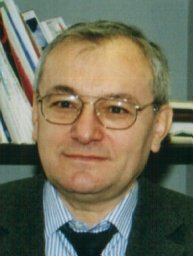 Vladimir S. Prassolov合作论文数Engelhardt Institute of Molecular Biology, Russian Academy of Sciences84
 Timofey Lebedev合作论文数49V. S. Prasolov合作论文数29P. M. Rubtsov合作论文数Engelhardt Institute of Molecular Biology, Russian Academy of Sciences28N. N. Orlova合作论文数Prokhorov General Physics Institute, Russian Academy of Sciences18Elmira Vagapova合作论文数16Carol Stocking合作论文数University of Hamburg16
 Anton Buzdin合作论文数I.M. Sechenov First Moscow State Medical University;European Organisation for Research and Treatment of Cancer;OmicsWay Corp;Institute of Bioorganic Chemistry12N. A. Nikitenko合作论文数10
 S N Kochetkov合作论文数Engelhardt Institute of Molecular Biology, Russian Academy of Sciences10
 Boris Fehse合作论文数University Medical Center Hamburg-Eppendorf;Dept. of Stem Cell Transplantation, Hamburg University;Research Dept. Cell and Gene Therapy, Hamburg University10V. I. Popenko合作论文数9Vladimir I. Popenko合作论文数Institute of Molecular Biology;Russian Academy of Sciences9
 Sorokin Maxim I合作论文数8A S Gornostaeva合作论文数7O. G. Leonova合作论文数7М. М. Прокофьева合作论文数7
 Elena Shyrokova合作论文数Department of Experimental Medicine, University of Lund7Suntsova Maria V合作论文数7Prokofjeva Maria M合作论文数7Morozov Alexey V合作论文数7Irina Yu. Petrushanko合作论文数Engelhardt Institute of Molecular Biology, Russian Academy of Sciences7
 Vladimir Mitkevich合作论文数Engelhardt Institute of Molecular Biology, Russian Academy of Sciences7
 Alexander A. Makarov合作论文数Utrecht University;Thermo Fisher Scientific7
 Riecken Kristoffer合作论文数Dept Stem Cell Transplantat, Univ Med Ctr Hamburg Eppendorf6Olga G. Leonova合作论文数6M. M. Prokofjeva合作论文数5D. Baskaran合作论文数5
 Alexander Burov合作论文数5
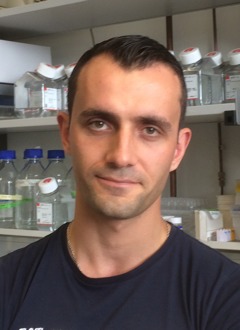 Sergey A. Dyshlovoy合作论文数Department of Internal Medicine II and Clinic, Hubertus Wald Tumor Center, Laboratory of Experimental Oncology, University Cancer Center Hamburg5
 Andrew Garazha合作论文数Group for Genomic Regulation of Cell Signaling Systems, Shemyakin-Ovchinnikov Institute of Bioorganic Chemistry, Moscow 117997, Russia;Laboratory of Bioinformatics, D. Rogachyov Federal Research Center of Pediatric Hematology, Oncology and Immunology;Shemyakin-Ovchinnikov Institute of Bioorganic Chemistry, D. Rogachyov Federal Research Center of Pediatric Hematology, Oncology and Immunology5
 Gunhild von Amsberg合作论文数5VL DRUTSA合作论文数5Maxim E. Zhidkov合作论文数5O. V. Orlova合作论文数4E. L. Igudin合作论文数4Valeria Vedernikova合作论文数4Karina Ivanenko合作论文数4
 Vadim Karpov合作论文数Engelhardt Institute of Molecular Biology, Russian Academy of Sciences4Ksenia M Burnysheva合作论文数4V. N. Senchenko合作论文数3O V Simonenko合作论文数3
 Polina Alekseevna Smirnova合作论文数3Alexey V Kantemirov合作论文数3D M Konovalov合作论文数3Olga Astashkova合作论文数Russian Acad Sci, Engelhardt Inst Mol Biol3Tobias Busenbender合作论文数3Alexander Roumiantsev合作论文数3Peter Groitl合作论文数3




