Cryopreservation of porcine blastocysts remains a significant challenge due to their high lipid content, which can lead to cryoinjury and reduced developmental competence. While liquid nitrogen (LN) vitrification causes significant molecular damage, LHe vitrification enhances oocyte viability, reduces cryoinjury, and better preserves ultrastructure and gene expression. Despite these advantages, the specific effects of LHe vitrification on porcine blastocysts are still underexplored. LHe vitrification significantly improved post-thaw survival rates (96.53 ± 4.27
We investigated whether combining human chorionic gonadotropin (hCG) with ketoprofen after timed artificial insemination (TAI) improves pregnancy rates and explored the underlying mechanisms through serum metabolomics and in vitro endometrial cell analysis. Dairy cows (n = 710) were allocated to three groups: hCG + ketoprofen (1500 IU hCG on day 7 and ketoprofen at 3 mg/kg on days 15-16 of TAI; n = 249), hCG only (1500 IU hCG on day 7 of TAI; n = 246), or control (n = 215). After adjusting for parity, days in milk, body condition score, and milk yield using multivariable logistic regression, the overall treatment effect was significant (χ² = 7.24, df = 2, P = 0.027). The hCG + ketoprofen group had a higher pregnancy rate than the control (adjusted OR = 1.63; 95% CI: 1.12-2.37; P = 0.028, Tukey-adjusted). However, the hCG-only group did not differ significantly from the control group (adjusted OR = 1.15; 95% CI: 0.79-1.68; P = 0.756, Tukey-adjusted). Serum metabolomic profiling by liquid chromatography-mass spectrometry (LC-MS) identified 17 significantly differential metabolites between hCG + ketoprofen and hCG only groups, with KEGG pathway enrichment revealing unsaturated fatty acid biosynthesis as the most prominently altered pathway. In lipopolysaccharide (LPS) challenged bovine endometrial epithelial cells (BEECs), ketoprofen significantly enhanced cell viability (0.5998 ± 0.0043 vs. 0.4498 ± 0.0037; p < 0.0001), reduced reactive oxygen species (ROS) accumulation, and suppressed PGF2α secretion by 36% (29.2 ± 2.77 vs. 45.6 ± 5.36 pg/mL; p < 0.0001). Additionally, ketoprofen downregulated the expression of key prostaglandin synthesis and steroidogenesis genes, including PTGS2, PLA2G4A, PTGFR, VEGFA, STAR, and CYP11A1. These findings suggest that hCG + ketoprofen treatment may enhance pregnancy rates through potentially complementary mechanisms, though these proposed mechanisms remain hypothetical and require in vivo confirmation.
Early postnatal nutrition is crucial for the growth and development of lambs, and artificial milk formulas are widely used as alternatives to breast milk in intensive sheep production. However, the molecular and microbial mechanisms underlying the differences between breast milk and formula feeding remain unclear. This study aimed to compare the fecal metagenomic and whole blood transcriptomic profiles of lambs fed breast milk (BF group) and commercial formula (FF group) from 4 to 45 days of age, to provide a theoretical basis for optimizing formula compositions. A total of 6 lambs were randomly divided into two groups (n = 3 per group), with body weight and body dimensions measured at 45 days of age, followed by fecal metagenomic sequencing and whole blood transcriptomic sequencing. The results showed that BF lambs had significantly higher body weight, body length, heart girth, and chest width than FF lambs. Metagenomic analysis revealed that at the phylum level, Bacteroidetes was enriched in FF lambs, whereas Firmicutes predominated in BF lambs. Differential abundance was also observed at the genus level (higher Desulfovibrio in FF lambs) and the pathway level, with BF lambs enriched in quorum sensing and FF lambs showing higher abundances of pathways related to ubiquinone and other terpenoid-quinone biosynthesis. Moreover, transcriptomic analysis identified 3290 differentially expressed genes (DEGs) between the two groups, with DEGs mainly enriched in metabolic pathways, mTOR signaling pathway, osteoclast differentiation, B cell receptor signaling pathway and MAPK signaling pathway. Collectively, compared with FF, BF enhanced lamb growth, optimized gut microbiome structure and modulated blood transcriptomic profiles related to metabolism, signaling and immunity. These findings highlight the key microbial taxa and functional pathways modulated by breastfeeding, providing valuable insights for the development of more effective milk formula alternatives.
Background: Sheep (Ovis aries) tail fat serves as a crucial energy reserve for adapting to harsh environments. However, excessive deposition can reduce farming efficiency and product quality. Elucidating the regulatory mechanisms of tail fat deposition is of great significance for genetic improvement in sheep. Methods: In this study, transcriptome sequencing was conducted on tail fat tissues from fat-tailed Kazakh sheep (KAZ), thin-tailed Suffolk sheep (SFK), and their F2 hybrid sheep (CSH) (3 individuals per group). Subsequently, qRT-PCR validation, Enrichr, and KEGG database analyses were performed to investigate the molecular pathways involved in tail fat deposition. Results: High-quality clean reads were obtained from sequencing, with a genome alignment rate ranging from 76.15% to 79.43% and good data reproducibility. Differential expression analysis revealed multiple differentially expressed genes (DEGs) between KAZ and CSH groups, KAZ and SFK groups, as well as SFK and CSH groups. Five core candidate genes (BDH1, EPHX1, BCAT2, FASN, ACACA) were identified, all enriched in the fatty acid synthesis pathway and highly expressed in fat-tailed sheep, which was confirmed by qRT-PCR. Additionally, 189 lncRNAs were identified to collectively regulate target genes (e.g., FABP family, AGPAT2), along with three common differentially expressed miRNAs (novel_120, novel_171, novel_440) targeting genes enriched in lipid transport and lipid droplet formation pathways. Conclusions: This study confirms that the lncRNA-mRNA-miRNA regulatory axis is a key pathway in tail fat formation, providing important theoretical support and molecular targets for genetic improvement of ovine tail fat deposition traits.
The objective of this study was to determine if the use of human chorionic gonadotrophin (hCG) and ketoprofen would affect the pregnancy percentages of dairy cows following timed artificial insemination (TAI). This experiment was conducted on a commercial dairy farm in China involving 799 healthy Holstein cows (2-4 parities) between October and December 2024. Cows were randomly assigned to three groups: hCG-3 = treated with (0 d: GnRH, 100 μg; 7-8 d: PGF2α, 0.4 mg; 52 h: GnRH, 100 μg; 14-16 h: TAI) + hCG 3 vials (300 µg)/cow on day 7 of TAI; hCG-2 group = TAI + 2 vials (200 µg) hCG/cow on day 7; and hCG+ketoprofen = TAI + 300 µg hCG/cow + ketoprofen 10 mL/cow on days 15 and 16 of TAI. Metabolomic profiling (untargeted and targeted) of 22 pregnant cows was conducted on serum collected on days 17 and 21 post-TAI. Results indicated greater pregnancy percentages in the cows of the hCG+ketoprofen-treated group compared to those in the other two groups (60.1% compared with 49.6% and 41.9%). The cows treated with hCG+ketoprofen had less oxidative stress markers, downregulation of arachidonic acid metabolism and upregulation of glycerophospholipid metabolism on day 17 after TAI, indicating that there was upregulation of tryptophan and serotonergic pathways, increased amino acid metabolism and continued anti-inflammatory effects on day 21 after TAI. These findings were confirmed by evaluation data collected by conducting the targeted metabolomic procedures, as indicated by the greater progesterone and melatonin and lesser 17-estradiol and 21-deoxycortisol concentrations. These findings indicate that combined hCG+ketoprofen administrations following the TAI treatment regimen improve pregnancy percentages in dairy cattle as a result of metabolic and endocrine milieu modulations.
In vitro production (IVP) of bovine embryos often results in lower developmental competence compared to in vivo-derived embryos. This disparity is further exacerbated by oocyte cryopreservation (vitrification). This study aimed to investigate the molecular basis of reduced embryo quality by profiling the transcriptomic impact of in vitro maturation (IVM) and vitrification on bovine blastocysts.Day 7 blastocysts from three groups were analyzed: (1) in vivo: control embryos produced entirely in vivo, (2) IVF: embryos from fresh oocytes subjected to IVM and in vitro fertilization, and (3) V_IVF: embryos from vitrified-thawed oocytes subjected to IVM and fertilization. Whole-blastocyst transcriptome profiling was performed using single-embryo RNA sequencing (SUPeR-seq; n = 3 blastocysts per group) and analyzed without cellular dissociation. Developmental competence was significantly reduced in the V_IVF group (~ 11.8% blastocyst rate) compared to the IVF group (~ 41.5%) and in vivo controls. Transcriptome analysis revealed 362 differentially expressed mRNAs in V_IVF embryos and 78 in IVF embryos compared to in vivo controls. Genes such as ATP1A1, GOT1L1, and QSOX1 were significantly altered, influencing pathways related to oxidative phosphorylation, lysosome activity, and chromatin organization. In addition, 694 DE-lncRNAs in the V_IVF group and 187 in the IVF group were identified. Exonic circRNAs (608) were detected with differential expression and enrichment in signaling and epigenetic pathways, including Hedgehog signaling and stem cell pluripotency regulation. Integrated network analysis revealed coordinated dysregulation across mRNA, lncRNA, and circRNA layers, suggesting disruption of multi-layered regulatory circuits. Overall, this study presents a detailed transcriptomic landscape of bovine blastocysts affected by IVM and vitrification.
Porcine testicular tissue cryopreservation underpins genetic resource banking and fertility preservation. However, conventional dimethyl-sulfoxide (DMSO) protocols recover only a fraction of viable spermatogonial stem cells (SSCs) and often compromise downstream function. This review synthesizes mechanistic evidence showing that cryoinjury is not solely a problem of ice formation and osmotic stress; it is amplified by a surge of reactive oxygen species that peroxidize the exceptionally polyunsaturated, low-cholesterol membranes of porcine germ cells. Together with high testicular iron flux, these features create conditions in which ferroptosis dominates freeze-thaw lethality. In contrast to previous reviews that chiefly attribute cryodamage to generic oxidative stress and advocate broad antioxidant supplementation, this review defines ferroptosis as the dominant mechanism of regulated cell death and proposes targeted antioxidant strategies accordingly. We summarize how glutathione peroxidase-4 (GPX4) insufficiency, labile Fe²⁺, and lipid radical propagation converge during cryostress, and we compile emerging evidence from large animals that targeting this pathway improves outcomes. Lipid-directed radical-trapping agents (ferrostatin-1, liproxstatin-1), vitamin-E analogs, selenium (to support GPX4 activity), and iron chelators each reduce post-thaw lipid peroxidation, preserve membrane and mitochondrial integrity, and enhance sperm/SSC performance. We discuss synergistic combinations and practical delivery considerations, including nano- and liposomal carriers for poorly soluble antioxidants. Translational sections integrate data from xenografting/autografting models showing that cryopreserved testicular tissue can reinitiate spermatogenesis and yield fertile gametes, underscoring the value of mechanism-informed media for both livestock and human fertility biobanking. Finally, we outline priorities for porcine-specific ferroptosis assays, standardized antioxidant-enriched freezing protocols, dose optimization, and long-term reproductive endpoints. Collectively, the evidence supports a paradigm shift: ferroptosis is a central driver of cryodamage in porcine testicular tissue, and ferroptosis-targeted, antioxidant-based cryoprotection offers a rational path to higher SSC survival, improved graft architecture, and better translational fertility outcomes.
Vulvar mesotherapy enables targeted drug delivery to the reproductive tract through the "first-pass effect". We administered human long-acting recombinant follicle-stimulating hormone (LArF) via vulvar mesotherapy (LArF-VM) for ovarian stimulation in pre-synchronized Holstein cows. The single-dose LArF-VM protocol was compared with the conventional intramuscular multi-injection short-acting porcine pituitary FSH (SApF-IM) protocol. The objectives were to optimize the LArF-VM dose, validate the optimal dose against the conventional protocol, and evaluate operational efficiency, costs, and welfare benefits. In the dose-optimisation experiment (n = 20), 150 µg LArF-VM gave significantly greater total follicles (18.6 ± 3.84), medium-sized follicles (12.8 ± 2.78), and retrieved oocytes (15.6 ± 2.70) compared to 100 µg or the SApF-IM protocol (P ≤ 0.05). The 150 µg dose outperformed all groups in oocyte recovery and produced a favourable follicle distribution (21.74% small, 69.57% medium, 8.70% large) versus SApF-IM (10.77% small, 49.23% medium, 40% large) (P ≤ 0.05). The LArF-VM protocol significantly reduced the number of handling rounds, injection time, diluent volume, and labour cost per donor cow (P ≤ 0.01). Validation experiment (n = 13) showed that follicular response (22.17 ± 7.9 vs 21.71 ± 9.4) and oocyte yield (15.67 ± 6.02 vs 15.86 ± 7.15) did not differ significantly between the LArF-VM and SApF-IM protocols (P > 0.05). The single-injection LArF-VM protocol offers a simpler, safer, less stressful, logistically superior, and cost-effective alternative for ovum pick-up while maintaining effective follicular stimulation and oocyte recovery in cattle.
Natural pigment lycopene (LYC), a carotenoid, possesses antioxidant, anti-apoptotic, anticancer, and immunoenhancing properties. During in vitro culture, this substance protects oocytes and early embryos from damage caused by reactive oxygen species (ROS), thereby enhancing the in vitro maturation (IVM) rate of oocytes and the developmental competence of early embryos. This study aimed to investigate the effects of supplementing different concentrations of LYC (0, 5, 10, and 15 μM) during in vitro culture of sheep oocytes and early embryos on their developmental competence. In contrast to the control group, the 5 μM LYC treatment group displayed a marked increase in the first polar body extrusion rate and the extent of cumulus cell expansion, as well as a significantly higher proportion of normal spindle assembly in sheep oocytes, but 15 μM LYC appeared to negatively affect oocyte maturation. Relative to all other experimental groups, the 5 μM LYC treatment group displayed significantly elevated rates of cleavage and blastocyst rate during early in vitro embryonic development. The levels of ROS in mature oocytes and early embryos were significantly decreased, whereas the GSH level was significantly elevated. Furthermore, LYC treatment significantly enhanced mitochondrial activity and markedly elevated the mitochondrial membrane potential (MMP) in mature oocytes and early embryos. Moreover, the total cell number of blastocysts was significantly increased. Moreover, in early embryos, the transcript levels of genes associated with both oxidative stress and apoptosis were favorably regulated. In conclusion, LYC supplementation boosted the rates of oocyte maturation and blastocyst formation in sheep, while elevating the developmental capacity of early embryos.
Background: Gut microbiota plays an important role in the digestive system of ruminants, affecting the health status of Hu sheep and their development and production traits. Factors such as gender, age, diet, and probiotic formulations may influence their compositions. Aim: This study aimed to investigate the morphological changes in the gastrointestinal tract and gut microbiota dynamics during postnatal development of Hu lambs and evaluate the effects of probiotic supplementation. Methods: During the 60-day experiment, rumen, duodenum, jejunum, and ileum samples were collected at 2 hours, 20 days, and 55 days after birth. High-throughput 16S rRNA gene sequencing was employed to analyze microbial diversity, and morphological analysis was conducted to compare differences in the digestive tract surface tissues. Results: The results indicated that the length of the rumen papillae and the height of the villi in the jejunum and ileum continued to increase with increasing age. The overall diversity of the gut microbiota exhibited a gradual upward trend. The microbial diversity indices were higher in the experimental group than in the control group at corresponding time points (p > 0.05). The number of beneficial bacteria, such as Ruminococci, increased, whereas the number of potentially pathogenic bacteria, such as Escherichia spp., decreased. Conclusion: Probiotic supplementation promotes early gastrointestinal tract development, enhances the colonization of beneficial bacteria, and improves microbial diversity in Hu lambs.
In vitro maturation (IVM) is essential for sheep oocyte competence in embryo production, yet oxidative stress during culture impairs both nuclear and cytoplasmic maturation. To address this, we investigated the effects of supplementing IVM medium with epigallocatechin-3-gallate (EGCG)-a green tea catechin with antioxidant and mitochondrial-protective properties-at concentrations ranging from 0 to 30 μmol/L. Supplementation with 10 μmol/L EGCG significantly improved IVM outcomes. Specifically, it increased first polar body extrusion and cumulus expansion, reduced ROS, and elevated glutathione (GSH) levels. EGCG also enhanced mitochondrial activity, membrane potential, and relative ATP levels, while attenuating Ca2+ overload. Furthermore, it reduced spindle-chromosome abnormalities and promoted cytoplasmic maturation, reflected in improved cortical granule distribution, enhanced endoplasmic reticulum activity, and elevated lysosomal function. Following parthenogenetic activation, EGCG-treated oocytes exhibited higher blastocyst rates. After IVF, these oocytes also showed higher cleavage and blastocyst rates. Blastocysts derived from these oocytes contained a higher total cell number, including both trophectoderm and inner cell mass (ICM), whereas the ICM:trophectoderm ratio remained unchanged. In summary, 10 μmol/L EGCG improved ovine IVM by supporting redox homeostasis, mitochondrial function, spindle-chromosome integrity, and cytoplasmic maturation.
Reproductive cryopreservation via vitrification is vital for livestock breeding and biodiversity conservation, as it enables ice-free storage of gametes and embryos. However, success increasingly depends on achieving rapid, uniform warming to avoid devitrification: the critical warming rate (CWR) required is often orders of magnitude higher than the critical cooling rate (CCR). Conventional convective thawing (e.g., water baths) produces edge-to-core thermal gradients that can lead to lethal ice formation in larger or more complex samples. Suboptimal warming disrupts cellular ultrastructure, leading to meiotic spindle collapse, mitochondrial depolarization, reactive oxygen species production, DNA damage, and apoptosis. These changes manifest as impaired embryo development and the formation of necrotic tissue cores. Notably, lipid-rich porcine oocytes and embryos are particularly susceptible to recrystallization during slow warming, with higher fragmentation and lower viability than their bovine and ovine counterparts. This review synthesizes thermophysical principles underlying the CWR requirement and biological evidence of the warming bottleneck across animal systems. This thermophysical imbalance means that rewarming, rather than cooling, is the decisive barrier to successful vitrification. We then discuss emerging volumetric rewarming technologies that uniformly deliver energy: magnetic nanoparticle-induced nanowarming, laser-driven photothermal heating, dielectric (radiofrequency/microwave) rewarming, and ultrafast Joule (ohmic) heating. These methods have demonstrably exceeded CWR thresholds in embryos, tissues, and organs, improving cell survival and function. We also highlight enabling tools such as microfluidic cryoprotectant (CPA) handling, automated vitrification platforms, artificial intelligence (AI)-guided protocol optimization, and isochoric (constant-volume) vitrification, which collectively enhance reproducibility and scalability of cryopreservation workflows. In conclusion, integrating volumetric heating modalities with these engineering innovations promises to transform animal cryopreservation: uniformly rapid warming will improve immediate post-thaw viability and preserve biological integrity, enabling routine, large-scale germplasm banking for livestock production and conservation.
Redox homeostasis plays a pivotal role in maintaining the oocyte's developmental capacity. However, accumulating evidence indicated that an excessive amount of reactive oxygen species (ROS) was produced during oocyte in vitro maturation (IVM). Therefore, it is necessary to develop strategies for further improving the anti-oxidative potential of IVM oocytes. This study aims to investigate the effects of hydroxyapatite (HA) nanoparticles on redox homeostasis maintenance during ovine oocyte IVM. Additionally, the mechanism underlying the protective role of HA against oxidative stress was systematically analyzed. In this study, HA nanoparticles were introduced at concentrations of 0.1%, 0.3%, and 0.5% during the IVM process. The survival rate of oocytes was assessed to identify the maximum concentration of HA nanoparticles without cytotoxicity after a 24 h culture period. The levels of ROS, glutathione (GSH), mitochondrial membrane potential, and early apoptosis were evaluated to determine the impact of HA nanoparticles on the antioxidant capacity of the oocytes. Furthermore, Smart-RNA sequencing technology was utilized to identify differentially expressed mRNAs (DEMs) induced by nanoparticles during the maturation process. The results indicated that 0.3% HA had no adverse effect on the oocyte survival rate, suggesting its potential applicability in IVM. What's more, antioxidant capacity assessments revealed that 0.3% HA nanoparticles significantly reduced ROS levels, increased GSH levels, and enhanced mitochondrial membrane potential; however, there was no difference in early apoptosis. Furthermore, a total of 928 DEMs were identified, including 377 upregulated and 551 downregulated mRNAs in oocytes treated with HA. Functional enrichment analysis of the DEMs using GO, KEGG, GSEA, and STEM revealed that HA nanoparticles enhanced oocyte maturation by modulating ROS through thermogenesis signaling pathway. Additionally, core genes such as ATP8, NDUFB10, PPARG, and COX5B were predominantly enriched in the thermogenic pathway.
Porcine embryo cryopreservation remains challenging due to high lipid content, oxidative stress, and ice recrystallization that compromise post-thaw survival and developmental competence. We evaluated an integrated vitrification approach combining antioxidants (berberine, melatonin), iron oxide (Fe3O4) nanoparticles, and antifreeze protein I (AFP I) with post-thaw interventions (glutathione and zona pellucida digestion) to synergistically improve cryosurvival and developmental competence of porcine parthenogenetic embryos. In vitro-matured parthenogenetic embryos were vitrified on Cryotop using a protocol that included berberine and melatonin in embryo culture, Fe3O4 nanoparticles and AFP I in cryoprotectant solutions, and post-warming treatment with glutathione and a brief zona pellucida digestion. Survival, hatching, adenosine triphosphate (ATP) content, reactive oxygen species (ROS) levels, cytoskeletal integrity, and the expression of BAX, BCL2, OCT4, and SOX2 genes were measured. Both the dual antioxidant (berberine + melatonin) and nanoparticle + AFP interventions produced greater improvements than individual additives. Fully integrating all components yielded the highest post-thaw viability, with ~94% survival and ~90% hatching, values statistically equivalent to those of fresh embryos. Treated embryos also showed significantly higher ATP levels, lower ROS accumulation (approaching levels in fresh embryos), and preserved microtubule structure (~91% normal). Vitrification alone upregulated BAX and downregulated BCL2, OCT4, and SOX2, whereas the integrated protocol restored their expression levels to near control levels. This multi-component antioxidant, nanoparticle, antifreeze strategy synergistically enhances the cryotolerance and developmental competence of vitrified porcine embryos by mitigating oxidative stress and cryoinjury. Post-thaw viability and molecular markers were restored to near-fresh conditions, demonstrating a promising approach to improve embryo cryopreservation outcomes in swine and potentially other species.
The success of assisted reproductive technology is contingent upon the growth potential of embryos post-vitrification process. When compared to in vivo embryos, it has been found that the high intracellular lipid accumulation inside the in vitro-derived embryos results in poor survival during vitrification. Based on this finding, the present study assessed the impact of incorporating forskolin and linoleic acid (FL) entering in vitro culture (IVC) on the embryos’ cryo-survival, lipid content, and viability throughout vitrification. Lipid metabolomics and single-cell RNA sequencing (scRNA-seq) techniques were used to determine the underlying mechanism that the therapies were mimicking. It was observed that out of 726 identified lipids, 26 were expressed differentially between the control and FL groups, with 12 lipids upregulated and 14 lipids downregulated. These lipids were classified as Triacylglycerol (TG), Diacylglycerol (DG), Phosphatidylcholine (PC), and so on. A total of 1079 DEGs were detected between the FL and control groups, consisting of 644 upregulated genes and 435 downregulated genes. These DEGs were significantly enhanced in the arachidonic acid metabolism, lipolysis, fatty acid metabolism, cAMP signaling pathway, and other critical developmental pathways. Based on the observation, it was concluded that forskolin and linoleic acid decreased the droplet content of embryos by modulating lipid metabolism, thus enhancing the vitrified bovine embryos’ cryo-survival.
Gut microbiota plays an important role in the digestive system of ruminants. It affects the health status of Hu sheep and their development and production rates. However, its composition may be influenced by several factors such as gender, age, and diet. In the animal husbandry industry, probiotic formulations have been widely used as alternatives to antibiotics, offering advantages such as non-toxicity, non-residue, and non-pathogenicity. This study aimed to investigate the morphological changes in the gastrointestinal tract and dynamics of gut microbiota during postnatal development of Hu lambs, and evaluated the effects of dietary supplementation with probiotic formulations on gut microbiota. Fifteen male Hu lambs were randomly divided into five groups, with three lambs per group. One blank control group did not receive food postnatally, while two control groups were fed according to standard farm practices with milk and a basic diet. Two experimental groups were administered 10 mL of probiotic formulations within 2 hours after birth, followed by the same feeding regimen as the controls. During the 60-day experiment, samples were collected from the rumen, duodenum, jejunum, and ileum at 2 hours, 20 days, and 55 days after birth. The collected samples included digestive tract tissues and their contents. High-throughput 16S rRNA gene sequencing was employed to identify and analyze microbial diversity, and morphological analysis was conducted to compare differences in the surface tissues of the digestive tract. The results indicated that, with increasing age, the length of the rumen papillae and the height of the villi in the jejunum and ileum of both control and experimental groups continued to increase. Additionally, the overall diversity of the gut microbiota exhibited a gradual upward trend. The microbial diversity indices in the rumen, duodenum, jejunum, and ileum of the experimental group were higher than those of the control group at corresponding time points, although the differences were not statistically significant (p > 0.05). The number of beneficial bacteria, such as Ruminococci, in the gastrointestinal tract of Hu lambs in the experimental group increased, while the number of potentially pathogenic bacteria, such as Escherichia spp., relatively decreased. The results suggest that the supplementation of probiotic formulations promotes early gastrointestinal tract development, enhances the colonization of beneficial bacteria, and improves microbial diversity in Hu lambs. This result provides valuable insights into the gastrointestinal development and microbial dynamics of Hu lambs, as well as the production of probiotic formulations for sheep.
IntroductionIn the evolutionary context of sheep, the development of fat tails represents an adaptive survival mechanism in response to varying food availability. Despite food resource instability, sheep store energy by accumulating tail fat to survive periods of famine. This energy storage function remains present in domesticated sheep, serving as a key evolutionary reason for the formation of sheep tail fat.MethodsHere, we conducted whole-genome resequencing of 555 sheep samples (30 samples were newly sequenced and 525 were retrieved from published data) globally to investigate selection signatures associated with fat-tailed traits using Fixation Index (FST), Nucleotide diversity (π), cross-population composite likelihood ratio (XP-CLR), and runs of homozygosity (ROH) methods.Result and discussionOur examination of selection signatures in Fat-tailed and Thin-tailed Sheep Populations identified 32 candidate genes, with 6 genes (PDGFD, BMP2, GLIS1, LIPE, MSRB3, and TBX15) implicated in fat accumulation and lipid metabolism. Notably, 8 significant Gene Ontology terms (mesenchymal cell differentiation, positive regulation of ERK1 and ERK2 cascades, hormone metabolic process, nucleocytoplasmic transport, regulation of hormone levels, response to growth factor, regulation of canonical Wnt signaling pathway, and tissue morphogenesis) may play a role in fat deposition and tail fat development. These results will provide molecular targets for low-fat sheep breeding and enhance economic returns in sheep farming.ConclusionThis study will play a crucial role in environmental adaptation and product development, comprehensively driving the development of the sheep farming industry and enhancing economic benefits.
Heat stress significantly impairs cattle fertility, particularly affecting oocytes and developing blastocysts. Developing strategies to improve embryonic development under heat stress is crucial for advancing the cattle breeding industry. This study evaluated the effects of a combined treatment with insulin-like growth factor 1 (IGF1), coenzyme Q10 (CoQ10), and melatonin (MT) (i.e., IGF1 + CoQ10 + MT) on oocyte competence and gene expression in the formed blastocysts. Bovine in vitro fertilization blastocysts were assessed under heat shock (41°C for 12 h) and control conditions (38.5°C). The combined supplementation of IGF1 (100 ng/mL), CoQ10 (50 μM), and MT (1 μM) during in vitro maturation and in vitro culture improved oocyte quality and competence by reducing reactive oxygen species levels and apoptosis, increasing mitochondrial membrane potential (ΔΨm), optimizing mitochondrial distribution, and upregulating quality-related genes. Heat shock decreased adenosine triphosphate (ATP) content, increased apoptosis, and affected gene expression related to development, apoptosis, ubiquitination, and autophagy, impacting blastocyst development. The combined treatment mitigated heat shock damage by enhancing ATP content, reducing apoptosis, and improving gene expression, thus improving blastocyst quality.
This study investigated Proanthocyanidin B2 (PCB2), a potent antioxidant flavonoid, using sheep oocytes as a model to assess its effects on oocyte developmental competence, oxidative stress levels, mitochondrial function, and early apoptosis. During in vitro maturation (IVM), supplementation with 5 μg/mL PCB2 significantly increased the first polar body extrusion rate compared to concentrations of 0, 2.5, and 7.5 μg/mL (P < 0.05). Further analysis revealed that 5 μg/mL PCB2 significantly reduced reactive oxygen species (ROS) levels while increasing glutathione (GSH) levels (P < 0.05). Additionally, this treatment enhanced mitochondrial activity, endoplasmic reticulum function, mitochondrial membrane potential, and cortical granule dynamics (P < 0.05). To elucidate the underlying mechanisms, Smart-seq RNA sequencing was employed to identify differentially expressed mRNAs (DEMs) in oocytes treated with 5 μg/mL PCB2. Compared to the control, 585 DEMs (293 upregulated and 292 downregulated) were detected. Transcriptomic analysis suggested that PCB2 promotes cytoplasmic maturation by enhancing EGFR-mediated activation of the PI3K/AKT pathway, thereby mitigating oxidative stress and preserving mitochondrial function.
Oleuropein (OLE), as the main effective active component in olive leaves, is a natural cyclic ether terpene polyphenolic compound found in plants of the genus Olea. It has antioxidant, anti-inflammatory and anti-apoptotic properties, and can reduce damage caused by reactive oxygen species. These characteristics indicate that it can enhance the maturation rate of oocytes and the developmental capacity of embryos—two key indicators in animal breeding. This study evaluated the effects of OLE on the in vitro maturation and early embryonic development of sheep oocytes. 20 μM OLE has the best promoting effect on the maturation rate of oocytes, and 30 μM OLE has the best increasing effect on the blastocyst rate. Compared with the control group, glutathione (GSH) level and mitochondrial membrane potential (MMP) level were significantly increased, ROS level was significantly decreased, the expression of antioxidant genes SOD1 and GPX3 was significantly elevated, and the expression of anti-apoptotic gene BCL2 was significantly elevated in the experimental group. In addition, during the in vitro development stage of early embryos, the expression level of the embryo development-related gene OCT4 significantly increased. The study has shown that OLE can effectively alleviate oxidative stress during in vitro culture, increase oocyte maturation rate and promote embryo development.
 Guoqing Shi (石国庆)合作论文数Xinjiang Academy of Agricultural and Reclamation Science;Shihezi University56
 Rong Dai (代蓉)合作论文数College of Animal Science and Technology, Shihezi University31
 JianHong Ni (倪建宏)合作论文数22
 Xiangwei Fu (傅祥伟)合作论文数College of Animal Science and Technology, China Agricultural University17
 Xueming Zhao (赵学明)合作论文数Institute of Animal Sciences of Chinese Academy of Agricultrual Sciences13LIU Changbin (刘长彬)合作论文数12Yonglin Yang合作论文数12
 Shouren Liu (刘守仁)合作论文数Xinjiang Academy of Agricultural and Reclamation Science;Shihezi University12Yanhua Guo合作论文数11ShouLiang Lu (卢守亮)合作论文数11WenHui Pi (皮文辉)合作论文数11Jun Li合作论文数10Jiachen Bai合作论文数9WenYan Shi (石文艳)合作论文数9
 Feng Guan (管峰)合作论文数College of Life Sciences, China Jiliang University9Liu Changbin合作论文数8Bai-Gao Yang合作论文数7LiangYuan Li (李良远)合作论文数7
 Lei Gao (高雷)合作论文数College of Life Sciences and Oceanography, Shenzhen University7GUO Hong (郭洪)合作论文数6WeiHuan Kong (孔维欢)合作论文数6
 Hua Yang (杨华)合作论文数新疆农垦科学院畜牧兽医研究所6Yucheng Liu合作论文数5Ping Zhou合作论文数5LiMin Wang (王立民)合作论文数5Ze-Dong Hao (郝泽东)合作论文数4Jingquan Yang合作论文数4Qingrui Zhuan合作论文数4Xingzhu Du合作论文数4Yao Yian (姚逸安)合作论文数4ZhenLiang Zhang (张振良)合作论文数4HongMei Pan (潘红梅)合作论文数4YunSheng Zhang (张云生)合作论文数4Min Shen (沈敏)合作论文数College of Animal Science and Technology, Shihezi University4
 Yunpeng Hou (侯云鹏)合作论文数College of Biological Sciences, China Agricultural University4Shenyuan Xiong (熊燊源)合作论文数4
 Wendell Q. Sun合作论文数Department of Biological Sciences;National University of Singapore4shenming ceng (曾申明)合作论文数3Aiju Liu合作论文数3Xiaoyi Feng合作论文数3LIU Yucheng (刘昱成)合作论文数3WANG Jingjing (王晶晶)合作论文数3
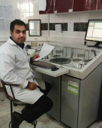 Mostafa Dehghani合作论文数Coll Anim Sci & Technol, China Agr Univ3Eer Hehua合作论文数Ningxia Acad Agr & Forestry Sci, Grass Feeding Livestock Engn Technol Res Ctr, Yinchuan, Peoples R China3
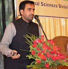 Muhammad Shahzad合作论文数Nuclear Institute for Agriculture and Biology, Faisalabad, Pakistan.3
 Hosein Salehian-Dehkordi合作论文数Chinese Acad Sci, Inst Zool, CAS Key Lab Anim Ecol & Conservat Biol, Beijing 100101, Peoples R China3Hongyu Liu合作论文数3
 Yunsheng Zhang (张云生)合作论文数Institute of Animal Sciences, Chinese Academy of Agricultural Sciences3




