4014 Background: Checkpoint inhibitors combined with anti-angiogenic therapy represents one of the standard first-line therapies for advanced hepatocellular carcinoma (aHCC). Nilvanstomig (ZG005) is a recombinant humanized anti-PD-1/TIGIT bispecific antibody. By blocking both pathways, it can synergistically activate T cells and enhance the anti-tumor activity of NK cells. This study evaluated ZG005 plus bevacizumab vs. sintilimab plus IBI305 (a bevacizumab biosimilar) as first-line therapy for aHCC. Methods: In this randomized, open-label, multicenter, phase 2 trial conducted in China, patients with aHCC who had not previously received systemic treatment were randomly assigned (1:1:1) to receive ZG005 10 mg/kg plus bevacizumab 15 mg/kg (Arm A); ZG005 20 mg/kg plus bevacizumab 15 mg/kg (Arm B); or sintilimab 200 mg plus IBI305 15 mg/kg (Arm C). All treatments were administered intravenously every 3 weeks until disease progression or unacceptable toxicity. Randomization was stratified by baseline AFP level ( < 400 vs. ≥400 ng/mL), macrovascular invasion or extrahepatic metastasis (presence vs. absence). The primary endpoint was IRC-assessed PFS per RECIST v1.1, with key secondary endpoints including PFS per mRECIST, ORR and DCR by both criteria, and OS. Results: As of the data cutoff (Nov 25, 2025), 95 patients were enrolled and received at least one dose of treatment (Arm A, n = 31; Arm B, n = 32; Arm C, n = 32). Baseline characteristics were well-balanced across the treatment arms. For all patients enrolled, the median age was 61 years (range, 37-75), with 85.3% of patients being male. Disease characteristics included BCLC stage B (28.4%) or C (71.6%), Child-Pugh score A5 (83.2%) or A6 (16.8%), baseline AFP ≥400 ng/mL in 45.3% of patients, and HBV positivity in 73.7%. Macrovascular invasion and/or extrahepatic metastasis were present in 71.6% (68/95) of patients. With a median follow-up of about 5 months, the IRC-assessed ORR was 32.3% in Arm A, 37.5% in Arm B, and 25.0% in Arm C per RECIST v1.1; the corresponding ORRs per mRECIST were 54.8%, 50.0%, and 34.4%. The IRC-assessed median PFS per RECIST v1.1 was not reached in Arm A or B, compared with 5.8 months in Arm C (Arm A vs. C: HR 0.40, 95% CI 0.15-1.05; Arm B vs. C: HR 0.28, 95% CI 0.10-0.79). The safety profiles were comparable across the three arms. No grade ≥3 hemorrhagic adverse events were reported in either Arm A or B. Conclusions: The combination of ZG005 and bevacizumab as first-line treatment in patients with aHCC demonstrated an early encouraging efficacy with an acceptable safety profile. Higher response rates and prolonged PFS were observed with the ZG005-based regimens. Longer follow up of the current study and future phase 3 trials are warranted to validate the efficacy and safety of ZG005 in combination with bevacizumab in aHCC. Clinical trial information: NCT06558227 .
ABSTRACT Precise tumor imaging is essential for accurate intraoperative decision‐making, thereby directly influencing patient prognosis. Optical molecular probes enabling non‐invasive, dynamic assessment of cancerous lesions are clinically crucial. However, current optical molecular probes face challenges in dodging false‐positive and false‐negative signals at once during imaging, limiting their clinical diagnostic use. Here, we introduce a class of lysosome‐targeted, activatable optical probes (Dx‐NH 2 ), leveraging the increased lysosomal abundance during tumor metabolic reprogramming. Lysosomal protonation retains probes for better single‐cell resolution and fewer false‐negative signals. To enable more high‐precision tumor imaging, we synthesized probe CN‐D‐GGT from CN‐D‐NH 2 by introducing a γ‐glutamyltransferase substrate, following our group's prior offensive and defensive integration strategy. Due to its smart design, CN‐D‐GGT, upon activation, shows marked changes in fluorescence and photoacoustic signals, enabling its application for multi‐modal imaging. It also has high specificity, distinguishing cancer cells in co‐culture (∼6‐fold) and clearly differentiating tumors from normal and inflamed tissues (T/N or T/I signal ratios > 3.5). Importantly, it can also differentiate cancerous from adjacent tissues in clinical samples. This work has developed a probe that can accurately light tumors in complex pathological environments, with the potential to assist in intraoperative resection decision‐making, avoid excessive or missed resection.
BACKGROUND:Intrahepatic cholangiocarcinoma (ICC) is a malignant tumour associated with a poor prognosis. Recent studies have suggested that ribonucleotide reductase subunit M2 (RRM2) could promote tumour progression. This study aims to explore the expression, function and mechanism of RRM2 in ICC by regulating ferroptosis through the HIF-1α signalling pathway. METHODS:The expression of RRM2 and HIF-1α was detected in ICC patients, and the relationship between RRM2 and prognosis was analysed. CCK-8, clone formation assay, wound healing assay, transwell invasion experiment, cell apoptosis detection and tumorsphere formation assay were conducted to evaluate the effect of RRM2 on the progression of ICC. Transcriptomics was used to explore the mechanism of RRM2 promotion of ICC. Ferroptosis inducer (Ferrostatin-1) and inducer (RSL3), HIF-1α inhibitor (LW6) and inducer (CoCl2) were applied to investigate the effects of RRM2 on reactive oxygen species (ROS), ferroptosis-related indicators and HIF-1α expression. The effects of RRM2 on tumour growth were validated using subcutaneous tumours in nude mice. RESULTS:RRM2 was significantly upregulated in ICC tissues and was associated with a poor prognosis in patients. Overexpression of RRM2 promoted the proliferation, colony formation, invasion, metastasis, stemness, and inhibited apoptosis of ICC cells. Transcriptomics clarified that the HIF-1α signalling pathway was a key downstream pathway. The expression of HIF-1α was significantly positively correlated with the expression of RRM2 and was associated with poor prognosis in patients. Overexpression of RRM2 upregulated HIF-1α and reduced ROS levels. RRM2 upregulated SLC7A11 and GPX4 and downregulated ACSL4 by regulating the HIF-1α signalling pathway, inhibiting intracellular iron ion accumulation and lipid peroxidation, thus suppressing ferroptosis. Under hypoxic conditions, the anti-tumour effect of knocking down RRM2 was weakened. HIF-1α inhibitor could reverse the ferroptosis inhibition caused by RRM2 overexpression, and the HIF-1α inducer could alleviate the enhanced ferroptosis caused by knocking down RRM2. In vivo experiments also confirmed that overexpression of RRM2 promoted tumour growth, upregulated Ki-67, GPX4, HIF-1α, and N-cadherin, and downregulated E-cadherin. CONCLUSIONS:RRM2 inhibited ferroptosis by regulating the HIF-1α signalling pathway, thereby promoting the progression of ICC. The RRM2/HIF-1α/ferroptosis axis may become a potential target for ICC therapy.
Precise tumor imaging remains challenging due to nonspecific activation and fluorophore diffusion of traditional probes. Therefore, novel tools capable of addressing these limitations are urgently needed. Here, we develop a "dual-locked" fluorescent nanoprobe NFL-NPs (near-infrared LAP-responsive fluorescent probe nanoparticles) to improve imaging accuracy by integrating leucine aminopeptidase (LAP) and tumor acidic microenvironment as substrates. Firstly, NFL, a small molecule probe, was constructed by introducing a leucine aminopeptidase (LAP)-cleavage group and a quinonemethide (QM) precursor into hemicyanine dye (HD), which was able to improve intracellular retention. Subsequently, to remain silence of NFL before reaching tumors, acid-sensitive polymers were used to encapsulate NFLs to construct NFL-NPs for hepatocellular carcinoma (HCC) imaging. Fluorescence images demonstrated sustained signal amplification in HCC-bearing mice livers (similar to 3-fold higher than healthy mice), with excellent correlation to both bioluminescence and H&E staining images. This "dual-locked" nanoprobe system optimizes false-signals limitations of traditional probes, offering a clinically translatable tool for precise HCC diagnosis.
e16222 Background: The management of advanced biliary tract cancers (BTC) remains a significant clinical challenge. Although the combination of immune checkpoint inhibitors (ICIs) with gemcitabine and cisplatin has established a new first-line standard of care, the clinical benefits are still limited. Surufatinib, a novel oral TKI, shows preclinical anti-tumor immune potential, but robust clinical and real-world evidence for its combination with ICIs/chemotherapy in BTC is lacking. regarding the efficacy and safety of surufatinib in combination with ICIs and chemotherapy for BTC is currently lacking. Methods: This single-center retrospective real-world study (ChiCTR 2500100586) enrolled patients (pts) with BTC who received surufatinib combined with ICIs and chemotherapy. Patients were required to have at least one measurable lesion per RECIST 1.1 criteria. The primary endpoints were objective response rate (ORR) and progression-free survival (PFS), Secondary endpoints included disease control rate (DCR), and overall survival (OS). Results: As of the latest data cut-off on June 1, 2026, a subset of the planned sample size had been enrolled. (N=12)The combination regimen showed promising anti-tumor activity, with a confirmed ORR of 41% (5/12), consisting of 0 complete responses (CR) and 5 partial responses (PR).The DCR was 66% (8/12),The preliminary median PFS was estimated to be 7.3 months .The safety profile of the regimen was generally consistent with the known profiles of the individual agents, with no new or unexpected safety signals identified. The most common grade ≥3 treatment-related adverse events (TRAEs) included hepatic impairment (8%); neutropenia, thrombocytopenia, hypertension, and proteinuria (all 0%). Most adverse events were manageable with dose modifications and supportive care. Conclusions: In this real-world study of first-line treatment for advanced BTC, surufatinib combined with ICIs plus gemcitabine-platinum chemotherapy demonstrated encouraging preliminary efficacy and manageable safety. Through synergistic effects (anti-angiogenesis + immunomodulation + cytotoxicity), this triple therapy is expected to break through the current efficacy bottleneck of conventional chemotherapy for advanced BTC. Larger sample size and longer follow-up are needed to validate these findings. Clinical trial information: ChiCTR2500100586. Best tumor response and treatment- related advers evevts. Best Tumor Response evaluated Treatment-related adverse events Study Endpoint N=12 Adverse events N=12 ORR,N(%)【95% CI】 5(41%)(15.24~72.38) AEs,N(%) 11(91%) 95% CI (61.51~99.79) DCR,N(%)【95% CI 】 8(66%) (34.99~89.43) TEAEs N(%) 11(91%)95% CI (61.51~99.79) CR,N(%)【95% CI】 0(0%)(0.00~26.55) SAEs,N(%) 0(0%)95% CI ( 0.00~26.55) Surgery,N(%)【95% CI 】 1(8%)(0.21~38.43) irAEs,N(%) 1(8%)95% CI (34.99~89.43) mPFS 7.3(months) Grade≥3,N(%) 1(8%)95% CI (34.99~89.43)
Supplementary Figure 1. Integrated spatial and single-cell analyses delineate molecular programs and cell composition of ECM-remodeling-associated subregions.
Hepatitis B virus (HBV) may alter immunotherapy responsiveness in HBV-positive hepatocellular carcinoma (HCC) patients. However, the underlying immune mechanisms remain unclear. To characterize the immune determinants underlying the enhanced immunotherapy response observed in HBV+ HCC patients, we comprehensively analyzed 528 HCC patients who received immunotherapy, encompassing diverse hepatitis infections. We performed an analysis incorporating single-cell RNA sequencing, spatial transcriptomics, and tissue microarray validation to map the tumor immune landscape. An adoptive T cell transfer combined with anti-programmed death-1 (PD-1) therapy in a syngeneic HCC mouse model was performed to validate key findings. HBV+ HCC patients exhibited superior responses to immunotherapy and prolonged overall survival. Remarkably, HBV+ HCC patients harbored an elevated proportion of exhausted CD8+ T cells, and these cells concurrently exhibited enhanced immune activity and cytotoxic potential. Our study spotlighted a novel subset of exhausted CD8+ T cells, termed PD-1+ CXCR6+ CD8+ T cells. In untreated cases, high levels of PD-1+ CXCR6+ CD8+ T cells correlated with poor prognosis. In contrast, among patients receiving immunotherapy, their enrichment was associated with markedly better outcomes. In vivo, adoptive transfer of CXCR6+ T cells markedly augmented the antitumor efficacy of anti-PD-1 therapy. Moreover, PD-1+ CXCR6+ CD8+ T cells demonstrated a prominent interaction with CXCL16+ macrophages in HBV+ HCC. Taken together, we identified a novel exhausted T cell subset, PD-1+ CXCR6+ CD8+ T cells, that are enriched in HBV+ HCC patients and maintained by CXCL16+ macrophages. The enrichment of PD-1+ CXCR6+ CD8+ T cells and their interaction with CXCL16+ macrophages may contribute to the enhanced immunotherapy response observed in HBV+ HCC.
Enzymes catalyze biochemical reactions in all living systems, and their subcellular localizations can profoundly influence distinct cellular functions. Yet, the physiological roles of subcellular enzyme activity remain largely unexplored, owing to the lack of tools that allow imaging of enzymatic activity with organelle-level precision in living cells. Here, we present a class of fluorogenic reporters for spatiotemporally resolved imaging of enzyme activity within specific subcellular compartments, including mitochondria (Mito), lysosomes (Lyso), endoplasmic reticulum (ER), and Golgi apparatus (Golgi). These fluorogenic reporters comprise a rhodol fluorophore whose fluorescence is quenched by an enzyme-specific substrate and a bioorthogonal cage group, with signal restoration occurring only upon sequential uncaging by an organelle-localized bioorthogonal activator and subsequent enzymatic activation. We applied this versatile strategy to visualize the activity of enzymes from different families in live cells, such as leucyl aminopeptidases (LAP), γ-glutamyl transferases (GGT), and monoamine oxidase A (MAO-A). Application to LAPs revealed distinct, compartment-dependent functions: Mito-LAP activity maintained redox homeostasis in stressed cells, favoring tumor cell survival, whereas ER-LAP activity promoted antigen presentation and immune-mediated tumor cell killing. Notably, drug profiling showed that cisplatin (CDDP) and camptothecin (CPT) concurrently amplified both Mito-LAP and ER-LAP activities, eliciting opposing influences on tumor progression. These findings also inspire the development of a mitochondria-targeted inhibitor to improve anticancer efficacy. This platform offers a genetically independent, broadly adaptable framework for dissecting compartmentalized enzymology in living cells.
Lipid metabolism reprogramming drives malignant proliferation and invasiveness in hepatocellular carcinoma (HCC). Beyond supplying energy and membrane components, lipids function as signaling molecules that modulate tumor cell epigenetics and the microenvironment. Accumulating research has clarified the implications of these metabolic alterations in HCC, providing a rationale for targeted therapies. This review summarizes key alterations in lipid metabolism within HCC and explores their mechanistic contributions to tumor progression. It further examines how lipid metabolic shifts in immune and stromal cells of the tumor microenvironment promote HCC advancement. Finally, we discuss the therapeutic potential of targeting lipid metabolism in liver cancer treatment.
Enzymes catalyze biochemical reactions in all living systems, and their subcellular localizations can profoundly influence distinct cellular functions. Yet, the physiological roles of subcellular enzyme activity remain largely unexplored, owing to the lack of tools that allow imaging of enzymatic activity with organelle-level precision in living cells. Here, we present a class of fluorogenic reporters for spatiotemporally resolved imaging of enzyme activity within specific subcellular compartments, including mitochondria (Mito), lysosomes (Lyso), endoplasmic reticulum (ER), and Golgi apparatus (Golgi). These fluorogenic reporters comprise a rhodol fluorophore whose fluorescence is quenched by an enzyme-specific substrate and a bioorthogonal cage group, with signal restoration occurring only upon sequential uncaging by an organelle-localized bioorthogonal activator and subsequent enzymatic activation. We applied this versatile strategy to visualize the activity of enzymes from different families in live cells, such as leucyl aminopeptidases (LAP), gamma-glutamyl transferases (GGT), and monoamine oxidase A (MAO-A). Application to LAPs revealed distinct, compartment-dependent functions: Mito-LAP activity maintained redox homeostasis in stressed cells, favoring tumor cell survival, whereas ER-LAP activity promoted antigen presentation and immune-mediated tumor cell killing. Notably, drug profiling showed that cisplatin (CDDP) and camptothecin (CPT) concurrently amplified both Mito-LAP and ER-LAP activities, eliciting opposing influences on tumor progression. These findings also inspire the development of a mitochondria-targeted inhibitor to improve anticancer efficacy. This platform offers a genetically independent, broadly adaptable framework for dissecting compartmentalized enzymology in living cells.
BackgroundHepatocellular carcinoma (HCC), the predominant pathological subtype of primary liver cancer, remains a major global health burden with poorly defined molecular mechanisms. Cell growth regulator 11 (CGR11), a novel secreted protein characterized by EF-hand motifs, has recently emerged as a potential extracellular signaling modulator in tumor biology. Although implicated in cancer cell proliferation and metastasis, its precise role and regulatory mechanisms in HCC progression have not been elucidated.MethodsWe integrated bioinformatics analysis with single-cell transcriptomic profiling and CellChat-based intercellular communication mapping. CGR11 expression and localization were validated in tissue microarrays, HCC cell lines, and tumor specimens using immunohistochemical staining, qRT-PCR, and Western blotting. In vitro experiments and both subcutaneous and orthotopic xenograft models were established to evaluate the biological effects of CGR11 overexpression and knockdown. RNA sequencing, LC3 fluorescence assay, and transmission electron microscopy were conducted to elucidate the underlying molecular mechanism.ResultsCGR11 expression was markedly increased in HCC tissues relative to adjacent non-tumorous liver tissues and correlated with poor patient prognosis. Functional and mechanistic analyses demonstrated that CGR11 promotes HCC cell proliferation, invasion and tumor growth by inhibiting autophagy levels through activation of the PI3K/AKT signaling. Conversely, CGR11 knockdown restored autophagy and significantly suppressed tumor progression in both cellular and animal models.ConclusionOur findings establish CGR11 as a novel oncogenic regulator that contributes to HCC progression by suppressing autophagy via PI3K/AKT activation. Targeting the CGR11-PI3K/AKT axis may therefore provide a promising avenue for precision therapeutic intervention in HCC.
Supplementary Figure 5. Spatial enrichment and CellChat analyses show strong interactions between FAP+ fibroblasts and hypoxic TAMs.
TPS11199 Background: Biliary tract cancer (BTC) is characterized by strong invasiveness and poor prognosis, with a metastasis and recurrence rate of up to 67% within 1 year after surgery and a 5-year overall survival rate of only 5%~15%. Among BTC subtypes, intrahepatic cholangiocarcinoma (ICC) accounts for a relatively high proportion, and its unique biological behaviors result in persistently high postoperative recurrence risk, which seriously endangers patients' life and health. Currently, major guidelines recommend that patients with potentially resectable ICC at high recurrence risk participate in clinical trials to explore optimal treatment strategies. For patients evaluated as "technically resectable but with high-risk recurrence" before surgery, clinical practice has shown that neoadjuvant therapy can effectively eliminate micrometastases and reduce the difficulty of R0 resection; postoperative adjuvant therapy can further prevent recurrence. Therefore, we innovatively propose a perioperative treatment model aimed at breaking through the existing therapeutic predicament. Methods: This study is a phase Ⅱ, single-arm, exploratory study with a fixed sample size of 20 patients. The inclusion criteria are intrahepatic cholangiocarcinoma (ICC) (TNM stage T1-4, N0-1, M0)based on preoperative assessment, initial technical resectability per imaging, and at least one high - risk recurrence factor. High-risk recurrence factors include: tumor diameter >5 cm, regional lymph-node positivity, satellite/multifocal lesions, radiological suspicion of diaphragmatic adhesion, CA19-9 >200 U/ml, and an anticipated surgical margin <1 cm on pre-operative imaging. Treatment involves two 21 - day cycles of GEMOX (gemcitabine 1000mg/m² on day 1 and 8, oxaliplatin 100mg/m² on day 1) with tislelizumab (200mg on day 1) and donafenib (0.2g orally twice daily). If investigators confirm patients meet surgical criteria (R0 resection expected), surgery follows 1 - 2 weeks post - treatment. Post - surgery, adjuvant therapy starts within 5 weeks. The regimen is capecitabine (1250mg/m² orally twice daily on days 1 -14) or S - 1 (40 -60mg orally twice daily on days 1 -14, weight - adjusted) plus tislelizumab (200mg on day 1) and donafenib (0.2g orally twice daily), repeated every 21 days for six cycles. Primary endpoint is event - free survival (EFS). Secondary endpoints are objective response rate (ORR) and disease control rate (DCR) per RECIST1.1, R0 resection rate, major pathological response rate(MPR, the proportion of patients who underwent radical surgery and had ≤10% residual viable tumor in the postoperative specimen), treatment completion rate, overall survival (OS), disease - free survival (DFS), and safety (treatment - related adverse events' incidence and severity). Clinical trial information: ChiCTR2500112067.
Abstract In hepatocellular carcinoma (HCC), stromal and immune components shape invasion and metastasis. To identify the cellular states that drive extracellular matrix (ECM) remodeling, define their organization at the tumor margin, and evaluate their association with prognosis, we profiled stromal regions at the tumor margin using single-cell RNA sequencing (scRNA-seq), spatial transcriptomics (ST), and multiplex immunofluorescence. Across scRNA-seq profiles from 79 patients, we identified a hypoxia-associated population of tumor-associated macrophages with high expression of a disintegrin and metalloproteinase domain 8 (ADAM8) and upregulation of ECM remodeling programs. ST and histology placed these ADAM8+ macrophages in stromal regions with disorganized collagen. They were often in hypoxic zones around necrosis and adjacent to FAP+ fibroblasts. Ligand–receptor analysis predicted engagement of the collagen–CD44 axis between the two cell types. In vitro, hypoxia induced expression of ADAM8 in macrophages. ADAM8 knockdown reduced the induction of TNFα, IL6, and IL1β and weakened the ability of macrophages to activate fibroblasts. In tissue microarrays, a higher fraction of ADAM8+ macrophages among CD68+ cells predicted shorter overall and recurrence-free survival. Consistently, in a murine HCC model, pharmacologic inhibition of ADAM8 decreased FAP+ fibroblast abundance. Overall, the data show that ADAM8+ macrophages and FAP+ fibroblasts form a hypoxia-linked ECM remodeling niche at the tumor boundary. This stromal remodeling unit associates with poor prognosis and suggests testable targets for stroma-focused therapy for HCC.
Background:Spontaneous tumor rupture is a unique and life-threatening presentation of hepatocellular carcinoma (HCC). The optimal postoperative management of patients with spontaneously ruptured HCC (srHCC) remains controversial. Hyperthermic intraperitoneal chemotherapy (HIPEC) has been proposed to reduce peritoneal dissemination, but its clinical benefit in srHCC is uncertain. Objectives:This study aimed to evaluate the survival benefit and safety of postoperative HIPEC combined with hepatic resection in patients with srHCC. Design:A retrospective multicenter cohort study was conducted, including patients with srHCC who underwent curative hepatectomy with or without postoperative HIPEC between 2018 and 2024. Methods:A total of 208 srHCC patients from 4 institutions were enrolled and categorized into the resection group (R) and the resection plus HIPEC group (R-HIPEC). Propensity score matching (PSM) and inverse probability of treatment weighting (IPTW) were applied to minimize baseline differences. The primary endpoint was recurrence-free survival (RFS), and the secondary endpoint was overall survival (OS). Survival outcomes were assessed using Kaplan-Meier analysis, Cox proportional hazards models, and subgroup analysis. Results:Across the primary, PSM, and IPTW cohorts, patients in the R-HIPEC group achieved significantly longer median RFS (mRFS) and OS than those in the R group. The median OS was 45.6 versus 26.4 months in the primary cohort (p = 0.025), 48.2 versus 26.4 months in the PSM cohort (p = 0.025), and 42.9 versus 26.5 months in the IPTW cohort (p = 0.012). The mRFS was 15.5 versus 7.7 months (p = 0.002), 18.2 versus 8.3 months (p = 0.002), and 14.7 versus 7.4 months (p = 0.014), respectively. Subgroup analysis indicated that patients with Barcelona Clinic Liver Cancer stage 0/A derived significantly greater RFS benefit from HIPEC than those with stage B/C (interaction p = 0.0264). For OS, a significant interaction was observed with postoperative immunotherapy (interaction p = 0.0054). The R-HIPEC group showed a lower incidence of peritoneal implantation metastasis, without an increase in perioperative complications. Conclusion:HIPEC combined with resection for srHCC can effectively prolong survival time. Resection combined with HIPEC and targeted therapy may be a promising strategy for srHCC.
Supplementary Figure 6. Analysis of TME signaling patterns and the effect of macrophage ADAM8 on fibroblast phenotype.
BACKGROUND:Hepatocellular carcinoma (HCC) is a leading cause of cancer death worldwide. Gemcitabine (Gem) is a commonly used drug against HCC, but its efficacy is limited by the development of resistance. Resveratrol (Res), a natural polyphenol with antitumor activity, may reverse Gem resistance in HCC, although the mechanism remains unclear. METHODS:The effects of Res on the proliferation, apoptosis, cell cycle, and invasion of Hep3B and HuH-7 cells were assessed via cell counting kit-8 (CCK-8), clonogenic, flow cytometry, and Transwell assays, respectively. Potential Res targets were predicted by network pharmacology, and markers of HCC prognosis were identified from the cancer genome atlas (TCGA) data. The interaction between Res and thymidylate synthase (TYMS) was validated by molecular docking and dynamics simulation. A Gem-resistant HuH-7 cell line (HuH-7/GR) was established, and when these cells were treated with Res combined with Gem, the effect on Gem sensitivity was detected by CCK-8 assay, clonogenic assay, and flow cytometry. Finally, a subcutaneous nude mouse model of HCC was used to evaluate the in vivo effects of Res combined with Gem. RESULTS:Res inhibited HCC cell proliferation, induced apoptosis and G2/M arrest, and suppressed invasion in a concentration-dependent manner. Network pharmacology and TCGA analysis identified TYMS as an important target gene for Res. TYMS was highly expressed in HCC tissues and correlated with poor prognosis. Res treatment reduced TYMS expression, while molecular docking and simulation showed stable binding of Res to TYMS. TYMS levels were elevated in HuH-7/GR resistant cells. Res combined with Gem was found to reverse drug resistance, inhibit proliferation and colony formation, and induce apoptosis. The Res + Gem combination group showed the smallest tumor volume in the in vivo model. CONCLUSION:By attenuating Gem resistance through TYMS inhibition, Res holds promise as a clinically viable adjunct to Gem-based chemotherapy, offering a potential strategy to improve outcomes in HCC patients.
Hepatitis B virus (HBV) may alter immunotherapy responsiveness in HBV-positive hepatocellular carcinoma (HCC) patients. However, the underlying immune mechanisms remain unclear. To characterize immune determinants underlying the enhanced immunotherapy response observed in HBV+ HCC patients, we comprehensively analyzed 528 HCC patients who received immunotherapy, encompassing diverse hepatitis infections. We performed an analysis incorporating single-cell RNA sequencing, spatial transcriptomics, and tissue microarray validation to map the tumor immune landscape. An adoptive T-cell transfer combined with anti-PD-1 therapy in a syngeneic HCC mouse model was performed to validate key findings. HBV+ HCC patients exhibited superior responses to immunotherapy and prolonged overall survival. Remarkably, HBV+ HCC patients harboured an elevated proportion of exhausted CD8+ T cells, and these cells concurrently exhibited enhanced immune activity and cytotoxic potential. Our study spotlighted a novel subset of exhausted CD8+ T cells, termed PD-1+ CXCR6+ CD8+ T cells. In untreated cases, high levels of PD-1+ CXCR6+ CD8+ T cells correlated with poor prognosis. In contrast, among patients receiving immunotherapy, their enrichment was associated with markedly better outcomes. In vivo, adoptive transfer of CXCR6+ T cells markedly augmented the antitumor efficacy of anti-PD-1 therapy. Moreover, PD-1+ CXCR6+ CD8+ T cells demonstrated a prominent interaction with CXCL16+ macrophages in HBV+ HCC. Taken together, we identified a novel exhausted T-cell subset, PD-1+ CXCR6+ CD8+ T cells, which are enriched in HBV+ HCC patients and maintained by CXCL16+ macrophages. The enrichment of PD-1+ CXCR6+ CD8+ T cells and their interaction with CXCL16+ macrophages may contribute to the enhanced immunotherapy response observed in HBV+ HCC.
Photodynamic therapy (PDT) has emerged as a promising approach for tumor treatment due to its non-invasiveness and high selectivity. However, the off-target activation of phototoxicity and the limited availability of tumor-specific biomarkers pose challenges for effective PDT. Here, we present the development of a novel ratiometric near-infrared-II (NIR-II) fluorescent organic nanoprobe, BTz-IC@IR1061, which responds specifically to hypochlorite (HClO) within tumors. This nanoprobe allows ratiometric fluorescence imaging to monitor and guide activated tumor PDT. BTz-IC@IR1061 nanoparticles were synthesized by codoping the small molecule dye BTz-IC, which generates reactive oxygen species (ROS), with the commercial dye IR1061. The presence of HClO selectively activates the fluorescence and photodynamic properties of BTz-IC while destroying IR1061, enabling controlled release of ROS for tumor therapy. We demonstrated the high selectivity of the nanoprobe for HClO, as well as its excellent photostability, photoacoustic imaging capability, and photothermal effects. Furthermore, in vivo studies revealed effective tumor targeting and remarkable tumor growth inhibition through tumor-activated PDT. Our findings highlight the potential of BTz-IC@IR1061 as a promising tool for tumor-specific PDT, providing new opportunities for precise and controlled cancer therapy.
Exocrine pancreatic insufficiency (EPI) is a major cause of maldigestion and malnutrition, resulting from primary pancreatic diseases or other conditions. As the prevalence of EPI continues to rise, accurate identification of its etiology has become critical for the diagnosis and treatment of pancreatic secretory insufficiency. EPI can result from both pancreatic and non-pancreatic disorders. Pancreatic disorders include acute and chronic pancreatitis, pancreatic tumors, cystic fibrosis, procedures that involve pancreatic resection, and other rare causes. Non-pancreatic disorders of EPI include diabetes mellitus, celiac disease, inflammatory bowel disease, gastrointestinal and esophagectomy surgery, as well as advanced patient age. This review aims to provide a comprehensive analysis of the literature on EPI etiology, with a thorough overview to support its consideration as a potential diagnosis.
 Chuang Peng (彭创)合作论文数湖南师范大学附属一医院(湖南省人民医院)67Yinghui Song合作论文数61Yuhang Li合作论文数19WeiMin Yi (易为民)合作论文数18Lianhong Zou (邹联洪)合作论文数湖南师范大学附属一医院(湖南省人民医院)17Zhihua Zhang合作论文数15
 Xiaobing Zhang (张晓兵)合作论文数College of Chemistry and Chemical Engineering, Hunan University14Zhangtao Yu合作论文数13
 Bo Jiang (江波)合作论文数Fine Arts Academy, Hunan Normal University13Guoyi Xia合作论文数10Xiehong Liu合作论文数10
 Guosheng Song (宋国胜)合作论文数College of Chemistry and Chemical Engineering, Hunan University10Nanhui Yu合作论文数9Zhiguo Tan合作论文数9
 Lin Yuan (袁林)合作论文数College of Chemistry and Chemical Engineering, Hunan University9Yuchen Qi合作论文数Zhejiang Univ, Inst Translat Med, Hangzhou 310009, Zhejiang, Peoples R China8PingZhou Yang (杨平洲)合作论文数8ZhenDong Zhong (钟振东)合作论文数8Junkai Huang合作论文数7
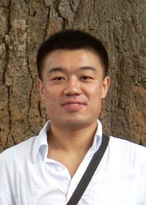 Bo Sun (孙博)合作论文数School of Life Science and Technology, ShanghaiTech University6Mengting Cai合作论文数6Yujing Zhang合作论文数6Xuepeng LI (李雪鹏)合作论文数6ZengPeng Sun (孙增鹏)合作论文数6Ranzhiqiang Yang合作论文数5Jiang Jin-qiong (江金琼)合作论文数5
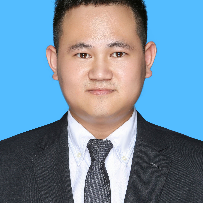 Le Wang合作论文数Department of Cancer Prevention, Zhejiang Cancer Hospital5Ou Li合作论文数5Ling Wu合作论文数5JianMing Liu (刘剑鸣)合作论文数5HongYing Tang (唐宏英)合作论文数5Xianhai Mao合作论文数5
 Zhong Cao (曹忠)合作论文数School of Chemistry and Chemical Engineering, Changsha University of Science & Technology;School of Food Science and Bioengineering, Changsha University of Science & Technology5
 Shuangyan Huan (宦双燕)合作论文数College of Chemistry and Chemical Engineering, Hunan University5Mengjun MO (莫梦军)合作论文数4Shayong Peng (彭沙勇)合作论文数4Xu Chen合作论文数4
 Wei Li (李伟)合作论文数Hunan University4
 Tianbing Ren (任天兵)合作论文数College of Chemistry and Chemical Engineering, Hunan University4
 Xing Yu (余星)合作论文数Hunan Normal University School of Medicine4Kashif Shakoor合作论文数3Jianwen Li合作论文数3Yang Shen合作论文数3Jiang Yu (蒋宇)合作论文数3Yu Jiang合作论文数3




