Intervertebral disc degeneration (IDD) is a leading cause of chronic low back pain worldwide. It involves degenerative changes in nucleus pulposus, annulus fibrosus, and cartilage endplate cells, driven by factors such as oxidative stress, inflammation, mechanical imbalance, and apoptosis. Mitochondrial dysfunction lies at the heart of IDD progression, marked by excessive reactive oxygen species, reduced ATP production, and compromised cellular homeostasis. Mitophagy, the selective autophagic clearance of damaged mitochondria, emerges as a key regulator of mitochondrial quality control. Canonical pathways including those involving PINK1–PRKN/Parkin, BNIP3 and BNIP3L/NIX, as well as non-canonical mechanisms involving PHB2 and cardiolipin, coordinate the removal of dysfunctional mitochondria. However, dysregulated mitophagy in IDD can spur persistent oxidative stress, chronic inflammation, and cell death in different disc cell types, thereby hastening tissue degeneration. Therapeutic strategies focusing on mitophagy modulation include small-molecule agents, gene therapy targeting key mitophagy regulators, and stem cell treatments that restore mitochondrial function. Despite promising preclinical findings, challenges remain, particularly in achieving specificity and safety, as well as translating these approaches into effective clinical interventions. Future efforts integrating multi-omics analyses, refined animal models, and gene-editing tools may deepen our understanding of mitophagy’s role in IDD and pave the way for novel, targeted therapies aimed at improving patient outcomes.
Ferroptosis, an iron-dependent form of programmed cell death driven by lipid peroxidation and reactive oxygen species (ROS), has emerged as a key mechanism in the progression of intervertebral disc degeneration (IDD). Mitochondria serve a central role in this process by regulating iron metabolism, ROS production and energy homeostasis. In IDD, mitochondrial dysfunction leads to increased lipid ROS levels, decreased glutathione peroxidase 4 (GPX4) activity and impaired antioxidant defenses, contributing to extracellular matrix degradation and nucleus pulposus cell death. The present review summarizes the core molecular mechanisms underlying ferroptosis and highlights the mitochondrial pathways that mediate ferroptosis in IDD. Furthermore, the advances in mitochondria-targeted therapeutic strategies are discussed, including antioxidants, iron chelators, GPX4 activators, mitophagy modulators and nanotechnology-based interventions. These approaches provide promising avenues for preventing ferroptosis-induced disc degeneration and preserving the viability of disc cells. Understanding the interplay between mitochondrial dysfunction and ferroptosis may offer novel insights for the development of precise and effective treatments for IDD.
Ossification of the posterior longitudinal ligament (OPLL) involving the C2 segment poses significant surgical challenges due to anatomical complexity and limited exposure. This report describes a novel application of the Expanded Shelter Technique to overcome C3-level exposure constraints in the management of continuous C2–C6 OPLL. This study used a clinical case report. A 60-year-old male presented with progressive neck/shoulder discomfort, right upper limb numbness, and weakness (MRC grade 2 right vs. grade 4 left). Imaging showed multi-level OPLL from C2 to C6, severe canal stenosis, and disc herniations. The Expanded Shelter Technique was performed. Neurological symptoms in the right upper limb resolved on the first postoperative day, with no new neurological symptoms emerging. At the six-month follow-up, neurological symptoms remained absent, and solid bony fusion was confirmed at the graft–vertebral interface. Direct spinal cord decompression was achieved. Subtotal resection of the C3 vertebral body effectively widened the surgical corridor and reduced procedural complexity. We report a case of continuous cervical OPLL spanning C2–C6 managed with the Expanded Shelter Technique. This approach successfully overcomes C3 exposure constraints through subtotal vertebral body resection and mesh reconstruction, providing a viable anterior decompressive solution for this challenging OPLL subtype.
Intervertebral disc degeneration (IDD) is a major pathological basis for spinal degenerative diseases, involving mechanisms such as abnormal mechanical loading, inflammatory responses, and genetic and environmental factors. The role of epigenetic regulation in IDD has gained attention as a potential therapeutic target. The present review systematically explores the contributions of DNA methylation, histone modifications, non‑coding RNAs (ncRNAs) and metabolic regulation to IDD progression, and elucidates their molecular mechanisms. Specific examples include: DNA methyltransferase 3β‑mediated DNA methylation promoting ferroptosis and oxidative stress in nucleus pulposus cells; enhancer of zeste homolog 2 (EZH2)‑mediated trimethylation of histone H3 lysine 27 modification inhibiting SOX9 expression, leading to cellular senescence and extracellular matrix degradation; and ncRNAs (such as microRNA‑143 and LINC01121) regulating gene transcription to affect inflammation and apoptosis. Additionally, metabolic products (such as NAD+, α‑ketoglutarate and lactate) interact with epigenetic pathways to influence IDD. Specifically, NAD+ acts as a cofactor for sirtuin deacetylases, thereby regulating histone and non‑histone protein acetylation; α‑ketoglutarate serves as a cofactor for TET DNA demethylases and Jumonji‑C histone demethylases, influencing DNA and histone demethylation; and lactate induces histone lactylation, which modulates gene transcription related to inflammation and extracellular matrix metabolism in IDD. Based on these mechanisms, novel therapies targeting epigenetics (such as DNA methylation inhibitors, EZH2 inhibitors and RNA interference) show therapeutic potential. Future research should further explore the crosstalk between epigenetic and metabolic regulation to advance the development of personalized and precision medicine strategies for IDD intervention.
Laminoplasty (LP) is a standard motion-preserving procedure for multi-segmental ossification of the posterior longitudinal ligament (OPLL), yet it may permit continued ossification progression due to retained segmental mobility. The anterior controllable antedisplacement and fusion (ACAF) technique offers ventral decompression with segmental stabilization, but its effect on OPLL fate remains unclear. To compare radiographic OPLL progression and neurological outcomes between ACAF and LP in patients with multilevel cervical OPLL. This retrospective study included 100 patients (50 ACAF, 50 LP) treated between 2019 and 2021. Radiographic parameters included OPLL thickness, length, width, cervical range of motion (ROM), and a novel morphological classification (Types 1–4). Neurological status was assessed using the Japanese Orthopaedic Association (JOA) score. Mean follow-up was approximately 34 months. ACAF demonstrated significantly smaller increases in OPLL thickness for Types 1, 2, and 3 lesions (all P < 0.05), with no significant difference in Type 4. ROM was markedly reduced in the ACAF group (7.32° ± 4.21° vs. 31.45° ± 6.23°, P < 0.001) and correlated with OPLL thickness progression (r = 0.612, P = 0.002). Neurological improvement was significantly higher in the ACAF group (70.12
Phenotypic transitions of nucleus pulposus (NP) cells are increasingly recognized as key drivers of intervertebral disc degeneration (IVDD), yet the differentiation fate of NP cells and its regulation by the immune microenvironment remain unclear. Using single-cell transcriptomic profiling with in vivo and in vitro validation, we identify an osteoblast-like NP cell subpopulation that emerges during IVDD and exhibits enhanced osteogenic differentiation capacity. Genetic disruption of NP cell osteogenic potential attenuates disc degeneration, supporting a causal role for this program in disease progression. We further show that GPNMB+ macrophages promote osteogenic differentiation and degeneration of NP cells through PDGF signaling and that inhibition of PDGF signaling reduces NP cell osteogenic reprogramming and alleviates disc degeneration. Together, these findings define immune-driven osteogenic reprogramming of NP cells as a key pathological mechanism in IVDD and highlight NP cell osteogenic differentiation as a potential therapeutic target.
Degenerative joint diseases, such as osteoarthritis (OA), intervertebral disc degeneration (IVDD) and rheumatoid arthritis (RA), cause pain and disability worldwide. Globally, OA affects >500 million individuals, IVDD affects 40-60% of adults and RA affects 0.5-1% of the global population. Current treatments (such as non-steroidal anti-inflammatory drugs and corticosteroids for OA, conservative management and spinal surgery for IVDD, and disease-modifying anti-rheumatic drugs/biologics for RA) focus on symptom relief and inflammation control, but they do not prevent disease progression nor restore damaged tissue. Furthermore, these treatments are often associated with risks of systemic side effects (such as gastrointestinal bleeding, cardiovascular events and immunosuppression) or surgical complications (such as infection and implant failure). Although accumulating evidence implicates mitochondrial dysfunction and excessive reactive oxygen species (ROS) in the pathogenesis of these disorders, strategies that directly target mitochondrial oxidative stress are yet to be developed and translated into the clinic. In the present study this gap in the knowledge was addressed by systematically reviewing mitochondria-targeted antioxidant therapies and mitochondrial quality-control mechanisms due to their potential as novel, disease-modifying approaches for degenerative joint diseases. The preclinical efficacy of mitochondria-directed antioxidants (such as mitoquinone, MitoTEMPO, 10-(6'-plastoquinonyl) decyltriphenylphosphonium and Szeto-Schiller-31) in alleviating ROS-induced cellular damage, inhibiting apoptosis/pyroptosis and preserving extracellular matrix integrity in OA, IVDD and RA models were summarized. Additionally, strategies to enhance mitophagy (such as through PTEN-induced kinase 1/Parkin), rebalance mitochondrial dynamics (such as through the dynamin-related protein 1/mitofusin 1/2) and activate antioxidant signaling pathways (such as nuclear factor erythroid 2-related factor 2 and sirtuin 3) were highlighted. The present study identified key translational challenges (such as optimal delivery systems, long-term safety and clinical validation) and suggested integrated therapeutic frameworks that combine targeted antioxidants with advanced drug carriers and adjunctive treatments. Mitochondria-focused interventions may have potential as the next generation of disease-modifying treatments for OA, IVDD and RA.
Background:Emerging evidence suggests that facet joint degeneration, disc pathology, and paraspinal muscle atrophy form a degenerative triad in young adults with lumbar disc herniation (LDH). However, the exact relationship remains unknown. This study aimed quantitatively to investigate their interrelationships through magnetic resonance imaging (MRI) morphometry. Methods:Axial T2-weighted MRI served as the assessment tool in this retrospective study enrolling 60 young patients (33 disc bulging and 27 disc herniation). Measurements obtained at the responsible segment level included: lumbar curvature (Cobb angle), Pfirrmann score, intervertebral space height, facet joint angle, mean width of the multifidus pure muscle cross-sectional area (PMCSA), and multifidus fatty infiltration grade. Parameters were statistically compared between the bulging and herniation groups. Subsequently, multivariate logistic regression assessed the correlation between LDH severity and the MRI parameters. Results:Intergroup analysis revealed statistically significant differences in age, sex distribution, treatment modality, Cobb angle, anterior/middle disc height, bilateral facet joint angles, and multifidus PMCSA (P < 0.05). Multivariate logistic regression demonstrated that facet tropism, Pfirrmann grade, and multifidus fatty infiltration grade were independently associated with LDH severity (P < 0.05). Conclusions:Facet tropism, intervertebral disc degeneration (quantified by Pfirrmann grade), and multifidus fatty infiltration represent established risk factors for LDH and likely contribute to its pathological cascade.
We present a case series of cervical radiculomyelopathy caused by calcification of the ligamentum flavum (CLF), with or without concomitant periodontoid calcification. A brief literature review is presented, highlighting a potential relationship between these 2 conditions. In this retrospective study, we reviewed medical records of 33 patients diagnosed with cervical CLF. And we propose the term "crowned dens sign" (CDSign) to describe the characteristic radiographic finding of circumferential calcification around the odontoid process of the axis. Among the 33 patients, 28 (84.8%) were female and 5 (15.2%) were male, with a mean age of 70.6 years. Based on the cervical computed tomography images, a total of 81 cervical segments of CLF were recorded. The most commonly involved levels were C4-5 and C5-6, demonstrating the characteristic pattern of mid-cervical predominance. The CDsign was identified in 26 cases (79%), demonstrating a high prevalence in this cohort. The patient cohort comprised the following treatment groups: 2 patients received conservative management, twenty-three underwent posterior surgical procedures, and 8 were treated with anterior cervical surgery. The coexistence of CDSign and CLF represents an exceptionally rare clinical occurrence. The correlation between these 2 conditions extends beyond mere coincidence. The coexistence of CLF and CDSign may represent a rare but distinct cervical manifestation of calcium pyrophosphate dihydrate deposition disease.
This study aimed to establish a novel classification system for lumbar intervertebral spaces based on anatomical morphology, to investigate the relationship between intervertebral space types and cage migration risk after transforaminal lumbar interbody fusion (TLIF), and to optimize fusion strategies tailored to different anatomical types. A retrospective cohort study was conducted on 103 patients who underwent TLIF between February 2022 and June 2023. Lumbar intervertebral spaces were classified into 3 fundamental types: lordotic, neutral and kyphotic and 2 modified types: normal and degenerative. Clinical outcomes (Visual Analogue Scale [VAS], Oswestry Disability Index [ODI]), and radiographic parameters (Intervertebral Space Angle [ISA], Intervertebral Space Height [ISH], Intervertebral Foramen Height [IFH]) were evaluated. Statistical analyses included t tests, ANOVA, chi-square tests, and logistic regression. VAS and ODI scores significantly improved postoperatively (P < 0.05). ISA, ISH, and IFH showed significant postoperative increases across all fundamental types. Cage migration occurred in 8 segments, predominantly in degenerative-type intervertebral spaces. Logistic regression revealed that the degenerative type was associated with a 17-fold increased risk of cage migration (OR = 17.02). No significant association was found between fundamental type and migration. Fusion rates exceeded 98
Intervertebral disc degeneration (IDD), a major contributor to chronic low back pain, involves inflammation, extracellular matrix (ECM) degradation, and nucleus pulposus cell apoptosis. Recent studies have highlighted the critical role of macrophage polarization, which influences inflammation, tissue repair, and disc homeostasis. Macrophages can polarize into pro-inflammatory M1 and anti-inflammatory M2 phenotypes. M1 macrophages exacerbate IDD by releasing cytokines such as IL-1β and TNF-α, while M2 macrophages promote tissue repair and ECM synthesis. The disc microenvironment-marked by hypoxia, acidity, and inflammatory mediators-critically shapes macrophage behavior. Key signaling pathways, including NF-κB, STAT, PI3K-Akt, and Keap1-Nrf2, regulate this polarization and affect IDD progression. Emerging therapeutic strategies aim to reprogram macrophage polarization, using stem cell-derived exosomes, miRNA/lncRNA-based gene therapy, anti-inflammatory drugs, and biomaterial-based delivery systems, to shift M1 to M2 phenotypes. This review summarizes current knowledge on macrophage polarization in IDD, the regulatory effects of the disc microenvironment, and associated molecular pathways. Furthermore, we discuss innovative immunomodulatory strategies targeting macrophage reprogramming. Future research should integrate multi-omics approaches, advanced biomaterials, and precise immune regulation to develop effective interventions for IDD.
Intervertebral disc degeneration (IDD) is a key contributor to lumbar degenerative diseases and chronic low back pain. Accumulating evidence indicates that Sequestosome 1 (SQSTM1/p62), a multifunctional adaptor protein, plays a pivotal role in IDD pathogenesis through its regulation of autophagy, oxidative stress, inflammation, and programmed cell death. This review summarizes the multifaceted functions of SQSTM1 in the context of IDD, including its involvement in the autophagy-lysosome pathway, antioxidant defense via the Keap1-Nrf2 axis, activation of the NF-κB signaling and NLRP3 inflammasome, and modulation of apoptosis, pyroptosis, and ferroptosis. Moreover, SQSTM1 contributes to extracellular matrix degradation by upregulating matrix metalloproteinases and downregulating their inhibitors. Given its dynamic expression during disc degeneration, SQSTM1 holds promise as both a biomarker for IDD progression and a therapeutic target. Potential strategies targeting SQSTM1 include the use of autophagy inducers, inflammatory pathway inhibitors, and ferroptosis/pyroptosis modulators. However, challenges remain in precisely modulating SQSTM1 activity and translating findings into clinical therapies. Future research leveraging advanced technologies such as single-cell RNA sequencing, proteomics, and organoid models is essential to unravel the complex, stage- and cell-specific roles of SQSTM1 in IDD. Understanding these mechanisms may open new avenues for effective treatment and improved patient outcomes in degenerative spinal disorders.
STUDY DESIGN:Retrospective study. OBJECTIVE:To develop and validate computed tomography (CT)-based classification schemes to eliminate ambiguity as much as possible and evaluate the adequacy and clinical value of its classification. BACKGROUND:There is no objective criteria for laminoplasty of more than one million Chinese patients with ossification of the posterior longitudinal ligament (OPLL) every year. CT imaging can accurately show the location, size, and shape of ossification, it is very important to propose a recognized simple classification of ossifications. PATIENTS AND METHODS:From 2016 to 2018, 100 patients with "moderate to severe" OPLL on CT were performed according to the following criteria. This study simply classifies the grade of the ossification as 1-2-3, the zone is A-B by the foramen facet spinal canal classification, and the interexaminer reliability is 96%. A prospective series of 60 patients for laminoplasty was performed between 2018 and 2019, and this classification scheme was verified according to the new standard. All patients with size 1 were selectively excluded from consideration for surgery. The Japanese Orthopedic Association scores from both series are superior to most published results for patients with OPLL. RESULTS:The first and second series reported good to excellent results of 89% and 93.3%, respectively, and 80% and 85% for 24 months. The difference in the incidence of C5 paralysis and axial pain was statistically significant among the different zones, and most of them recovered within 6 months. The most common size and location types are 2-AB, 3-AB, and 2A. The most severe type is 3-AB. CONCLUSIONS:The foramen facet spinal classification of OPLL is a simple and reliable method for objectively evaluating the ossification of patients with OPLL based on CT research. LEVEL OF EVIDENCE:Level III.
Background: Intervertebral disc degeneration (IDD) is a complex phenomenon and a multifactorial degenerative disease that creates a heavy economic burden on health systems globally. Currently, there is no specific treatment proven to be effective in reversing and delaying the progression of IDD. Method: This study consisted of animal and cell culture experiments. The role of DNA methyltransferase 1 (DNMT1) on regulating the M1/M2 macrophages polarization and pyroptosis, as well as its effect on Sirtuin 6 (SIRT6) expression in an IDD rat model and in tert-butyl hydroperoxide (TBHP)-treated nucleus pulposus cells (NPCs) were explored. Rat models were constructed, followed by transfection with lentiviral vector to inhibit DNMT1 or overexpress SIRT6. The NPCs were treated with THP-1-cells conditioned medium, and their pyroptosis, apoptosis, and viability were evaluated. Western blot, histological and immunohistochemistry staining, ELISA, PCR, and flow cytometry were all used to evaluate the role of DNMT1/ SIRT6 on macrophage polarization. Results: Silencing DNMT1 inhibited apoptosis, the expression of related inflammatory mediators (e.g., iNOS) and inflammatory cytokines (e.g., IL6 and TNF-α). Moreover, silencing DNMT1 significantly inhibited the expression of pyroptosis markers IL- 1β, IL-6, and IL-18 and decreased the NLRP3, ASC, and caspase-1 expression. On the other hand, M2 macrophage specific markers CD163, Arg-1, and MR were overexpressed upon silencing DNMT1 or SIRT6 overexpression. At the same time, silencing DNMT1 exerted a regulatory effect on increasing the SIRT6 expression. Conclusions: DNMT1 may be a promising potential target for IDD treatment due to its ability to ameliorate the progression of the disease.
BACKGROUND:New-onset neurological symptoms such as numbness and pain in lower extremities might appear immediately after conventional lumbar interbody fusion (LIF) surgery performed in patients with lumbar spinal stenosis.METHODS AND ANALYSIS:This is a multicenter, randomized, open-label, parallel-group, active-controlled trial investigating the clinical outcomes of modified LIF sequence versus conventional LIF sequence in treating patients with lumbar spinal stenosis. A total of 254 eligible patients will be enrolled and randomized in a 1:1 ratio to either modified LIF sequence or conventional LIF sequence group. The primary outcome measure is the perioperative incidence of new-onset lower extremity neurological symptoms, including new adverse events of pain, numbness, and foot drop of any severity. Important secondary endpoints include visual analogue scale (VAS) pain score and lumbar Japanese Orthopaedic Association (JOA) recovery rate. Other safety endpoints will also be evaluated. The safety set used for safety data analysis by the actual surgical treatment received and the full analysis set for baseline and efficacy data analyses according to the intent-to-treat principle will be established as the two analysis populations in the study.CONCLUSION:This study is designed to investigate the clinical outcomes of modified LIF sequences in patients with lumbar spinal stenosis. It aims to provide clinical evidence that the modified "fixation-fusion" sequence of LIF surgery is effective in treating lumbar spinal stenosis.TRIAL REGISTRATION:http://www.chictr.org.cn/index.aspx ID: ChiCTR2100048507.
Abstract Background: Calcification of the cervical ligamentum flavum(CLF) and periodontoid calcification are two rare diseases in the orthopaedic clinic. Only few reports have described these coexisting conditions. Objectives: We present our cases of cervical radiculomyelopathy caused by CLF combined with or without periodontoid calcification, and the relationship between the two clinical conditions are briefly discussed on the review of data of the literature. Methods: We retrospectively reviewed 33 patients with CLF. In our case series, the clinical and radiological characteristics of patients are described and analyzed. The calcification surrounding the dens is termed as crowned dens sign(CDSign) in our study. And the relationship between the two conditions of CLF and CDSign are discussed. Results:There were 28 women and 5 men aged between 56 and 86 years. Neurological symptoms and neck pain were presented in most patients. Calcification sites on axial CT images were described in 33 cases with 81 levels, C4-5 and C5-6 were attacked most frequently, and multiple- rather than single-level involvement could be observed in our series. CDSign was identified in 26 cases, and the the prevalence were 79%. Following evaluation, 23 patients received posterior surgery, and 8 patients underwent anterior cervical surgery. Conclusions: The coexistence of CDSign and CLF is an uncommon phenomenon. The association of these two diseases is stronger than a coincidence. It is possible that the coexistence of CLF with CDSign may be a rare form of the cervical manifestation of calcium pyrophosphate dihydrate (CPPD) deposition disease.
STUDY DESIGN:A retrospective study. OBJECTIVE:To develop a new MRI scoring system to assess patients' clinical characteristics, outcomes and complications. METHODS:A retrospective 1-year follow-up study of 366 patients with cervical spondylosis from 2017 to 2021. The CCCFLS scores (cervical curvature and balance (CC), spinal cord curvature (SC), spinal cord compression ratio (CR), cerebrospinal fluid space (CFS). Spinal cord and lesion location (SL). Increased Signal Intensity (ISI) were divided into Mild group (0-6), Moderate group (6-12), and Severe group (12-18) for comparison, and the Japanese Orthopaedic Association (JOA) scores, visual analog scale (VAS), numerical rating scale (NRS), Neck Disability Index (NDI) and Nurick scores were evaluated. Correlation and regression analyses were performed between each variable and the total model in relation to clinical symptoms and C5 palsy. RESULTS:The CCCFLS scoring system was linearly correlated with JOA, NRS, Nurick and NDI scores, with significant differences in JOA scores among patients with different CC, CR, CFS, ISI scores, with a predictive model (R2 = 69.3%), and significant differences in preoperative and final follow-up clinical scores among the 3 groups, with a higher rate of improvement in JOA in the severe group (P < .05), while patients with and without C5 paralysis had significant differences in preoperative SC and SL (P < .05). CONCLUSION:CCCFLS scoring system can be divided into mild (0-6). moderate (6-12), severe (12-18) groups. It can effectively reflect the severity of clinical symptoms, and the improvement rate of JOA is better in the severe group, while the preoperative SC and SL scores are closely related to C5 palsy. LEVEL OF EVIDENCE:III.
OBJECTIVE:It is clinically important for pedicle screws to be placed quickly and accurately. Misplacement of pedicle screws results in various complications. However, the incidence of complications varies greatly due to the different professional titles of physicians and surgical experience. Therefore, physicians must minimize pedicle screw dislocation. This study aims to compare the three nail placement methods in this study, and explore which method is the best for determining the anatomical landmarks and vertical trajectories.METHODS:This study involved 70 patients with moderate idiopathic scoliosis who had undergone deformity correction surgery between 2018 and 2021. Two spine surgeons used three techniques (preoperative computed tomography scan [CTS], visual inspection-X-freehand [XFH], and intraoperative detection [ID] of anatomical landmarks) to locate pedicle screws. The techniques used include visual inspection for 287 screws in 21 patients, preoperative planning for 346 screws in 26 patients, and intraoperative probing for 309 screws in 23 patients. Observers assessed screw conditions based on intraoperative CT scans (Grade A, B, C, D).RESULTS:There were no significant differences between the three groups in terms of age, sex, and degree of deformity. We found that 68.64% of screws in the XFH group, 67.63% in the CTS group, and 77.99% in the ID group were placed within the pedicle margins (grade A). On the other hand, 6.27% of screws in the XFH group, 4.33% in the CTS group, and 6.15% in the ID group were considered misplaced (grades C and D). The results show that the total amount of upper thoracic pedicle screws was fewer, meanwhile their placement accuracy was lower. The three methods used in this study had similar accuracy in intermediate physicians (P > 0.05). Compared with intermediate physicians, the placement accuracy of three techniques in senior physicians was higher. The intraoperative detection group was better than the other two groups in the good rate and accuracy of nail placement (P < 0.05).CONCLUSION:Intraoperative common anatomical landmarks and vertical trajectories were beneficial to patients with moderate idiopathic scoliosis undergoing surgery. It is an optimal method for clinical application.
随着近年来我国脊柱外科的飞速进展,各医院都加强了对脊柱外科人才的培养与储备,脊柱外科进修学习的需求不断增加.根据脊柱外科的特殊性以及进修医师的现状与需求,上海长征医院骨科在进修医师的岗前培训、临床教学、科研能力培养等多方面引入交互式教学新模式,使理论知识及相关的实践经验密切关联,以期在较短的进修培养时间内培养出更多优秀的脊柱外科医生,提高带教医生教学水平,扩大医院影响力,实现"双赢".
BACKGROUND CONTEXT:Anterior controllable antedisplacement and fusion (ACAF) is a novel surgical technique for the treatment of ossification of the posterior longitudinal ligament (OPLL) but the surgical learning curve for this technique has not been previously characterized.PURPOSE:The aim of this study was to quantify a surgeon's learning curve for ACAF and the effect of surgeon experience on postoperative outcomes.STUDY DESIGN:Prospective study of a single institution and single surgeon experience with ACAF surgery.PATIENT SAMPLE:A total of 70 consecutive patients with OPLL undergoing ACAF surgery by a single, non-ACAF trained surgeon between 2017 and 2020 were analyzed.OUTCOME MEASURES:Intraoperative and postoperative outcomes (blood loss, operative time, errors of surgical procedure, length of hospital stay, Japanese Orthopedic Association (JOA) scoring system, and surgical complications) were assessed.METHODS:We prospectively reviewed the first 70 ACAF procedures between 2017 and 2020 performed by a single, non-ACAF trained surgeon. The function relationship between the operative time and case number was fitted using a locally weighted scatterplot smoothing (LOESS) plot. Spearman's correlation analysis was performed to determine factors affecting the operative time. The operative time-related learning curve for ACAF was established and difficulty of each procedure was assessed using a cumulative sum (CUSUM) model. The association between the specific errors of surgical procedures and surgeon experience was further analyzed. A modified CUSUM model was also used to establish the surgical procedure-related learning curve, and thus whether these two learning curves matched with each other was observed. Postoperative outcomes in relation to surgeon experience was compared using a Wilcoxon rank sum test and Chi-squared test.RESULTS:Operative time presented a specific pattern of fewer patient-dependent changes as the case number increased. Spearman's correlation analysis showed the operative time was more affected by the case number (r=-0.73) than the complexity of condition and number of levels hoisted. The operative time-related CUSUM model identified the early (first 29 cases) and late phase (late 41 cases) of the learning process, which was also confirmed by a modified CUSUM model based on surgical procedure. The critical point of the CUSUM model for bilateral osteotomies was at case number 29, and time reduction after the early phase was approximately 34 minutes. Length of hospital stay and blood loss were less during the late phase than during the early phase (p<.05). Although no significant difference was observed in postoperative JOA scores between two phases, patients in the late phase obtained higher recovery rates of neurologic function than those in the early phase (p<.01). There was significant difference in the number of specific errors between the two phases (p=.02). There were no significant differences in overall complication rates between two phases, but a higher incidence of certain complications caused by specific errors was observed in the early phase (p=.02), including CSF leakage, C5 palsy and incomplete decompression.CONCLUSIONS:We described, for the first time, a detailed learning curve for ACAF surgery. About 29 cases were needed to achieve mastery of ACAF. Once mastered, the surgeon could deal with various OPLL presentations in a universal way regardless of condition complexity and number of surgical levels. Bilateral osteotomies were the most difficult part of ACAF and produced the greatest reduction in time after mastery. We found a close association between specific errors of surgical procedure for ACAF and surgeon experience. Furthermore, certain complications caused by these errors should be on the alert during the early phase of learning ACAF, including CSF leakage, C5 palsy and incomplete decompression.
 Jiangang Shi (史建刚)合作论文数Shanghai Changzheng Hospital97
 Sun Jing-Chuan合作论文数65
 Deyu Chen (陈德玉)合作论文数Department of Orthopaedics, Changzheng Hospital, Second Military Medical University58Haisong Yang合作论文数38
 Wen Yuan (袁文)合作论文数Shanghai Changzheng Hospital38Xu Ximing合作论文数34Wang Yuan合作论文数32Guodong Shi合作论文数31Yang Hou合作论文数26YaKe Meng (孟亚轲)合作论文数22
 Kaiqiang Sun合作论文数21Yu Chen合作论文数20Qing-Jie Kong合作论文数18Bing Zheng合作论文数17
 Xinwei Wang (王新伟)合作论文数Shanghai Changzheng Hospital17
 Lianshun Jia (贾连顺)合作论文数Shanghai Changhai Hospital17ZhiMin He (何志敏)合作论文数16Yan Liu (刘岩)合作论文数16Shunmin Wang合作论文数14Guohua Xu合作论文数13
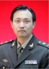 Xuhua Lu (卢旭华)合作论文数中国人民解放军第二军医大学13
 Aimin Chen (陈爱民)合作论文数Shanghai Fourth People's Hospital Affiliated to Tongji University;School of Medicine, Tongji University13Wang Hai-Bo合作论文数11Xiaoqiu Yuan合作论文数10Jialin Jiang合作论文数10XinWei Wang (王新伟)合作论文数10
 Bin Zhang合作论文数Sch Informat Resource Management, Renmin Univ9Le Huan合作论文数8Xiaofei Sun合作论文数8DingLin Zhao (赵定麟)合作论文数8LiangYi Wang (王良意)合作论文数8ZhenYu Cao (曹振羽)合作论文数8LaiGen Wang (王来根)合作论文数8
 Haijun Tian合作论文数7Yong Yang合作论文数7Dan Han合作论文数7YangYuePing Ou (欧阳跃平)合作论文数7
 Sanhuai Gou (苟三怀)合作论文数上海长征医院7MEI Zi-jian (梅子健)合作论文数6
 Xi Luo合作论文数6HanLin Zou (邹翰林)合作论文数6
 Bin Ni (倪斌)合作论文数上海长征医院骨科6Jian Zhu (朱健)合作论文数5Wei-cheng PAN (潘伟成)合作论文数5Lei Shi合作论文数5HongLiang Shen (沈宏亮)合作论文数5JianWei Xu (徐建伟)合作论文数5




