BACKGROUND AND STUDY AIMS:Superficial esophageal cancer (SEC) within a diverticulum is extremely rare and poses a high risk of perforation during endoscopic submucosal dissection (ESD), presenting unique technical challenges. This study aims to evaluate the feasibility, safety, and efficacy of ESD for treating SEC in diverticula. PATIENTS AND METHODS:The retrospective study was conducted on patients diagnosed endoscopically with SEC in a diverticulum who subsequently underwent ESD with histological confirmation of esophageal cancer. Data on demographics, closure methods, incidence of adverse events, histopathological characteristics, and prognostic outcomes were collected from medical records and video recordings of endoscopic procedures and analyzed. RESULTS:Twelve patients were enrolled, and R0 resection was achieved in all cases. Post-ESD wound management varied, tailored to the defect characteristics: titanium clips were used to close wounds with a preserved superficial muscular layer in five patients (5/12, 41.7%), while endoscopic hand-suturing-developed by our research team primarily for closing large mucosal defects after endoscopic resection of colorectal tumors-was performed in three patients (3/12, 25.0%) with defects in the intrinsic muscular layer. Four patients (4/12, 33.3%) with small defects in the muscularis propria were left untreated. None of the patients experienced serious adverse events, such as post-operative bleeding or perforation, and there were no cases of recurrence or metastasis during follow-up. CONCLUSION:This study provides evidence supporting ESD as a feasible, effective, and safe treatment for SEC in diverticula. Tailoring closure techniques to the defect characteristics is crucial for preventing complications and achieving optimal clinical outcomes.
Abstract Background Diagnosing biliary strictures (BSs) presents significant challenges. Brush cytology (BC), a common clinical method, has a suboptimal diagnostic yield. Transpapillary forceps biopsy (TPB) can improve diagnostic accuracy but is technically demanding and has a relatively low success rate, partly due to a lack of effective biopsy forceps. To address this, we have developed a novel type of guidewire-introduced bile duct biopsy forceps. This study retrospectively evaluates the clinical value of this new forceps combined with bile duct brush cytology (BC) compared with BC alone. Methods During the endoscopic retrograde cholangiopancreatography (ERCP), bile duct biopsy was attempted first using the new forceps under fluoroscopic guidance. If feasible, BC was performed afterward. The obtained samples were sent for pathological and cytological examination. If biopsy was unsuccessful, only BC was performed. Results A total of 262 patients were enrolled. The sensitivity of bile duct biopsy was 60.8%, slightly lower than that of BC (66.1%). However, combining TPB with BC significantly increased the diagnostic sensitivity for malignant biliary stricture to 77.7%. Conclusion The newly developed guidewire-introduced bile duct biopsy forceps effectively reduce the procedural difficulty and enhance the success rate of bile duct biopsy. It is a safe and effective tool for diagnosing biliary stricture. Furthermore, combining bile duct biopsy with BC significantly increases the diagnostic accuracy of malignant biliary stricture.
Hereditary multiple osteochondromas (HMO), previously known as hereditary multiple exostoses (HME), is a congenital skeletal developmental anomaly characterized by multiple osteochondromas that commonly grow outward from the metaphyses of long bones. Hereditary multiple exostoses is an autosomal dominant genetic disease characterized by multiple cartilage growth disorders, which affect the long bones of the limbs, scapula, and ribs, among others. Consensus on the diagnosis and treatment of spinal involvement is relatively lacking. For such patients, experience in terms of clinical screening, preoperative evaluation, surgical intervention indications and timing, surgical expertise and lessons learned, and follow-up is needed and critical. This review focuses on the clinical evaluation and orthopedic surgical treatment of HME patients with spinal involvement. It summarizes the patients' diagnosis, clinical features, and treatment strategies based on evidence from the published literature. The clinical manifestations, location of onset, and surgical intervention are summarized in detail in this review. The above contents will help improve the clinical diagnosis and treatment level of HME patients with spinal involvement internationally.
Tumor-induced osteomalacia (TIO) is a rare paraneoplastic disorder characterized by an insidious onset, while its underlying pathogenesis has not yet been fully explored. Identifying and accurately localizing the causative tumors in TIO remains highly challenging in clinical practice. Although complete surgical excision following precise diagnosis is currently considered the most effective treatment approach, surgical management strategies still require optimization, and recurrence may occur even after tumor resection. TIO is primarily recognized as a metabolic bone disease driven by excessive secretion of fibroblast growth factor 23 (FGF23) by tumors; however, its detailed etiological features and molecular pathogenic mechanisms remain unclear. Besides FGF23, other phosphate-regulating hormones and pathogenic genes are also believed to participate in disease development. A clearer understanding of FGF23-mediated phosphate regulation, including hormone secretion, circulation, transport, and interactions with target organs, is critical for early diagnosis and the development of effective therapeutic strategies. Further investigation into the mechanisms responsible for refractory and recurrent TIO is also needed. Integrated multi-omics approaches are expected to provide deeper insight into the complex pathogenic basis of TIO, supporting the development of improved diagnostic and therapeutic strategies. This review summarizes recent progress in understanding the etiology and pathogenesis of TIO, intending to improve disease comprehension and facilitate more effective clinical diagnosis and treatment.
Background: Osteosarcoma is a relatively rare malignant tumor characterized by high aggressiveness and poor prognosis. Traditional diagnostic approaches have several limitations, particularly in early detection and disease monitoring. In recent years, exosome-derived RNAs have attracted increasing attention in cancer research because of their potential relevance to intercellular communication and tumor-related molecular alterations. However, the molecular characteristics associated with plasma exosomal RNAs in osteosarcoma remain poorly understood. Methods: In this study, RNA expression in plasma exosomes from osteosarcoma patients was investigated using differential expression, enrichment, immune infiltration, and competing endogenous RNA (ceRNA) network analyses, followed by in vitro assessment of both RNA and protein expression levels. Results: ceRNA network analysis revealed 7 candidate core molecules and 2 putative ceRNA axes—hsa_circ_0015475/hsa-miR-4753-3p/retinoic acid receptor-related orphan receptor alpha (RORA) and growth arrest-specific 5 (GAS5)/hsa-miR-10a-5p/RORA—which converge on the common messenger RNA RORA. In vitro assessment revealed that RORA messenger RNA and protein expression levels were reduced in osteosarcoma cell lines. A pan-cancer analysis further revealed that RORA was differentially expressed across multiple cancer types. Conclusion: This study revealed 2 potential RORA-related ceRNA axes, hsa_circ_0015475/hsa-miR-4753-3p/RORA and GAS5/hsa-miR-10a-5p/RORA, that may be associated with osteosarcoma-related molecular alterations. The reduced expression of RORA in osteosarcoma cell lines suggests its potential relevance to osteosarcoma biology.
BACKGROUND Endoscopic treatment is the primary therapy for type I gastric neuroendocrine tumors (G-NETs), but it may not address the underlying pathogenesis, increasing the risk of progression. AIM To investigate the effectiveness of endoscopic treatment and identify progression risk factors. METHODS This retrospective study involved 128 patients with type I G-NETs treated between January 2009 and May 2024. The patients were categorized into non-progressive (n = 87) and progressive (n = 41) groups. Baseline characteristics, treatment details, and follow-up data were analyzed using univariate and multivariate Cox regression analyses to identify prognostic variables. RESULTS The baseline characteristics analysis showed no significant differences between the groups. The median follow-up time was 25.5 months (14.00-58.50 months). The univariate and multivariate analyses confirmed that endoscopic treatment combined with adjuvant somatostatin analogs (SSAs) was associated with a lower risk of progression (hazard ratio = 0.38, 95% confidence interval: 0.17-0.90, P = 0.027), whereas a neutrophil-to-lymphocyte ratio (NLR) of ≥ 2 indicated a higher risk (hazard ratio = 2.14, 95% confidence interval: 1.08-4.26, P = 0.030). Kaplan-Meier analysis confirmed NLR ≥ 2 and adjuvant SSA use as independent prognostic variables. CONCLUSION Combining endoscopic treatment with SSAs is effective for managing type I G-NETs. SSAs and NLR were identified as independent prognostic factors, highlighting their potential to reduce recurrence risk and improve outcomes.
BACKGROUND:Neuroendocrine tumors (NETs) are relatively rare in the duodenum. We aimed to compare the clinical outcomes of endoscopic submucosal dissection (ESD) and laparoscopic endoscopic cooperative surgery (LECS) for non-ampullary duodenal NETs. MATERIALS AND METHODS:This single-center retrospective study included patients with non-ampullary duodenal NETs treated with ESD or LECS between 2010 and 2024. Data on patient demographics, surgical outcomes, and short- and long-term prognoses were collected. RESULTS:We enrolled a total of 29 patients. No significant differences were observed between the ESD and LECS groups with regard to sex, age, height, weight, or tumor location. The median tumor diameter was significantly larger in the LECS group than in the ESD group (12.00 mm vs. 5.00 mm). The tumors in the LECS group were located deeper (submucosa or muscularis propria) than those in the ESD group. Both groups achieved high rates of en bloc resection (100% for ESD and 100% for LECS) and R0 resection (100% for ESD and 93.3% for LECS). There were no significant differences in postoperative complications, such as bleeding, stenosis, or abdominal infection between groups. The median duration of hospitalization was longer in the LECS group than in the ESD group (11.00 vs. 7.00 days). No recurrence or metastasis was observed in either group during follow-up. CONCLUSION:Both ESD and LECS demonstrated high en bloc resection rates and R0 resection rates, along with favorable short- and long-term prognoses for non-ampullary duodenal NETs. LECS appears to be a safe and effective treatment option with an efficacy comparable to that of ESD and may be suitable for larger or deeply infiltrating lesions, potentially helping avoid the need for more invasive surgical procedures.
BACKGROUND:Lesions manifesting after radiotherapy are difficult to treat. Significant inflammations often manifest at the rectal wall after radiotherapy, the rectal wall often experiences significant inflammation, and the intestinal cavity is typically in an edematous state. METHODS:This retrospective study included 22 patients who had previously undergone radiotherapy or chemoradiotherapy and subsequently developed rectal lesions. All patients underwent either endoscopic mucosal resection (EMR) or endoscopic submucosal dissection (ESD) for lesion removal. Data on demographics, treatment modalities, time to lesion detection, lesion characteristics, and clinical outcomes were collected. The safety and efficacy of the endoscopic treatments were evaluated, with follow-up information gathered through in-person interviews and telephone calls. RESULTS:The mean age of 22 patients was 62.1 years, and the mean time to lesion detection after initial treatment was 90.6 months post-initial treatment. Most patients (77.3%) had a single lesion, and EMR or ESD were successfully performed with no major postoperative complications. Most lesions were adenomas (90.3%). 72.7% of patients were alive during the entire follow-up period, and 13.6% survived for more than 5 years. No significant complications, such as perforation or bleeding, were reported. CONCLUSION:Endoscopic resection with EMR and especially ESD appears feasible and safe for selected patients with small rectal lesions after radiotherapy. These preliminary findings highlight the importance of endoscopic surveillance for early detection and timely management. Larger multicenter studies are warranted to validate the feasibility and safety of this approach and to better define its long-term outcomes.
Percutaneous endoscopic gastrostomy provides a safe and effective minimally invasive surgical route, thus ensuring long-term enteral nutritional support therapy [1]. Patients with esophageal cancer may develop scars from long-term radiotherapy, causing complete esophageal obstruction and difficulties in replacing gastrostomy tubes. Our center utilizes a new approach to replacing gastrostomy tubes by incising the atretic segment of the esophagus through transoral and transgastric fistula bimodality ([Video 1]).
This study presents a preoperative prediction model for tumor-induced osteomalacia (TIO) surgery outcomes on the basis of patient characteristics. The model, which was validated in 309 patients, identifies key risk factors and aids in clinical decision-making to optimize treatment strategies, reduce the number of unnecessary surgeries, and improve patient care. Tumor-induced osteomalacia (TIO) should be curable by complete removal of the causative tumor. Knowledge of the prognosis of surgery is lacking. This study aimed to establish a prediction model that uses the preoperative characteristics of patients to predict the surgical treatment outcomes of patients with TIO. This was a single-center, retrospective, case-control study. The main outcome was the surgical outcomes of patients with TIO. Patients with TIO who underwent surgical treatment were divided into a training set and a validation set. A nomogram was established in the training set, and the model was evaluated by the C-index, calibration curve, and clinical impact curve and verified in the validation set. A total of 309 patients with TIO were included, with 222 in the training set and 87 in the validation set. The C-index of the nomogram was 0.864 (p < 0.001). The model had high goodness of fit—which is suggested by the calibration curve, and clinical benefit is indicated by the decision curve analysis and clinical impact curve. In the validation set, the area under the curve of the prediction model was 0.782 (p < 0.001), and decision curve analysis and clinical impact curve also suggested the existence of clinical benefit. This study established a prognostic model for the preoperative prediction of surgical outcomes for TIO. This model can be used as a reference in clinical practice for the development of individualized treatment strategies.
Inflammatory myofibroblastic tumor (IMT) is a rare spindle-cell neoplasm. IMT currently suffers from a paucity of standardized diagnostic and therapeutic guidelines. The Chinese expert consensus committee on the diagnosis and treatment of IMT formed an "Expert consensus on the diagnosis and treatment of inflammatory myofibroblastic tumor". This consensus was developed through a comprehensive synthesis of expert opinions, an extensive review of the literature, and a series of offline and online deliberations. The committee aspires that this consensus will enhance the therapeutic outcomes and prognosis for patients with IMT in the future.
The debate over the optimal reconstruction technique following proximal gastrectomy continues. Transhiatal tunnel valvuloplasty (ThTV) is a novel esophagogastrostomy method. This study aimed to evaluate the feasibility and safety of ThTV. A cohort with upper early gastric cancer or Siewert type II tumors who underwent laparoscopic proximal gastrectomy was retrospectively reviewed in a single center. The ThTV esophagogastrostomy procedure involved placing a lengthy gastric tube into the lower mediastinum and firmly binding it to the esophagus. Demographic and surgical morbidity data were extracted from the medical records. Between March 2023 and November 2023, 15 patients underwent laparoscopic proximal gastrectomy using ThTV. The cohort consisted of 13 males and 2 females, with a median age of 68 years (range 49-77). The median operative time was 213 minutes (range 171-370). The median times for tunnel construction and anastomosis were 7 minutes (range 4-30) and 17 minutes (range 10-29), respectively. The median tumor size was 2.0 cm (range 1.0-5.0), and the median number of lymph nodes dissected was 29 (range 13-49). TNM staging revealed 9 cases of stage I, 5 cases of stage II, and 1 case of stage III. As of January 1, 2025, the median follow-up duration was 16.8 months (range 13.8-22.2). No tumor recurrence was observed. No patients reported severe reflux symptoms (Visick score ≥III). Gastroscopy confirmed reflux esophagitis (Los Angeles classification Grade A) in one patient, and an anastomotic stricture requiring endoscopic balloon dilation was observed in another patient. Transhiatal tunnel valvuloplasty is a simple and reliable anti-reflux method following laparoscopic proximal gastric surgery. The further verification of the esophageal function is warranted.
Background:The effectiveness of endoscopic screening for upper gastrointestinal (UGI) tract cancers in high-risk areas of China has been well-established. However, the practicality of extending this screening to a wider geographical area remains uncertain. To bridge this gap, we have conducted a hospital-based opportunistic endoscopic screening (OpENS) program for UGI cancers since 2018. Our objectives were to elucidate the implementation process of the OpENS program and assess its effectiveness. Methods:875 hospitals from 710 districts/counties have participated in the OpENS program during 2019-2023. The endoscopic specialists and pathologists participating in the program were mandated to take annual training programs to acquire the fundamentals of screening techniques. Eligible patients who underwent endoscopic examinations were screened for UGI cancers. Patients diagnosed with high-grade intraepithelial neoplasia (HGIN), carcinoma in situ (CIS) and tumors in esophagus or/and stomach were defined as positive cases. Patients with HGIN and CIS were defined as early cases. All hospitals were required to submit screening data via the program's platform, with both the quality of the submitted data and the hospitals' performance being subject to a comprehensive evaluation. The age-standardized incidence rates (ASIRs) for the districts/counties where the participating hospitals were situated were derived from the cancer registry data for the year 2020. Districts/counties with ASIRs for UGI cancers over 22.0/105 were classified as high-risk areas. The positive detection rate (PDR) and early diagnosis rate (EDR) were calculated. Findings:After data cleaning, we included 808 hospitals from 616 districts/counties, with a collective participation of 7,066,892 individuals during 2019-2023. The overall PDR and EDR across all sites were 2.35% and 19.77%. The PDRs and EDRs were 1.02% and 23.18% in esophagus, and were 1.37% and 17.80% in stomach. The PDR was higher among males compared to females (3.59% vs 1.20%), and was increasing with age. The EDR was higher among females compared to males (20.66% vs 19.45%), peaking in the age group of 60-64 years. The PDRs and EDRs were higher in high-risk areas of UGI cancers (p < 0.05). After adjusting for age, sex, province, year of screening, regional UGI cancer incidence level and hospital tier, the hospitals that consecutively participated in the program for five years demonstrated higher PDRs and EDRs when compared to other hospitals (p < 0.05). Among the consecutively participated hospitals, tertiary-level hospitals demonstrated positive associations with the PDRs for both the esophagus and stomach when compared to secondary-level hospitals (p < 0.001). However, the tertiary-level hospitals showed a negative association with the EDR for the esophagus (OR = 0.91, 95% CI: 0.86-0.96, p = 0.001), but exhibited a positive association with the EDR for the stomach (OR = 1.11, 95% CI: 1.05-1.17, p < 0.001). Interpretation:The OpENS program has rapidly expanded across the country. The program has demonstrated high PDRs and EDRs, with hospitals that performed well exhibiting significantly better screening outcomes. Among these well-performed hospitals, secondary-level hospitals can achieve comparable results to tertiary-level hospitals in screening for esophageal lesions, albeit not for stomach lesions. Considering the current hospital capacities in China, the training-guided opportunistic UGI endoscopy screening exhibits significant feasibility and effectiveness across areas with varying level of UGI risks. Funding:CAMS Innovation Fund for Medical Sciences (No. 2021-I2M-1-061, 2021-I2M-1-023).
CONTEXT:Phosphate homeostasis was compromised in tumor-induced osteomalacia (TIO) due to increased fibroblast growth factor 23 (FGF23) secretion. Nevertheless, the glucose metabolic profile in TIO patients has not been investigated. OBJECTIVES:This work aimed to clarify the glucose metabolic profiles in TIO patients and explore their interaction with impaired phosphate homeostasis. METHODS:20 TIO patients, 20 individuals with normal glucose tolerance, and 20 patients with type 2 diabetes mellitus (DM) were enrolled and underwent an oral glucose tolerance test (OGTT). Serum phosphate and FGF23 concentration were monitored during OGTT. RESULTS:In patients with TIO, 60% (12/20) exhibited impaired glucose tolerance (IGT) and 5% (1/20) had type 2 DM. Those with IGT or type 2 DM experienced more ambulatory difficulties (69.2% vs 42.9%), lower phosphate concentrations (0.43 ± 0.10 vs 0.53 ± 0.10, P = .042), and lower calcium concentrations (2.20 ± 0.08 vs 2.30 ± 0.40, P = .001) compared to TIO patients without these conditions. According to correlation analysis, serum phosphate levels were negatively correlated with plasma glucose levels at 60 minutes (P < .001), fasting plasma insulin levels (P < .05), and homeostasis model assessment for insulin resistance (P < .05). Those with high FGF23 levels had a higher glucose level at 60 minutes (10.5 [9.3, 12.3] vs 7.3 [6.4, 10.1], P = .048) than that of low group. After glucose loading, both FGF23 and phosphate levels exhibited a decreasing trend. CONCLUSION:The development of diabetes in TIO patients may be predisposed by ambulatory issues, low phosphate, and elevated FGF23 levels. Dysglycemia might further aggravate hypophosphatemia.
Objective: To evaluate the efficacy and safety of neoadjuvant chemotherapy (NAC) compared to NAC combined with immune checkpoint inhibitors (ICI) in patients with muscle-invasive bladder cancer (MIBC). Propensity score matching (PSM) was employed to assess the impact of these two treatment regimens on the pathological complete response rate (pCR) and overall survival (OS). Methods: A retrospective analysis was conducted on 320 MIBC patients treated at the Cancer Hospital affiliated to Sun Yat-sen University Gansu Hospital between January 2017 and June 2022. Patients were categorized into the NAC group (n=194) and the NAC+ICI group (n=126) based on their treatment regimens. After PSM, 154 patients were included, with 77 in each group. Baseline characteristics, clinical efficacy, and prognosis were analyzed using various statistical methods. Results: Before PSM, significant differences were observed between the groups in baseline characteristics, including tumor diameter, tumor number, and adjuvant treatment (all P<0.05). After PSM, these differences were no longer statistically significant (all P>0.05). The NAC+ICI group demonstrated a significantly higher pCR rate both before and after PSM (both P<0.001). Similarly, pathological downstaging rates were higher in the NAC+ICI group before and after PSM (both P<0.001). However, there was no significant difference in disease control rates between the two groups before (P=0.057) and after PSM (P=0.240). Logistic regression analysis identified the treatment regimen (before PSM: P<0.001, OR=0.161; after PSM: P<0.001, OR=0.141) and complications (before PSM: P=0.005, OR=2.339; after PSM: P=0.019, OR=2.753) as independent risk factors for pCR. Cox regression analysis revealed that age (before PSM: P<0.001, HR=1.059; after PSM: P=0.011, HR=1.066), pretreatment T stage (before PSM: P<0.001, HR=2.342; after PSM: P<0.001, HR=3.244), tumor diameter (before PSM: P=0.005, HR=1.810; after PSM: P=0.025, HR=2.077), and treatment outcome (before PSM: P<0.001, HR=1.722; after PSM: P=0.020, HR=1.444) were independent prognostic factors for OS. Conclusion: NAC combined with ICI significantly improves pCR and pathological downstaging rates in MIBC patients. Independent prognostic factors affecting OS include age, pretreatment T stage, tumor diameter, and treatment outcome.
Currently, due to the invasive nature of colonoscopy and the associated pain, people avoid undergoing the procedure, making it difficult to detect the majority of potential early stage colorectal carcinoma/precancerous lesions or advanced adenoma. Advanced colorectal adenoma is the main precursor to the development of colorectal carcinoma. Therefore, improving advanced colorectal adenoma detection rate can significantly decrease the development and morbidity of colorectal carcinoma. Accordingly, a non-invasive method to screen high-risk people for colonoscopy in clinical practice is urgently needed. With the development of medical technology, screening methods for colorectal carcinoma are emerging rapidly, and diverse non-invasive methods are being developed. Cell-free DNA (cfDNA), commonly referred to as liquid biopsy, has promising application prospects as a minimally invasive strategy for early screening of colorectal cancer. CfDNA has already been applied in the field of prenatal diagnosis, advanced carcinoma, and organ transplantation, and the application cfDNA in advanced colorectal adenoma is at the cutting-edge of current research. Thus, this review summarizes the progress in research on different biological characteristics of cfDNA and its utility in the screening of advanced colorectal adenoma, including sizes of cfDNA molecules, end signature of cfDNA (preferred ends, end motifs, jagged ends), nucleosomal footprints, cfDNA topology, cfDNA methylation, and cfDNA integrity. We hope that this review will advance this promising research field.
 Xi Zhou合作论文数97Shuzhong Liu合作论文数84
 Guiqi Wang (王贵齐)合作论文数Cancer Hospital Chinese Academy of Medical Sciences, Shenzhen78
 Yipeng Wang (王以朋)合作论文数Peking Union Medical College Hospital62Shun He (贺舜)合作论文数57
 Lizhou Dou (窦利州)合作论文数Cancer Hospital Chinese Academy of Medical Sciences56Yueming Zhang合作论文数51An Song合作论文数48Zhen Huo (霍真)合作论文数39
 Weibo Xia (夏维波)合作论文数Peking Union Medical College Hospital26
 Liyan Xue (薛丽燕)合作论文数国家癌症中心/中国医学科学院肿瘤医院病理科24
 Yan Ke (柯岩)合作论文数中国医学科学院肿瘤医院22Xudong Liu (刘旭东)合作论文数18
 Guixing Qiu (邱贵兴)合作论文数Peking Union Medical College Hospital18
 Mei Li (李眉)合作论文数Institute of Medicinal Biotechnology, Chinese Academy of Medical Sciences16
 Wei Yu (余卫)合作论文数Peking Union Medical College Hospital, Chinese Academy of Medical Science15
 Jianguo Zhang (仉建国)合作论文数Peking Union Medical College Hospital15
 Yan Jiang (姜艳)合作论文数Peking Union Medical College Hospital15
 Xisheng Weng (翁习生)合作论文数Peking Union Medical College Hospital15
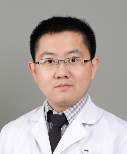 Huan-Wen Wu (吴焕文)合作论文数Peking Union Medical College Hospital14
 Xiaoping Xing (邢小平)合作论文数Peking Union Medical College Hospital14Xiang Li合作论文数12Siyuan Yao合作论文数12Lian Zhou合作论文数12
 Li Huo (霍力)合作论文数Peking Union Medical College Hospital12Ning Lu合作论文数12
 Ou Wang (王鸥)合作论文数Peking Union Medical College Hospital12Ruizhi Jiajue合作论文数11Yue Chi合作论文数11
 Zhihong Wu (吴志宏)合作论文数Peking Union Medical College Hospital11Xinying Yu合作论文数10
 Wei Lv (吕威)合作论文数Peking Union Medical College Hospital10A Song合作论文数9
 Qianqian Pang合作论文数Chinese Academy of Medical Sciences, Peking Union Medical College Hospital9Deli Zhao合作论文数9Yumeng Liu合作论文数9Zhengqi Li合作论文数9
 Yu Xia (夏宇)合作论文数Peking Union Medical College Hospital9
 Lijia Cui合作论文数8Y-P Wang合作论文数8Xiaolin Ni合作论文数8YueMing Zhang (张月明)合作论文数8
 Zhihao Chen合作论文数School of Earth Sciences and Engineering, Hohai University7
 Dongbing Zhao (赵东兵)合作论文数Cancer Hospital Chinese Academy of Medical Sciences7Xunwu Meng (孟迅吾)合作论文数7Xiu-min Qin合作论文数6Yiyi Gong合作论文数6Xiao Liu合作论文数6
 Sanford M. Dawsey合作论文数Division of Cancer Epidemiology & Genetics, National Cancer Institute6




