Background Perineural invasion (PNI) is a hallmark of malignancy in solid tumors, including oral squamous cell carcinoma (OSCC), and is closely associated with poor prognosis. Emerging evidence suggests a critical association between PNI and the tumor immune microenvironment (TME). Methods In this study, we used spatial transcriptomics, in vitro and in vivo experiments, and clinical specimen validation to systematically study the interplay between neural infiltration and immune modulation in OSCC. Results Our results suggest that intratumoral nerves may enhance the recruitment of regulatory T cells (Tregs) through C-C motif chemokine ligand 5 (CCL5)-mediated chemotaxis and further promote the acquisition of an immunosuppressive phenotype in Tregs by activating the RAMP1 signaling pathway. Through these coordinated mechanisms, neural components in the TME contribute to immune suppression and facilitate tumor progression. Therapeutically, both combination of CCL5 blockade with anti-CTLA-4 antibody and the combination of RAMP1 blockade with anti-PD-1 antibody exhibited significantly enhanced anti-tumor efficacy. Conclusions This study highlights a previously underappreciated neural-Treg axis in the TME and provides new insights into potential combinatorial strategies for cancer immunotherapy.
BACKGROUND:Pain is the most frequent complication in patients with oral squamous cell carcinoma (OSCC) and is associated with poor prognosis. Calcitonin gene-related peptide (CGRP), abundantly expressed in sensory neurons, has been implicated in multiple pain disorders; however, its role in cancer-associated pain remains unclear. We investigated the relationship between CGRP and OSCC-associated pain and evaluated the analgesic potential of Rimegepant, a CGRP-receptor antagonist approved for migraine. METHODS:Plasma CGRP was measured in OSCC patients and correlated with preoperative pain scores and clinical outcomes over a 30-month follow-up. Immunohistochemistry quantified CGRP within tumor-infiltrating nerve fibers. In a unilateral orthotopic tongue OSCC mouse model, we compared the analgesic effects of Rimegepant with carprofen and tramadol. RNA-sequencing of ipsilateral trigeminal ganglia from treated mice was performed to explore potential mechanisms. RESULTS:Plasma CGRP levels were positively correlated with patient-reported pain severity and were associated with reduced overall survival. Increased CGRP expression in tumor-infiltrating nerves was linked to greater pain intensity and lymph node metastasis. In an OSCC mouse model, both systemic and local administration of Rimegepant significantly attenuated facial mechanical hypersensitivity. Transcriptomic analysis of trigeminal ganglia, together with RT-qPCR validation, revealed suppression of innate immune and neuroinflammatory pathways following Rimegepant treatment. CONCLUSIONS:Our findings link sensory-nerve-derived CGRP to OSCC-associated pain and support further evaluation of CGRP receptor antagonism as a potential analgesic strategy in cancer.
The clinical role of thermal ablation in thyroid disease management has broadened substantially over the past decade as imaging guidance and minimally invasive technologies have advanced. Although thermal ablation is increasingly used in selected thyroid conditions, global practice varies considerably in patient eligibility, treatment indications, and procedural standardization. Countries with extensive clinical experience in thyroid thermal ablation have helped shape the evolving use of this technology. In China, thermal ablation has been widely implemented in high-volume centers for benign thyroid diseases, low-risk thyroid carcinoma, and metastatic cervical lymph nodes, generating a substantial body of real-world evidence. This experience has highlighted the need for structured, consensus-based recommendations that harmonize clinical practice and clarify appropriate indications. To address this need, the Interventional Ultrasound Committee of the Chinese College of Interventionalists, together with relevant academic societies and multidisciplinary experts in surgery, interventional medicine, ultrasound, endocrinology, oncology, pathology, and nuclear medicine, developed this consensus through a formal methodology combining a modified Delphi process, expert panel discussions, and iterative revisions. The consensus integrates available evidence with collective clinical experience to define key indications, contraindications, and procedural considerations for thyroid thermal ablation. This multidisciplinary expert consensus represents an updated synthesis of practice-oriented recommendations derived from extensive clinical application in China. By outlining standardized approaches and identifying areas requiring further investigation, it serves as a practical reference for clinicians and contributes to ongoing international dialogue on the appropriate use of thermal ablation in thyroid disease management.
Immunotherapy has revolutionized the treatment of malignant tumors and is now recognized as a first-line option for various cancers. In resectable locally advanced oral squamous cell carcinoma (OSCC), neoadjuvant immunotherapy has been integrated into clinical practice, representing significant progress. While neoadjuvant immunochemotherapy shows promising efficacy in this setting, several critical challenges remain unresolved. These include defining optimal endpoints and response evaluation methods, identifying and managing hyperprogression, determining surgical strategies for patients with significant tumor reduction, assessing the feasibility of de-escalating postoperative adjuvant therapy in those achieving pathological complete response, and managing immune-related adverse events. This consensus addresses these challenges by integrating current evidence with pressing clinical questions and incorporating multidisciplinary expert insights from the OSCC field. The objective is to establish a standardized, unified framework for evaluating and managing these complex issues in resectable locally advanced OSCC.
Background: Neutrophil extracellular traps (NETs) are network structures composed of DNA, histones, and antimicrobial proteins,released by activated neutrophils to trap and eliminate extracellular pathogens. Recent research has demonstrated a strong correlation between NETs and various diseases, including immune dysregulation, thrombosis, and malignancies. This review synthesizes current research on NETs, focusing on its biological role in oral squamous cell carcinoma (OSCC) and explores its potential in treating. Methods: A literature review in the PubMed database was conducted to examine the impact of NETs on the homeostasis of oral microbiota and the involvement in the development of oral microbiota-related carcinogenesis. Results: Various microorganisms, including Porphyromonas gingivalis, Fusobacterium nucleatum, Streptococcus spp., along with Candida albicans, as well as certain viruses such as Human papillomavirus (HPV), Human herpes virus 8 (HHV-8), and Herpes simplex virus-1 (HSV-1)are regulated by NETs during oral colonization and proliferation and have been identified as contributors to the pathogenesis of oral squamous cell carcinoma. NETs have been shown to play a dual role in the carcinogenic process of oral microbiota in humans. At the initial stage of tumor formation, NETs inhibit tumorigenesis by eliminating tumorigenic bacteria that infiltrated the tumor; however, following tumor establishment, various cytokines and chemokines that promote tumor progression are released by neutrophils during the NETs formation. Conclusion: This article reviews the oncogenic mechanisms of NETs in the oral microbiota, with potential implications for early tumor detection and the development of microbe-targeted therapies.
e18098 Background: Locally advanced squamous cell carcinoma of the head and neck (HNSCC) is associated with high risks of local recurrence and distant metastasis, resulting in poor prognosis. Neoadjuvant immunochemotherapy (NICT) has emerged as a promising strategy to improve outcomes. This study aimed to explore the safety and efficacy of penpulimab (PD-1 blockade) plus chemotherapy as neoadjuvant treatment for patients with HNSCC. Methods: This phase II trial enrolled patients with untreated, locoregionally advanced, resectable HNSCC (Stage III/IVa). Patients received three cycles of NICT consisting of penpulimab (200 mg), nab-paclitaxel (260 mg/m²), and cisplatin (75 mg/m²) on day 1 of each 21-day cycle, followed by surgery. The primary endpoint was the major pathological response rate (MPR). Postoperative patients achieving MPR were randomized 1:1 to receive either alternative adjuvant treatment (Penpulimab for low-to-intermediate recurrence risk, radiotherapy + Penpulimab for high recurrence risk) or standard treatment. Those not achieving MPR received standard treatment. Secondary endpoints included 2-year DFS, ORR, local & distant MFS, OS, pCR, and safety. Results: As of January 15, 2025, 63 patients who met the inclusion and exclusion criteria were enrolled, with a median age of 59 years, 79.4% male, 92.1% with an ECOG score of 0, 92.1% with oral cancer, and 46.0% and 54.0% in Stage III and IVa, respectively. A total of 59 patients completed neoadjuvant treatment, achieving an ORR of 86.4% and a DCR of 98.3%. Among them, 50 patients underwent surgery, all achieving R0 resection. Pathological evaluations revealed that 33 patients (66.0%) met the MPR criteria. In terms of safety, 88.9% of patients experienced all-grade treatment-related adverse events (TEAEs), with 27.0% reporting ≥Grade 3 TEAEs, mainly leukopenia (4.8%) and bone marrow suppression (4.8%). Immune-related adverse events (irAEs) occurred in 38.1% of patients, with 6.3% experiencing ≥Grade 3 irAEs. Conclusions: Neoadjuvant penpulimab combined with chemotherapy demonstrated high ORR and MPR rates, with an acceptable safety profile in resectable HNSCC. These results support further investigation of this regimen in larger, randomized trials. Clinical trial information: NCT06081673 .
e18005 Background: Targeting vascular endothelial growth factor receptor (VEGFR) may synergistically enhance the antitumor effects of immune checkpoint inhibitors (ICIs). This study evaluated the efficacy and safety of camrelizumab with chemotherapy (Arm A) or apatinib (Arm B) as first-line treatment in patients with recurrent or metastatic head and neck squamous cell carcinoma (R/M HNSCC). Methods: This open-label, double-cohort, multicenter, phase II study (NCT05156970) enrolled patients with recurrent or metastatic HNSCC who had not received prior systemic therapy for metastatic or recurrent disease. In Arm A, patients received camrelizumab (200 mg intravenous, day 1), followed by docetaxel (75 mg/m²) and cisplatin (75 mg/m²) or carboplatin (area under the curve 5) on day 2 every 3 weeks for up to six cycles, followed by camrelizumab monotherapy (200 mg intravenous, day 1, every 3 weeks). In Arm B, patients received camrelizumab (200 mg intravenous, day 1, every 3 weeks) plus oral apatinib (250 mg daily). The primary endpoint was overall survival (OS). Secondary endpoints included objective response rate (ORR), progression-free survival (PFS), and safety. Results: Between July 2021 and May 2024, 81 patients were enrolled (41 in Arm A and 40 in Arm B). The median follow-up was 11.93 months. The median OS was 16.40 months (95% confidence interval [CI], 4.13-28.67) in Arm A and 21.00 months (95% CI, 16.63-25.37) in Arm B. The median PFS was 4.57 months (95% CI, 1.11-8.03) in Arm A and 4.20 months (95% CI, 0.00-8.54) in Arm B. The ORR was 46.3% (95% CI, 32.0%-61.0%) in Arm A and 50.0% (95% CI, 35.0%-65.0%) in Arm B. Grade 3 or higher treatment-related adverse events occurred in six patients (14.6%) in Arm A and seven (17.5%) in Arm B. Conclusions: Camrelizumab combined with chemotherapy or apatinib demonstrated encouraging survival outcomes, with a favorable safety profile. These findings support further investigation of camrelizumab-based regimens as first-line treatments for patients with recurrent or metastatic HNSCC. Clinical trial information: NCT05156970 .
Oral squamous cell carcinoma (OSCC) presents a substantial global health threat with high incidence and mortality rates. Locoregional recurrence remains a major challenge. Recent advancements in near-infrared (NIR) fluorescence-guided surgery (FGS), particularly using Indocyanine Green (ICG), shows promise in enhancing both diagnostic precision and surgery guidance. ICG exploits the enhanced permeability and retention (EPR) effect, allowing it to accumulate in tumor tissue and guide surgical resection. However, the application of fluorescence imaging with ICG in the surgical treatment of OSCC is still in its early stages, lacking standardized protocols in indications, procedural guidelines, and delivery precautions. Herein, we summarized collaborative consensus among experts from multiple medical institutions and references from both domestic and international literature, as well as multicenter clinical studies on ICG-guided surgery for OSCC, aiming to provide guidance for clinical practices in OSCC surgical treatment with ICG. This consensus advocates for the widespread adoption of ICG-guided surgery to improve surgical navigation, tumor margin delineation and efficiency of pathological assessment in OSCC treatment.
Whether and how cancer exploits distant organs to escape immune surveillance remains largely unknown. Using clinical data from head and neck squamous cell carcinoma (HNSCC) patients and three murine oral cancer models, we find that cancer cells under immune pressure secrete slit guidance ligand 2 (SLIT2) through an activating transcription factor 4 (ATF4)-dependent pathway, which activates tumor-innervating nociceptive neurons and aggravates cancer-induced pain. This activation then stimulates tumor-draining lymph-node (TDLN)-innervating nociceptive neurons and increases calcitonin gene-related peptide (CGRP) secretion, remodeling TDLNs into an immune-suppressed state. Consequently, decreased CCL5 secretion from immune-suppressed TDLNs promotes M2-like polarization of tumor-associated macrophages, facilitating tumor growth and reducing immune checkpoint blockade (ICB) efficacy. Targeting nociceptive neurons or the ATF4-SLIT2-CGRP axis restores immune activity, alleviates cancer-induced pain, and improves ICB responses. Our findings reveal an inter-organ neuroimmune circuit co-opted by cancer to escape immune surveillance, suggesting potential therapeutic strategies to enhance immunotherapy.
Oral squamous cell carcinoma (OSCC) was the most common malignant type of head and neck squamous cell carcinoma (HNSCC) with a low survival rate. The microbiota in oral cavity or tumor tissues may play a critical role in the OSCC. In this study, we characterized the microbiota from oral cancer tissues, oral swabs and saliva of patients with OSCC using 16S rRNA sequencing. We found differential profiles and amounts of microbiota in oral cancer tissues compared with adjacent tissues, as well as in oral swabs and saliva from OSCC patients compared with healthy individuals. Fusobacterium nucleatum and Porphyromonas endodontalis were found increased in cancer tissues and saliva from OSCC patients. Prevotella melaninogenica was found increased in the saliva and oral swabs from OSCC patients. These data suggested that microbiota varied according to different samples. Kyoto Encyclopedia of Genes and Genomes (KEGG) analysis indicated an important role of metabolic pathways in the interaction between microbiota and cancers. Then we analyzed the metabolites from cancer tissues and saliva of OSCC patients by liquid chromatograph-mass spectrometry/mass spectrometry (LC-MS/MS) and gas chromatograph-mass spectrometry (GC-MS). Differential profiles of metabolites were also observed in the cancer tissues compared with adjacent tissues and in the saliva from OSCC patients compared with healthy individuals. It showed that denticulaflavonol was significantly increased while D-mannose was significantly decreased in both cancer tissues and saliva of OSCC patients. Taken together, these results suggested an association between microbiota/metabolites (such as Fusobacterium and mannose) and OSCC, in which the molecular mechanism need further investigated.
Head and neck squamous cell carcinoma (HNSCC) is a common malignant tumor that affects the head and neck area. HNSCC typically has complex clinical symptoms and is detected at a late stage. Therefore, it is quite difficult to give patients the best possible care and treatment. Faced with these challenges, nanomaterials are receiving increasing attention in the biomedical field due to their unique structural and physicochemical properties. The application of nanomaterial therapeutics has produced innovative HNSCC treatment options that go beyond the constraints of conventional surgery, radiation therapy, and chemotherapy. In this review, we first reviewed the pathological mechanisms of HNSCC. Subsequently, a detailed introduction to nanomaterials was given, including their classification and definition. Then, the use of nanomaterials in the diagnosis and therapy of HNSCC was discussed, and incorporating the perspective of organoids and clinical trials to increase the feasibility of nanomaterial applications. In addition, we also reviewed the advantages and disadvantages of nanomaterials, proposed the idea of nanomaterial design, looking at how these properties may be used to improve our understanding and treatment of HNSCC. The present approaches for detecting and treating HNSCC can be improved by integrating the special qualities of nanomaterials with the available therapeutic modalities. This will eventually enhance patient outcomes in the battle against this highly invasive malignant tumor.
BACKGROUND:Previous studies have highlighted the critical role of the Yes associated protein 1 - TEA domain transcription factor (YAP1-TEAD) interaction in the progression of oral cancer. However, there remains a lack of direct disruptors targeting the YAP1-TEAD protein interaction and a detailed investigation of their effects on oral cancer. Therefore, we aimed to rationally design peptide YAP1-TEAD protein interaction disruptors through computational methods and explore the effects of direct disruption of the YAP1-TEAD interaction in oral squamous cell carcinoma (OSCC). METHODS:Peptide-based computational residue scanning was employed to design peptide disruptors of YAP1-TEAD interaction by analyzing the effects of mutations on binding affinity and stability. Molecular dynamics (MD) simulations were performed to assess the binding affinity and stability from a dynamic perspective. Surface plasmon resonance (SPR) assays were used to assess the in vitro binding affinities. After three rounds of iterative optimization, two with the highest affinities of this series of disruptors were applied to OSCC cell lines to investigate the primary effects of direct disruption of the YAP1-TEAD interaction. RESULTS:The expression of YAP1, TEADs, and four downstream transcriptional targets were significantly higher in OSCC tissues compared to paired normal tissues. The design and evaluation of YAP1-TEAD disruptors were conducted using computational residue scanning, MD simulations, and SPR assays. After three rounds of optimization, YTPD9 and YTPD11 demonstrated the highest affinities, with binding affinities 10 times more than that of the wildtype YAP1. When applied to OSCC cell lines, the disruptors exhibited a limited effect on cell proliferation, but they were able to inhibit the abilities of migration and invasion effectively. CONCLUSION:This study developed a series of peptide disruptors targeting YAP1-TEAD protein interaction through computational simulations and in vitro experiments. The biological evaluation of two of these disruptors in OSCC confirmed that direct disruption of the YAP1-TEAD primarily affects the migration and invasion ability of oral cancer.
Background/purpose: Discoid lupus erythematosus (DLE) serves as an interdisciplinary disease involved in dermatology and stomatology in many cases. The purpose of this study was to analyze the scientometric characteristics and research trends of DLE. Materials and methods: All the papers on DLE were comprehensively retrieved from the Scopus database. Discipline comparison (dermatology versus others) and chronological comparison (before 2005 versus 2005-2024) were performed, so that the number of papers can be to some extent compared in the analysis. Results: Among all the 1239 papers on DLE, 738 (59.6 %) were published by dermatologists. Diagnosis aspect, skin manifestation, facial dermatoses, scar formation, hair loss, epiluminescence microscopy, dermoepidermal junction, arthralgia, anamnesis, young adult, Ro antibody, and dermoscopy were distinctive keywords for dermatologists; While Mouth mucosa, mouth diseases, lip neoplasms, eyelid disease, blepharitis, conjunctivitis, autoantibody, blood, neutrophil, fever, sex difference, preschool child were distinctive keywords for other scholars. Treatment response, dapsone, etretin, clobetasol, mycophenolate mofetil, glucocorticoid, triamcinolone, and drug safety were therapeutic keywords for dermatologists. Whereas quinacrine, alpha tocopherol, hydrocortisone, adverse drug reaction were therapeutic keywords for other scholars. The trend has changed to complication, disease duration, young adult, dermatitis, pruritus, antibody titer, Ro antibody, anamnesis, dermoscopy, arthralgia, dermoepidermal junction, keratosis, and African American, drug withdrawal, tacrolimus, mycophenolate mofetil, etretin, clobetasol, cyclophosphamide, and drug safety after 2005. Conclusion: This scientometric study elucidated the current scenario and research trends of DLE, and would help in improving in reciprocal collaboration and communication for investigations on this disease. 2025 Association for Dental Sciences of the Republic of China. Publishing services by Elsevier B.V. This is an open access article under the CC BY-NC-ND license (http://creativecommons. org/licenses/by-nc-nd/4.0/).
Vascularized fibula free flap (FFF) grafts are frequently used to reconstruct mandibular defects. However, the current planning methods for osteotomy, splicing, and fibula placement present challenges in achieving satisfactory facial aesthetics and restoring the original morphology of the mandible. In this study, we propose a novel two-step framework for automated preoperative planning in FFF mandibular reconstruction. The framework is based on mandibular shape completion and morphometric descriptors. Firstly, we utilize a 3D generative model to estimate the entire mandibular geometry by incorporating shape priors and accounting for partial defect mandibles. Accurately predicting the premorbid morphology of the mandible is crucial for determining the surgical plan. Secondly, we introduce new two-dimensional morphometric descriptors to assess the quantitative difference between the planning scheme and the full morphology of the mandible. We have designed intuitive and valid variables specifically designed to describe the planning scheme and constructed an objective function to measure the difference. By optimizing this function, we can achieve the best shape-matched 3D planning solution. Through a retrospective study involving 65 real tumor patients, our method has exhibited favorable results in both qualitative and quantitative analyses when compared to the planned results of experienced clinicians using existing methods. This demonstrates that our method can implement an automated preoperative planning technique, eliminating subjectivity and achieving user-independent results. Furthermore, we have presented the potential of our automated planning process in a clinical case, highlighting its applicability in clinical settings.
OBJECTIVE:To investigate the cellular components and immunological characteristics of the head and neck rhabdomyosarcoma (RMS) microenvironment. METHODS:We conducted single-cell transcriptomics to analyze the cellular components of the RMS microenvironment. CellChat was utilized for analyzing intercellular interactions. The cancer genome atlas database was used for validation. CIBERSORT was applied for immune infiltration profiling. Functional enrichment analyses were performed using the Kyoto Encyclopedia of Genes and Genomes. Gene set scores were calculated using single-sample gene set enrichment analysis. Subcutaneous allograft models and bulk RNA sequencing were used for validation. Flow cytometry and immunohistochemistry were used to identify M2-like macrophages. RESULTS:Our findings revealed an extremely low presence of neutrophils in RMS samples compared with normal sample. RMS sample with high Schwann cell infiltration exhibited an increase in M2-like macrophage infiltration. Receptor-ligand pairs, specifically MIF-CD74 and PTN-SDC3, were identified between Schwann cells and M2-like macrophages. In the RMS sample characterized by significant Schwann cell infiltration, M2-like macrophages demonstrate robust expression of axon guidance factors and are enriched in the axon guidance pathway. CONCLUSIONS:Our study provides valuable insights into the microenvironment and immunological characteristics of RMS, offering crucial information for further research and potential therapeutic strategies.
Biomaterials can modulate the local immune microenvironments to promote peripheral nerve regeneration. Inspired by the spatial orderly distribution and endogenous electric field of nerve fibers, we aimed to investigate the synergistic effects of electrical and topological cues on immune microenvironments of peripheral nerve regeneration. Nerve guidance conduits (NGCs) with aligned electrospun nanofibers were fabricated using a polyurethane copolymer containing a conductive aniline trimer and degradable L-lysine (PUAT). In vitro experiments showed that the aligned PUAT (A-PUAT) membranes promoted the recruitment of macrophages and induced their polarization towards the pro-healing M2 phenotype, which subsequently facilitated the migration and myelination of Schwann cells. Furthermore, NGCs fabricated from A-PUAT increased the proportion of pro-healing macrophages and improved peripheral nerve regeneration in a rat model of sciatic nerve injury. In conclusion, this study demonstrated the potential application of NGCs in peripheral nerve regeneration from an immunomodulatory perspective and revealed A-PUAT as a clinically-actionable strategy for peripheral nerve injury.
AbstractPatients with locally advanced head and neck squamous cell carcinoma (LA‐HNSCC) have poor survival outcomes. The real‐world efficacy of nimotuzumab plus intensity modulated radiotherapy (IMRT)‐based chemoradiotherapy in patients with LA‐HNSCC remains unclear. A total of 25,442 HNSCC patients were screened, and 612 patients were matched by propensity score matching (PSM) (1:1). PSM was utilized to balance known confounding factors. Patients who completed at least five doses of nimotuzumab were identified as study group. The primary end point was 3‐year overall survival (OS) rate. Log‐rank test examined the difference between two survival curves and Cloglog transformation test was performed to compare survival at a fixed time point. The median follow‐up time was 54.2 (95% confidence interval [CI]: 52.7–55.9) months. The study group was associated with improved OS (hazard ratio [HR] = 0.75, 95% CI: 0.57–0.99, p = 0.038) and progression‐free survival (PFS) (HR = 0.74, 95% CI: 0.58–0.96, p = 0.021). Subgroup analysis revealed that aged 50–60 year, IV, N2, radiotherapy dose ≥ 60 Gy, without previous surgery, and neoadjuvant therapy have a trend of survival benefit with nimotuzumab. Nimotuzumab showed favorable safety, only 0.2% had nimotuzumab‐related severe adverse events. Our study indicated the nimotuzumab plus chemoradiotherapy provides survival benefits and safety for LA‐HNSCC patients in an IMRT era.
OBJECTIVES:To analyze the clinicopathological features, locoregional or distant metastasis, and prognosis of adenoid cystic carcinoma of submandibular gland (SMG-AdCC). METHODS:The clinicopathological data of 80 patients with SMG-AdCC from January 2005 to December 2017 were analyzed retrospectively, and the relationships between different parameters of SMG-AdCC and its locoregional or distant metastasis or prognosis were analyzed. RESULTS:As of December 2019, 41 patients (51.25%) were tumor-free, while 20 patients were found to be living with tumors. The locoregional metastasis rate of grade II-III SMG-AdCC were found to be significantly higher than that of grade I. The five-year DFS and OS rates were 70.8% and 87.1%, respectively. Univariate analysis showed that clinical size, extraglandular extension, pathological grade, pathological node (pN) status, and perineural invasion were correlated with DFS. Multivariate Cox regression analysis showed that pathological grade and extraglandular extension were independent prognostic factors for DFS; pN status and extraglandular extension were independent prognostic factors impacting OS. CONCLUSION:The pathological grade is a risk factor for locoregional metastasis of SMG-AdCC. Pathological grade, pN status, and status of extraglandular extension are independent prognostic factors for DFS/OS in SMG-AdCC patients.
Bone tissue engineering scaffolds may provide a potential strategy for onlay bone grafts for oral implants. For determining the fate of scaffold biomaterials and osteogenesis effects, the host immune response is crucial. In the present study, bredigite (BRT) bioceramic scaffolds with an ordered arrangement structure (BRT-O) and a random morphology (BRT-R) were fabricated. The physicochemical properties of scaffolds were first characterized by scanning electron microscopy, mechanical test and micro-Fourier transform infrared spectroscopy. In addition, their osteogenic and immunomodulatory properties in an onlay grafting model were investigated. In vitro, the BRT-O scaffolds facilitated the macrophage polarization towards a pro-regenerative M2 phenotype, which subsequently facilitated the migration and osteogenic differentiation of bone marrow-derived mesenchymal stem cells. In vivo, an onlay grafting model was successfully established in the cranium of rabbits. In addition, the BRT-O scaffolds grafted on rabbit cranium promoted bone regeneration and CD68 + CD206 + M2 macrophage polarization. In conclusion, the 3D-printed BRT-O scaffold presents as a promising scaffold biomaterial for onlay grafts by regulating the local immune microenvironment.
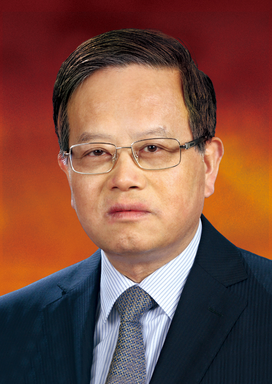 Zhiyuan Zhang (张志愿)合作论文数School of Medicine, Shanghai Jiaotong University;Shanghai Ninth People’s Hospital111
 Tong Ji (季彤)合作论文数Department of Oral Maxillofacial-Head Neck Oncology, Shanghai Ninth People’s Hospital, School of Medicine, Shanghai Jiao Tong University;College of Stomatology, Shanghai Jiao Tong University88
 Jian Sun (孙坚)合作论文数Department of Oral Maxillofacial - Head Neck Oncology, Shanghai Ninth People’s Hospital, School of Medicine, Shanghai Jiao Tong University67
 LiQun Xu (徐立群)合作论文数59
 Laiping Zhong (钟来平)合作论文数Shanghai Ninth People's Hospital, School of Medicine, Shanghai Jiao Tong University;Huashan Hospital, Fudan University58
 Wenjun Yang (杨雯君)合作论文数Shanghai Ninth People's Hospital, Shanghai Jiao Tong University School of Medicine57
 Yongjie Hu (胡永杰)合作论文数Shanghai Ninth People's Hospital, Shanghai Jiao Tong University School of Medicine54
 Li jiang (李江)合作论文数National Clinical Research Center for Oral Diseases, Ninth People's Hospital, Shanghai Jiaotong University School of Medicine52Lizhen Wang合作论文数48
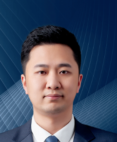 Jiannan Liu (刘剑楠)合作论文数Shanghai Ninth People’s Hospital,Shanghai JiaoTong University School of Medicine44
 Hanguang Zhu (竺涵光)合作论文数Shanghai Ninth People’s Hospital, Shanghai JiaoTong University School of Medicine43
 Min Ruan (阮敏)合作论文数Shanghai Ninth People’s Hospital, Shanghai Jiao Tong University School of Medicine43
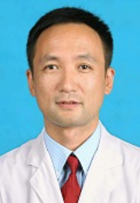 Yue He (何悦)合作论文数Shanghai Ninth People's Hospital, School of Medicine, Shanghai Jiao Tong University41
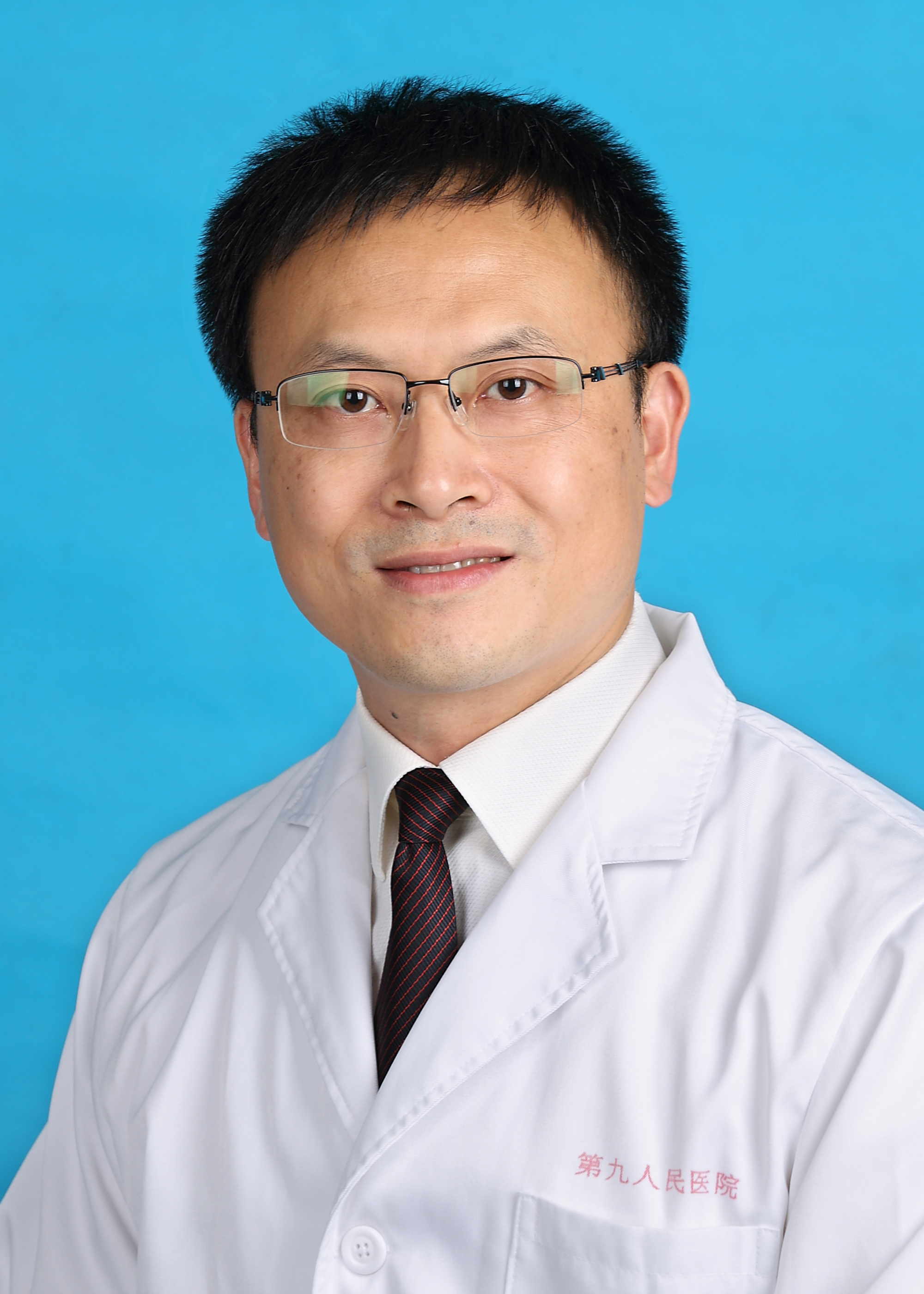 Siyi Li (李思毅)合作论文数Shanghai Ninth People's Hospital, Shanghai Jiao Tong University School of Medicine39
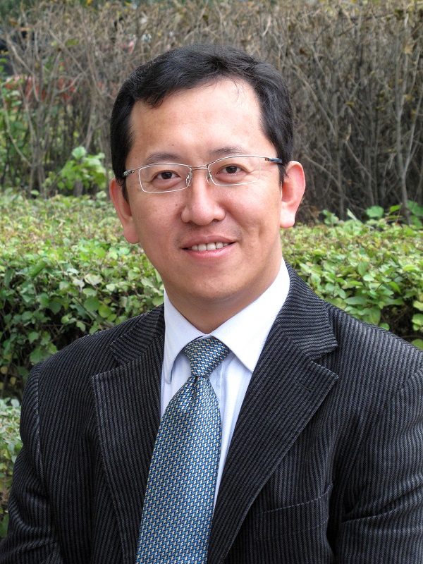 Xingzhou Qu (曲行舟)合作论文数Shanghai Ninth People's Hospital, Shanghai Jiao Tong University School of Medicine30
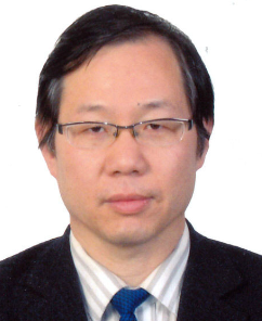 Wantao Chen (陈万涛)合作论文数Shanghai Ninth People's Hospital, Shanghai Jiao Tong University School of Medicine29
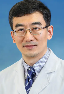 Yiqun Wu (吴轶群)合作论文数Shanghai Ninth People’s Hospital, Shanghai JiaoTong University School of Medicine25
 Xi Yang (杨溪)合作论文数‘School Of Stomatology, Hainan Medical University;Shanghai Ninth People's Hospital, Shanghai Jiao Tong University School of Medicine24Dongwang Zhu合作论文数22
 Weimin Ye (叶为民)合作论文数Department of Oral and Maxillofacial Surgery, Ninth People's Hospital, College of Stomatology, Shanghai Jiao Tong University School of Medicine19
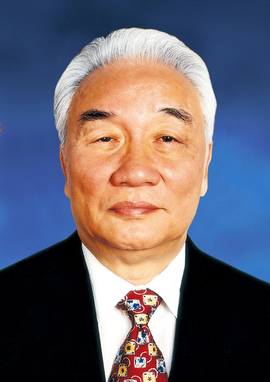 Weiliu Qiu (邱蔚六)合作论文数College of Stomatology, Shanghai Jiao Tong University19Shengwen Liu合作论文数18Ma Chunyue合作论文数18
 Guoxin Ren (任国欣)合作论文数Shanghai Ninth People's Hospital, School of Medicine, Shanghai Jiao Tong University17Ying Liu (刘莹)合作论文数17Xuelai Yin合作论文数17Yun Zhu (朱云)合作论文数16Tian Zhuowei合作论文数15
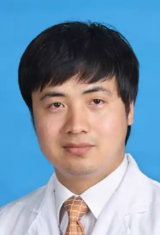 Wei Cao (曹巍)合作论文数Shanghai Ninth People's Hospital, Shanghai Jiao Tong University School of Medicine15
 Zhen Tian (田臻)合作论文数Shanghai Ninth People's Hospital, Shanghai Jiao Tong University School of Medicine14
 Xingjun Qin (秦兴军)合作论文数Shanghai Ninth People’s Hospital, School of Medicine, Shanghai Jiao Tong University14
 Jun Li (李军)合作论文数Department of Oral and Maxillofacial Surgery, Ninth People's Hospital, College of Stomatology,Shanghai Jiao Tong University School of Medicine14Shanghui Zhou合作论文数14Nannan Han合作论文数13Ren Zhen-hu合作论文数13
 Jingzhou Hu (胡镜宙)合作论文数Department of Oral Maxillofacial-Head and Neck Oncology, Shanghai Ninth People's Hospital, School of Medicine, Shanghai Jiao Tong University13
 Zhengxue Han (韩正学)合作论文数Beijing Stomatological Hospital, Capital Medical University13Chengzhe Yang合作论文数12Jinye Fu合作论文数12
 Wei Guo (郭伟)合作论文数Shanghai Ninth People’s Hospital, Shanghai Jiao Tong University School of Medicine12
 Wei Liu (刘玮)合作论文数The First People' s Hospital of Shanghai Affiliated to Shanghai Jiaotong University School of Medicine12
 Jiawei Zheng (郑家伟)合作论文数Shanghai Red Cross Ninth People's Hospital;College of Stomatology, Shanghai Jiao Tong University12Wanlin Xu合作论文数12Chunye Zhang合作论文数11Ong Hui Shan合作论文数11Yiran Tan合作论文数11
 Guofang Shen (沈国芳)合作论文数Shanghai University of Medicine & Health Sciences10
 Yunong Wu (吴煜农)合作论文数Stomatological College, Nanjing Medical University10




