BackgroundPARP(Poly(ADP-ribose) polymerase) inhibitors are established as effective treatment for ovarian cancer; however, there have been no systematic studies of PARP inhibitor use in older patients with ovarian cancer.MethodsThis study was conducted at Peking University People’s Hospital. Archival data from patients with advanced age (≥ 65 years) with ovarian cancer between March 2018 and September 2023, who received olaparib or niraparib as maintenance therapy following either first-line platinum-based chemotherapy or platinum-sensitive recurrence (PSR) chemotherapy and achieved complete (CR) or partial response (PR). The two clinical scenarios (first-line maintenance and PSR maintenance) were analyzed separately.ResultsOf 56 included patients, 20 (35.7%) and 34 (60.7%) received olaparib and niraparib, respectively, while 2 (3.6%) switched from olaparib to niraparib due to intolerance. The mean age was 70.14 ± 4.63 years. Median progression-free survival (mPFS) was 24 months in 35 (62.5%) patients receiving PARP inhibitors as first-line maintenance therapy(1L-group). In the 1L group, CA125 level, whether CR was reached, BRCA mutation status, and R0 at initial surgery before PARP inhibitor therapy were associated with PFS in univariate analysis, and the latter three factors were independently associated with PFS on multivariate analysis. In the PSR group, among patients with PSR ≥12 months (n=13), mPFS was not reached; among those with PSR 6–12 months (n=7), mPFS was 9.6 months. Anemia (22.7%) was the most frequent grade 3–4 adverse event in the olaparib group, whereas thrombocytopenia (11.1%) was more common in the niraparib group. Both groups of patients experienced dose reduction, interruption, and discontinuation within the first 6 months. Niraparib had a lower termination rate than olaparib, and long-term PARP inhibitor use was generally well-tolerated.ConclusionsPARP inhibitor (olaparib and niraparib) maintenance therapy yields favorable clinical outcomes for elderly ovarian cancer patients. Three clinical factors, attainment R0(no residual disease) after initial surgery, BRCA mutation, and CA-125 ≤10 U/mL before PARP inhibitor treatment, were predictive of longer PFS in elderly ovarian cancer patients undergoing first-line maintenance therapy with PARP inhibitors.
Objective: We investigated the clinical value of a novel circulating tumor cell (CTC) detection method-subtraction enrichment combined with immunostaining and fluorescence in situ hybridization (SEiFISH)-in ovarian cancer (OC). This study evaluated the diagnostic and prognostic significance of chromosome 8 aneuploidy in CTCs and circulating tumor endothelial cells (CTECs) for preoperative diagnosis, treatment efficacy assessment, and recurrence monitoring. Methods: A total of 331 patients were enrolled, including 56 with newly diagnosed primary OC, 265 with benign ovarian tumors, and 10 with borderline tumors. Peripheral blood CTCs and CTECs were detected using SEiFISH; their quantity and ploidy characteristics were analyzed in relation to clinical indicators. To assess dynamic CTC changes during disease progression and treatment response, 72 patients were followed longitudinally, of whom 19 experienced recurrence. Results: The CTC detection rate in OC patients was 92.9%, with significantly higher counts than that in the benign tumor group (median 5 vs. 2). Receiver operating characteristic analysis demonstrated good diagnostic performance for total CTCs [area under the curve (AUC)=0.699], with triploid CTCs achieving the highest efficacy (AUC=0.792), surpassing carbohydrate antigen 125 (CA125) (AUC=0.702). Postoperative follow-up showed that 70% of patients exhibited concurrent decreases in CTCs and CA125 levels, indicating disease improvement. In 30% of patients, CTC levels did not correlate with changes in CA125 levels. Individual case evidence suggests that CTC alterations may serve as an early indicator of recurrence or metastasis. Among the 19 recurrent cases, 73.7% showed elevated CTCs at recurrence that decreased following treatment. In four patients, CTCs reflected disease progression earlier than CA125, indicating higher sensitivity for recurrence monitoring. Conclusions: CTCs with chromosome 8 aneuploidy demonstrate significant clinical value in the preoperative diagnosis, treatment efficacy evaluation, and recurrence monitoring of OC. Dynamic CTC changes may serve as a more sensitive indicator than CA125 for disease surveillance, supporting the translational potential of CTC-based biomarkers in OC.
BACKGROUND:Large-scale studies of germline variants in hereditary cancer susceptibility genes among Chinese epithelial ovarian cancer (EOC) patients remain limited. This study assessed the prevalence and clinical significance of germline variants in 21 genes relevant to hereditary breast and ovarian cancer. METHODS:In this multicenter prospective cohort (February 2017-December 2018), 961 unselected EOC patients underwent germline testing for 21 genes. Variant frequencies were compared with international data, and associations with clinicopathologic characteristics and survival outcomes were evaluated. RESULTS:Pathogenic or likely pathogenic (P/LP) variants were identified in BRCA1 (17.79%), BRCA2 (6.35%), and other homologous recombination (HR)-related genes (2.71%). P/LP variants in non-HR-related genes were rare (0.1%). BRCA1 and BRCA1/2 P/LP variant carriers were more likely to respond to platinum-based chemotherapy (p = .002 and p < .001). Variants of uncertain significance or higher (VUS+) in HR-related genes were associated with better overall survival (hazard ratio, 0.57; p = 0.004) and progression-free survival (hazard ratio, 0.75; p = 0.02), with a trend more pronounced than that observed for BRCA1/2 VUS + carriers. CONCLUSIONS:Over 25% of Chinese EOC patients carry germline HR-related gene variants, which are associated with better treatment response and survival. Broad genetic testing is critical, and the prognostic value of VUS warrants further investigation.
Early detection of asymptomatic ovarian cancer (asym-OC) remains a critical challenge, the failure of which underlies its high mortality. Performing serum peptidomic profiling of 843 participants in the cohort SOCFCP, we distill 1,081 initial features into a 7-marker panel for asym-OC detection via a biology-informed machine-learning (ML)-based feature selection strategy. Three markers significantly revert toward non-OC levels after surgery. Integrating the panel with age, CA125, and HE4, we develop and externally validate (n = 159) a LightGBM model, ProMS+. For early-stage OC detection, ProMS+ shows a specificity of 92.6% at 95.0% sensitivity, outperforming CA125 (44.7%), HE4 (11.2%), and Risk of Ovarian Malignancy Algorithm (ROMA) (24.0%), with an area under the curve (AUC) of 0.993. In a simulated high-risk population (n = 100,000; OC prevalence = 1%), ProMS+ yields a high AUC (0.983) and a higher positive predictive value than CA125, HE4, and Age + CA125 + HE4 combined model (0.201 vs. 0.027, 0.090, and 0.064). ProMS+ offers a promising, non-invasive, and interpretable approach for the early detection of asym-OC.
Endometriosis is a common gynecological disorder characterized by the ectopic growth of endometrium-like tissue outside the uterine cavity and is associated with pelvic pain, infertility, and reduced quality of life. Although several theories have been proposed, the mechanisms by which endometrial cells disseminate and establish ectopic lesions remain incompletely understood. Here, we developed a dual-color pseudo-menstrual mouse model that enables real-time in vivo tracking of circulating endometrial cells (CECs) while distinguishing their tissue origin as uterine-derived or lesion-derived. Using in vivo flow cytometry and two-photon intravital microscopy, we found that both uterine-derived and lesion-derived CECs can enter the circulation and are detected at higher levels during menstruation-like breakdown, potentially in association with local vascular remodeling as suggested by intravital imaging observations. These cells could localize to distant organs, including the lungs and other extrapelvic organs. In addition, both CEC populations exhibited three distinct daily peak periods, and CEC dynamics were significantly associated with fluctuations in serum prolactin levels. In conclusion, these findings provide in vivo support for hematogenous dissemination in endometriosis and suggest that hormonal rhythmicity may regulate CEC dynamics, with potential diagnostic and therapeutic implications.
BACKGROUND:Cross-resistance is observed between platinum and Poly (ADP-ribose) polymerase inhibitors (PARPi). We aim to propose the definition of PARPi resistance and demonstrate the best therapeutic strategy for patients with PARPi resistance. METHODS:A retrospective analysis was performed on patients diagnosed with epithelial ovarian cancer from October 2015 to November 2022. Patients were treated with PARPi for more than 6 months and received chemotherapy after progression. RESULTS:Totally, 41 patients were enrolled, with 21 receiving PARPi for 6 to 12 months and 20 for more than 12 months. The median duration of PARPi was 12 months, and the median time to second progression (TTSP) was 3.45 months (range, 1.0-20.2 months). The Kaplan-Meier and Cox analysis revealed a significantly shorter TTSP for patients who received PARPi for more than 12 months compared to those for 6 to 12 months. After PARPi resistance, 34 (82.9%) received platinum-based chemotherapy, with an overall response rate (ORR) of 26.5% (9/34). Seven patients (17.1%) received arsenic trioxide (ATO)-based chemotherapy, with an ORR of 57.1% (4/7). During subsequent chemotherapy, 12/34 patients switched to ATO-based chemotherapy due to progression, of which five cases were evaluated as effective (41.7%). CONCLUSION:PARPi resistance has a negative impact on the subsequent chemotherapy. The progression of the disease beyond 6 to 12 months should be considered as acquired resistance. Non-platinum chemotherapy, such as ATO-based combined sequential chemotherapy, may emerge as the preferred option for patients with PARPi resistance.
Platinum resistance remains a major obstacle to effective treatment and improved prognosis in ovarian cancer. Although 5-methylcytosine (m5C) RNA modification has been implicated in chemoresistance, its precise functional role in ovarian cancer remains unclear. In this study, we integrated RNA-Seq and single-cell transcriptomic data from cisplatin-resistant ovarian cancer cell lines and patient samples, identifying the m5C reader protein ALYREF as a key regulator of platinum resistance. Functional studies using ALYREF and NSUN2 knockdown, overexpression, and mutant constructs—combined with multi-omics analyses (RNA-Seq, m5C-BIS-Seq, and RIP-Seq)—revealed that ALYREF binds to m5C-modified LGR4 mRNA, enhancing its stability and promoting activation of the Wnt/β-catenin signaling pathway. Critically, this regulatory mechanism is dependent on NSUN2-mediated m5C modification of LGR4 mRNA. Together, our findings demonstrate that the NSUN2/ALYREF/LGR4 axis mediates platinum resistance through m5C-dependent stabilization of LGR4 and downstream Wnt signaling activation. Thus, targeting ALYREF may represent a promising strategy to overcome platinum resistance in ovarian cancer.
Ovarian cancer (OC) poses a significant threat to women’s health due to its insidious onset, tendency to metastasize, and high recurrence rate. These characteristics significantly impede early diagnosis and effective interventions. The current lack of effective screening and prognostic tools necessitates urgent solutions. The Shanghai Ovarian Cancer and Family Care Project (SOCFCP) is an ongoing cohort study aiming to address these challenges by identifying factors and biomarkers associated with OC onset, treatment, and prognosis. The ultimate goal is to develop predictive models and assessments crucial for diagnosing, treating, and prognosing the disease. During the study period from 2023 to 2028, the SOCFCP aims to recruit over 2000 patients with ovarian lesions, 900 high-risk individuals, and 3000 healthy controls in Shanghai, China, with long-term follow-up. The study comprises three cohorts: (1) a bidirectional patients cohort, comprising those diagnosed with ovarian disease and treated at the department of gynecologic oncology at Shanghai first maternity and infant hospital since 2013; (2) a prospective high-risk cohort, consisting of family members of ovarian and/or breast cancer patients, or individuals with related gene mutations; and (3) a healthy control cohort, drawn from women undergoing health examination at hospitals or participating in community gynecologic cancer screenings. Participants will attend regular visits for questionnaire surveys, anthropometric measurements, and the collection of biological specimens, such as blood, fecal, and tissue samples. Collected data encompass baseline information, sociodemographic details, hospital admission records, and biological and clinical parameters, alongside treatment information. Primary outcomes for the patient cohort include mortality, tumor recurrence, major complications, and changes in health-related quality of life post-diagnosis. For the high-risk and healthy cohorts, ovary-related diseases will serve as the primary outcome. Various predictive models, such as multivariate logistic models, Cox regression, and linear mixed-effects regression models, will be employed to analyze various endpoints appropriately. This extensive cohort study integrates a diverse range of information, including lifestyle factors, environmental influences, genetic characteristics, and clinical features, alongside an extensive collection of biological samples. The resources generated by this project will provide valuable foundations for a comprehensively understanding of the etiology, treatment, and prognosis of OC. The SOCFCP is poised to foster cutting-edge multidisciplinary research on OC, spanning both fundamental and clinical studies. NCT06118307 in ClinicalTrials.gov. Registration Date: November 7, 2023.
ABSTRACT Background In patients with locally advanced cervical cancer (LACC) undergoing concurrent chemoradiotherapy (CCRT), the high incidence of radiation‐induced lymphopenia significantly affects prognosis. There are significant variations in lymphocyte count (ALC) recovery patterns among patients, and their impact on prognosis remains unclear. This study aims to quantify the lymphocyte recovery patterns by the lymphocyte recovery index (LRI) and evaluate its prognostic value. Methods This study reviewed patients with LACC who had ALCs available within 6 months post‐CCRT. Lymphopenia was graded using CTCAE 5.0, and lymphocyte recovery patterns were quantified using LRI (the ratio of ALCs at 6 months post‐treatment to baseline ALCs). Cox regression analysis was conducted to assess the correlation between LRI, other clinical factors, and survival. The dose–volume of bone marrow (BM) following pelvic radiotherapy was collected, and measurements of spleen standardized uptake value (SUV) and spleen‐to‐liver SUVmax ratio (SLR) were obtained from pre‐treatment 18F‐FDG PET/CT. Logistic regression analysis was used to identify independent risk factors for LRI. Results A total of 180 patients were included retrospectively. During CCRT, 53 patients (29.4%) experienced G4 lymphopenia. The median LRI was 53.4% (range 13.2%–159.4%). Multivariable analysis revealed that LRI, G4 lymphopenia, and FIGO stage were associated with progression‐free survival (PFS) and overall survival (OS). Subgroup analysis revealed that the degree of lymphopenia (G4 and G1‐3) did not affect the correlation between LRI and PFS (P: 0.001 and 0.011) or OS (P: 0.003 and 0.043). Regarding FIGO stage, the impact of LRI on PFS (p < 0.001) and OS (p < 0.001) was primarily observed in patients with FIGO stage > II. Logistic analysis identified BM‐V10 > 96.0% and SLR > 0.90 as independent risk factors for LRI. Conclusion In patients with LACC after CCRT, the LRI is associated with prognosis. Splenic metabolism and BM irradiation are associated with lymphocyte recovery.
OBJECTIVE:Assessment of whether contrast-enhanced ultrasound (CEUS) can be used to differentiate diverse cervical lesions. METHODS:A retrospective analysis of ultrasonographic reports was conducted for patients with different cervical lesions, including 18 cases of chronic cervicitis, 28 cases of cervical intraepithelial neoplasia grade I (CIN1), 46 cases of cervical intraepithelial neoplasia grade II (CIN2), 100 cases of cervical intraepithelial neoplasia grade III (CIN3), 7 cases of carcinoma in situ (CIS), and 26 cases of cervical cancer (CC). Timing began with the contrast agent injection via the elbow vein, recorded separately when the myometrium and cervix began to be enhanced. The intensity of enhancement of the cervix was observed and recorded as hyper-enhancement, iso-enhancement, or hypo-enhancement with respect to the myometrium. The rate of regression of the cervix enhancement was also analyzed and classified as fast, synchronous, or slow relative to the myometrium. Quantitative data were analyzed using either one-way ANOVA or the Kruskal-Wallis H test, while qualitative data were analyzed using Fisher's exact test. Significant differences were further analyzed using post-hoc tests, logistic regression models, and ROC curves. RESULTS:Menopausal status affected longitudinal, anteroposterior, and transverse diameters of the cervix, in addition to cervical volume. Moreover, the time when the myometrium and cervix began to image was also different between the menopause group and the pre-menopause group (P < 0.05), whereas there was no significant variance of pulsatility index (PI), resistance index (RI), enhancement intensity of the cervix, or rate of the cervix fading. The study population was grouped according to their menopausal status, and then we discovered that in the pre-menopausal group, the cervical anteroposterior diameter (APD) differed in the six cervical lesion groups (P = 0.006), especially in CIN1 & CIN2, CIN1 & CIN3, and CIN1 & CC (P = 0.014, 0.045, 0.021, respectively). The enhancement start time (EST) of the cervix differed among the cervical lesion groups (P = 0.02). In particular, there was a significant difference in CC & CIN2 and CC & CIN3 (P = 0.04, 0.03, respectively). The subjects of the study were divided into two groups: those with cervical cancer and those with precancerous lesions. The discrepancy of cervical EST was still significant along with the sensitivity of 0.62, the specificity of 0.76 using a cutoff of 14.895 s, and the accuracy was 0.74. CONCLUSION:The cervical EST can serve as an indicator of malignancy, and CEUS can be a complementary tool for cervical cancer screening. However, it should be noted that qualitative CEUS analysis alone was unable to differentiate among various precancerous lesions.
Background It is crucial to identify the high-risk factors associated with the recurrence and metastasis of endometrial cancer (EC) in order to implement more precise clinical stratification and management strategies for EC patients. Methods A total of 336 patients with stage I-III EC were retrospectively analyzed. According to the recurrence site, they were divided into locoregional recurrence (LR) and poor-prognosis recurrence (PPR). The factors that may affect the prognosis of EC were analyzed and the subgroups were analyzed. Results Among the no recurrence (NR), LR and PPR groups, 5-year OS were 89.4%, 60.2% and 46.8%, 5-year RFS were 100%, 15.4% and 6.4%. The FIGO stage, molecular classification, lymphovascular space invasion (LVSI) and smoking history were independent risk factors affecting 5-year OS and 5-year RFS in EC patients (p < 0.05). Pathological type and progesterone receptor (PR) were independent risk factors affecting 5-year OS (p < 0.05). Histologic Grade and adjuvant therapy were independent risk factors affecting 5-year RFS (p < 0.05). Myometrial invasion, LVSI and FIGO stage were independent risk factors in the LR subgroup (p < 0.05), FIGO stage, ER and PR were independent risk in the PPR subgroup (p < 0.05). Conclusions Patients with myometrial invasion ≥ 1/2 and substantial LVSI may be more likely to have LR, while patients with positive ER and PR are more likely to have PPR. We need to pay attention to these factors to help us judge the prognosis of EC patients.
5546 Background: Early diagnosis is crucial for improving the prognosis of ovarian cancer (OC). However, most patients (pts) are diagnosed at advanced stages due to subtle symptoms and traditional biomarkers' limitations. We aimed to develop an serum panel using multi-omics data for cost-effective detection of asymptomatic OC (asym-OC). Methods: Participants were recruited from the Shanghai Ovarian Cancer and Family Care Project ( NCT06118307 ) involving five centers. A total of 843 individuals were included: 135 asym-OC pts and 708 non-OC individuals (290 pts with benign lesions and 418 healthy controls). Fasting serum samples (1 μL each) were analyzed using MALDI-TOF MS to generate proteo-metabolic (pro-met) data. For each sample, the original MS have ~43,900 data points from 100-13,000 Da. Preoperative data from three centers (N=680) were used to develop Light Gradient Boosting Machine (LGBM) models to identify key signals differentiating OC from non-OC. An independent external validation set (N = 163) was assembled by the other two centers. To develop a biologically interpretable and generalizable panel, the model was further refined to minimize biomarkers while maintaining efficacy. Validation of the panel was conducted using postoperative pro-met data from 42 asym-OC pts, transcriptomic data from 89 with asym-OC and 39 with benign lesions, supported by bioinformatic analyses. Results: Ten biomarkers, which are involved in coagulation, complement system, carcinogenesis, epithelial-mesenchymal transition, and the Warburg effect, were selected in the panel. The panel achieved an AUC of 0.90 (95% CI: 0.82-0.98) in the external validation set. The enhanced model that integrating the panel, age, BMI and HE4 achieved an AUC of 0.94 (95% CI: 0.90-0.99). In subgroup analyses, the enhanced model outperformed CA125, HE4, and ROMA, with AUCs of 0.96 (early-stage OC vs. non-OC) and 0.96 (OC vs. endometriosis) (See Table). Moreover, postoperative levels of the biomarkers in the panel approached those of the non-OC (p < 0.05). Transcriptomic data of the tissues corresponded to the pathophysiological alterations associated with OC development or progression. Conclusions: This study introduces a novel, non-invasive, and cost-effective serum panel that demonstrates high sensitivity and specificity for detecting asym-OC, offering a promising tool for early diagnosis of the disease. Early-stage OC vs. Non-OC (N=58/708) OC vs. Endometriosis (N=135/80) Enhanced model: 10-biomarker Pro-Met panel + BMI +age + HE4 0.96 [0.94-0.99] 0.93 [0.90-0.97] CA125 0.87 [0.82-0.93] 0.77 [0.71-0.83] HE4 premenopausal 0.73 [0.63-0.83] 0.91 [0.80-0.94] postmenopausal 0.67 [0.58-0.74] 0.88 [0.76-0.86] ROMA premenopausal 0.75 [0.60-0.81] 0.91 [0.80-0.93] postmenopausal 0.89 [0.67-0.84] 0.87 [0.53-1.00]
This study investigated BRCA1/2 and homologous recombination repair (HR) pathway gene variants in Chinese epithelial ovarian cancer (EOC) patients. Germline and somatic variants in 21 HR-related genes were analyzed in 229 patients using a 21-gene ovarian panel and in 141 patients using a 508-gene pan-cancer panel. BRCA1, BRCA2, and HR-related gene mutation rates were 17.9%, 3.5%, and 23.1%, respectively, with TP53 as the most frequent somatic mutation (66.4%). Combined germline and somatic BRCA1/2 mutation rates rose to 23.6 and 6.1%. Survival analysis (n = 200) demonstrated longer overall survival (OS) in patients carrying BRCA1/2 or HR mutations. Notably, strategies including likely pathogenic (LP) and variants of uncertain significance (VUS) showed improved OS, especially in BRCA2 and BRCA1/2 somatic carriers. These findings suggest that integrating germline, somatic, and VUS data enhances survival prediction and guides treatment decisions in Chinese EOC patients.
Although some evidence suggests that Chinese and white women might have different pelvic floor anatomy such as levator complex and connective tissue support for pelvic organs, it is unknown if these differences affect the mechanisms of pelvic organ prolapse. We sought to determine whether differences exist in MRI-based structural failure patterns between Chinese and American white women with prolapse. This is a case–control study in different racial cohorts recruited in China and the USA. The Chinese cohort included 19 women with anterior-predominant prolapse and 24 controls with normal pelvic support. The American white cohort included 30 women with anterior-predominant prolapse and 30 controls. Both cohorts underwent the same clinical evaluation and MRI protocol. Three structural support systems were quantified on stress 3D MRI at maximal Valsalva: vaginal factors (length, width); connective tissue attachment (cervix, lateral paravaginal height); and hiatus factors (urogenital hiatus size, major levator ani injury). Abnormal structural support was defined as any measurement outside the normal range defined as the 5th to 95th percentile in controls from the respective cohort. The percentages of the women with abnormal support in the two cohorts were compared. Among those with prolapse, Chinese women were more likely than white women to have abnormally long vaginal length and width (90
Background: To identify the metastatic lymph nodes and remove them accurately, the fluorescent surgical navigation ability of the ovarian cancer-specific fluorescent probe COC183B2-800 was assessed to verify the metastatic lymph nodes in the nude mouse model. Methods: The nude mouse model related to lymph node metastases in human ovarian cancer was established using the SKOV3-ip1 cell line. Besides, the COC183B2-800 probe (IRDye800CW Ester conjugated COC183B2 antibody) was fabricated. Moreover, in vivo fluorescence imaging was performed to determine the ability of the COC183B2-800 fluorescent probe to identify metastatic lymph nodes in the nude mouse model. Results: The nude mouse model related to lymph node metastases in human ovarian cancer was successfully established. In vivo fluorescence imaging was performed 30 hours after the injection of the COC183B2-800 fluorescent probe (25 μg) into the animal model, which can achieve specific imaging of metastatic lymph nodes. All metastatic lymph nodes were detected in vivo and in vitro (8/8), and only 1 negative lymph node with reactive enlargement showed a false positive fluorescent signal. Conclusions: The targeted fluorescent probe COC183B2-800 can be employed to identify metastatic lymph nodes in the nude mouse model related to lymph node metastases in human ovarian cancer with high specificity and sensitivity. Targeted fluorescence imaging using COC183B2-800 is expected to become a method to achieve precise lymphadenectomy.
Near-infrared fluorescence (NIRF) imaging is an excellent choice for image-guided surgery due to its simple operation and non-invasiveness. Developing tumor-specific fluorescent molecular probes is key to fluorescence imaging-guided surgery. EGFR (epidermal growth factor receptor) is closely related to the proliferation and growth of tumor cells and is highly expressed in epithelial ovarian cancer (EOC). The study aims to construct a NIR fluorescent molecular probe using cetuximab (an EGFR monoclonal antibody) and investigate its feasibility for targeting EOC in vivo through fluorescence imaging. We determined the expression of EGFR in EOC. NIR fluorescent molecular probe with cetuximab (cetuximab-Cy7) was chemically engineered and identified. The subcutaneous xenografted tumor model of EOC was induced using SKOV3-Luc cell line with positive expression of EGFR. Cetuximab-Cy7 was used for in vivo fluorescence imaging, and phosphate-buffered saline, free Cy7 dye and mouse isotype immunoglobulin G-Cy7 were used as controls. NIRF imaging system was performed to study the distribution and targeting of the probes. Tumors were imaged in situ and ex vivo, and fluorescent intensity was quantified. Resected specimens were analyzed to confirm diagnosis, and immunohistochemical (IHC) staining was used to identify EGFR expression. EGFR expression was increased in EOC tissues than fallopian tube tissues. The high expression of EGFR was significantly correlated with well-differentiation, residual lesions ≤ 1 cm, no recurrence and increased survival. NIRF imaging showed that the cetuximab-Cy7 enabled detection of tumor lesions in EOC-bearing mice with the optimal dose of 30 µg. The suitable imaging time window may be 24–96 h post-injection. Ex vivo fluorescence imaging indicated that fluorescent signal was mainly detected in the tumor and the lung. IHC results confirmed that xenografts were EGFR positive. Cetuximab-Cy7 can specifically target the tumors of EOC xenografted nude mice. This research lays the foundation for future studies on EOC surgery navigation.
Extracellular matrix protein 1 (ECM1) is a glycoprotein that may be a key player in tumorigenesis and tumor progression. However, knowledge regarding the role of ECM1 in endometriosis (EM) is still lacking. Microarray analyses were performed to compare the mRNA expression patterns between paired EU tissues and ectopic endometrial (EC) tissues (n = 4) from EM patients. ECM1 expression was significantly increased in the eutopic endometrial (EU) tissues than paired EC tissues of endometriotic patients and normal endometrial (NE) tissues of controls without EM. Blocking ECM1 with siRNA attenuated the migration and invasion of hEM15A cells and modified the distribution of the F-actin cytoskeleton. We conducted microarray analyses and bioinformatics analyses to investigate the differentially expressed genes (DEGs) and related pathways regulated by ECM1. A total of 161 DEGs between the siECM1 and the negative control (siNC) treatments were identified, consisting of 79 downregulated genes and 82 upregulated genes. Enriched DEGs were associated with 9 gene ontology (GO) terms. Moreover, a protein-protein interaction (PPI) network was constructed for the hub genes and modules. Radixin (RDX) was the second most downregulated gene in the siECM1 group compared with the siNC group. ECM1 knockdown significantly decreased the expression of RDX, RhoC, ROCK1, N-cadherin and β-catenin but not ROCK2. ECM1 showed high tissue-specific expression in EU tissues from EM patients, and may contribute to the migration, invasion and reorganization of the F-actin cytoskeleton in eutopic endometrial stromal cells via the RhoC/ROCK1 signaling pathway in EM.
This study aimed to develop and evaluate radiomics models to predict CD27 expression and clinical prognosis before surgery in patients with serous ovarian cancer (SOC). We used transcriptome sequencing data and contrast-enhanced computed tomography images of patients with SOC from The Cancer Genome Atlas (n = 339) and The Cancer Imaging Archive (n = 57) and evaluated the clinical significance and prognostic value of CD27 expression. Radiomics features were selected to create a recursive feature elimination-logistic regression (RFE-LR) model and a least absolute shrinkage and selection operator logistic regression (LASSO-LR) model for CD27 expression prediction. CD27 expression was upregulated in tumor samples, and a high expression level was determined to be an independent protective factor for survival. A set of three and six radiomics features were extracted to develop RFE-LR and LASSO-LR radiomics models, respectively. Both models demonstrated good calibration and clinical benefits, as determined by the receiver operating characteristic (ROC) curves, calibration curves, and decision curve analysis. The LASSO-LR model performed better than the RFE-LR model, owing to the area under the curve (AUC) values of the ROC curves (0.829 vs. 0.736). Furthermore, the AUC value of the radiomics score that predicted the overall survival of patients with SOC diagnosed after 60 months was 0.788 using the LASSO-LR model. The radiomics models we developed are promising noninvasive tools for predicting CD27 expression status and SOC prognosis. The LASSO-LR model is highly recommended for evaluating the preoperative risk stratification for SOCs in clinical applications.
High malignancy is a prominent characteristic of epithelial ovarian cancer (EOC), emphasizing the necessity for further elucidation of the potential mechanisms underlying cancer progression. Aneuploidy and copy number variation (CNV) partially contribute to the heightened malignancy observed in EOC; however, the precise features of aneuploidy and their underlying molecular patterns, as well as the relationship between CNV and aneuploidy in EOC, remain unclear. In this study, we employed single-cell sequencing data along with The Cancer Genome Atlas (TCGA) to investigate aneuploidy and CNV in EOC. The technique of fluorescence in situ hybridization (FISH) was employed using specific probes. The copy number variation within the genomic region of chromosome 8 (42754568-47889815) was assessed and utilized as a representative measure for the ploidy status of individual cells in chromosome 8. Differential expression analysis was performed between different subgroups based on chromosome 8 ploidy. Gene ontology (GO), Kyoto Encyclopedia of Genes and Genomes (KEGG), protein–protein interaction (PPI), and hub–gene analyses were subsequently utilized to identify crucial genes involved. By classifying enriched tumor cells into distinct subtypes based on chromosome 8 ploidy combined with TCGA data integration, we identified key genes driving chromosome 8 aneuploidy in EOC, revealing that PRKDC gene involvement through the mediated non-homologous end-joining pathway may play a pivotal role in disease progression. Further validation through analysis of the GEO and TCGA database and survival assessment, considering both mRNA expression levels and CNV status of PRKDC, has confirmed its involvement in the progression of EOC. Further functional analysis revealed an upregulation of PRKDC in both ovarian EOC cells and tissues, with its expression showing a significant correlation with the extent of copy number variation (CNV) on chromosome 8. Taken together, CNV amplification and aneuploidy of chromosome 8 are important characteristics of EOC. PRKDC and the mediated NHEJ pathway may play a crucial role in driving aneuploidy on chromosome 8 during the progression of EOC.
Background and ObjectivesTo compare the feasibility and diagnostic value of sentinel lymph node (SLN) mapping with carbon nanoparticles (CNPs) and indocyanine green (ICG) in endometrial cancer (EC). MethodsThis was a single-center, open-label, randomized controlled trial. Between August 1, 2020 and April 30, 2022, patients with early-stage EC were assessed for enrollment. All patients underwent SLN mapping with ICG or CNPs +/- pelvic and/or para-aortic lymphadenectomy. The detection rate (DR), its impact factors, the sensitivity and the negative predictive value (NPV) of SLN mapping were analyzed. ResultsIn total, 206 patients, with 103 per group, were included. The overall and bilateral DRs of the two groups were comparable without significant differences. There was no difference regarding the distribution of mapped SLNs. The sensitivity was 66.7% for both groups, and the NPVs were not significantly different. Furthermore, the sensitivity and NPV reached 100% when calculated either per hemipelvis or only in patients with bilateral SLN detection. ConclusionsSLN mapping by CNPs in EC is feasible with high DRs and diagnostic accuracy compared with ICG. CNPs might be an alternative to ICG for SLN mapping when near-infrared imaging equipment is lacking, especially in stage IA patients.
 Heng Cui (崔恒)合作论文数Peking University People's Hospital83
 Xiaohong Chang (昌晓红)合作论文数Peking University People's Hospital35
 Honglan Zhu (祝洪澜)合作论文数Peking University People's Hospital30
 Xue Ye (叶雪)合作论文数24
 Lihui Wei (魏丽惠)合作论文数Peking University People′s Hospital23
 Jianliu Wang (王建六)合作论文数Peking University People's Hospital20
 Y. Cheng合作论文数18
 Danhua Shen (沈丹华)合作论文数Peking University People's Hospital;Peking University Health Science Center12Sha Dou合作论文数11Jie Feng合作论文数Center for Information Science, Peking University11
 Xiuli Sun (孙秀丽)合作论文数Peking University People's Hospital10
 Xudong Liang (梁旭东)合作论文数Department of Gynecology, People's Hospital,Peking University9
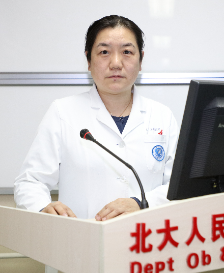 Yue Wang (王悦)合作论文数Peking University People's Hospital9
 Xiaoping Li (李小平)合作论文数Peking University People's Hospital8Chong Li合作论文数7
 Zhiqi Wang (王志启)合作论文数Peking University People's Hospital7Yuanfen Wang合作论文数6Ruiqiong Ma (马瑞琼)合作论文数6
 Xiangyang Guo (郭向阳)合作论文数Beijing Haidian Hospital6
 Haibo Wang (汪海波)合作论文数Peking University Clinical Research Institute6
 Yunong Gao (高雨农)合作论文数Beijing Cancer Hospital6Shang Wang合作论文数5Shiyan Wang合作论文数5TianYun Fu (付天云)合作论文数5HongWu Wen (温宏武)合作论文数5Chen Zhang (张晨)合作论文数School of Life Sciences, Peking University5
 Shijun Wang (王世军)合作论文数Department of Obstetrics and Gynecology,Peking University People's Hospital,Beijing ,China5
 Xin Yang (杨欣)合作论文数Peking University People's Hospital5Zujuan Zhang合作论文数4Chongyuan Zhu合作论文数4He-Nian QIAN (钱和年)合作论文数4Ting-Ting Cao合作论文数4Junchen Chen合作论文数4QiuBo Lv (吕秋波)合作论文数4Wang Sha合作论文数4Yan Zhao (赵彦)合作论文数4
 Jin-song Han合作论文数3Yingchao YANG (杨莹超)合作论文数3Huiping Liu合作论文数3Minmin Liu合作论文数3
 Luan Wenqing合作论文数3Bei LIU (刘蓓)合作论文数3Yarong Song合作论文数3Zhi-Jian Tang合作论文数3Jinku Zhang合作论文数3Na Yu合作论文数3Xing Kang合作论文数3YanXiu Guo (郭延秀)合作论文数3SuMei Wang (王素美)合作论文数3




