The spindle apparatus is essential for both mitotic and meiotic cell division. In contrast to its mitotic counterpart, the meiotic spindle of mammalian oocytes lacks centrosomes and thus relies on acentrosomal microtubule organizing centers to assemble the spindle. In addition, the positioning of spindle in oocytes is mediated by the F-actin network, not the astral microtubules (MTs). Overall, the role of astral MTs in meiotic spindle positioning has been overlooked due to the lack of centrosomes. In this study, we present an optimized method for staining and super-resolution imaging of astral MTs during meiosis. Though lacking centrosomes, both interpolar and astral MTs were present in the spindle through meiosis. The growth of astral MTs was inhibited by CDK1 and actin-related protein 2/3. Premature extension of astral MTs at metaphase I impaired the positioning of meiotic spindle, resulting in symmetric division of oocytes after meiosis I. Collectively, these results provide novel insights into the regulation of astral MT growth to facilitate the positioning of meiotic spindle in oocytes.
Human oocyte meiosis utilises a specialised translational control strategy to coordinate meiotic progression, mediated through dynamic regulation of mRNA stores. While germ cell-specific RNA-binding proteins (RBPs) are known to orchestrate this post-transcriptional programme, the mechanistic basis of RBP-mediated cell fate specification remains elusive. Here, we demonstrate that BOLL, a Deleted in Azoospermia (DAZ) family protein, forms protein aggregates during meiotic prophase to drive translational reprogramming in human oogenesis. We determined that BOLL enhances the translation efficiency of cell cycle regulators, as demonstrated by integrative translatome-transcriptome analysis combined with RNA immunoprecipitation sequencing. We also revealed the functional interaction network of BOLL with core translation machinery components through its conserved DAZ-containing domain. Crucially, we identified SDS-resistant protein aggregates as a structural signature of BOLL in human oocyte-like cells, demonstrated by semi-denaturing electrophoretic analysis. Using human foetal ovarian tissues and an hESC-derived oogenesis model, we delineate a paradigm wherein BOLL-containing aggregates exert spatiotemporal control over cell cycle genes during meiosis prophase. These findings reveal that protein aggregates of gametogenesis-specific RBPs constitute an evolutionarily conserved mechanism in mammalian reproductive regulation.
IntroductionAcupuncture has been explored as a potential intervention for POR; however, high-quality evidence is limited. This multicenter randomized trial evaluated the effect of acupuncture on the number of oocytes retrieved following controlled ovarian hyperstimulation (COH) in women with POR.MethodsThis multicenter, randomized, controlled study was conducted at nine tertiary hospitals in China between August 2018 and March 2023, with follow-up extended through March 2024. A total of 140 women aged ≤ 40 years, who met the Bologna criteria and were eligible for the antagonist ovulation induction protocol, were recruited and randomly assigned to either an acupuncture group or a control group. The acupuncture group received 36 acupuncture sessions prior to COH, while the control group received in vitro fertilization (IVF) only. The primary outcome was the number of oocytes retrieved. Secondary outcomes included embryological parameters, ovarian reserve markers, and clinical pregnancy and live birth rates.ResultsThe intention-to-treat population included 140 participants. Following intervention, the number of oocytes retrieved did not differ significantly between the acupuncture group (median [IQR]: 2.00 [1.00-3.00]) and control group (median [IQR]: 2.00 [1.00-4.00]), median between-group difference: 0.00, 95% CI [-1.00, 0.00], p = 0.283). Among secondary outcomes, the cleavage rate was higher in the acupuncture group than in the control group (100% vs. 87.39%; between-group difference: 12.61%; 95% CI [6.64%, 18.57%]; p < 0.001). Basal follicle-stimulating hormone (FSH) levels were lower in the acupuncture group compared to the control group (median [IQR]: 9.08 [6.53-12.8] vs. 11.31 [8.23-16.53]; between-group difference: -2.40; 95% CI [-4.76, -0.37]; p = 0.019). There were no statistically significant differences between groups in clinical pregnancy rate (34.29% vs. 21.43%; p = 0.090), live birth rate (21.43% vs. 15.71%; p = 0.385) and other prespecified outcomes. Results from the per-protocol (PP) analysis were consistent with the ITT findings. No serious adverse events were observed.ConclusionsThis study did not find evidence that acupuncture significantly improves the number of oocytes retrieved in patients with POR. While it was associated with a significantly higher embryo cleavage rate and lower basal FSH levels, acupuncture did not significantly improve clinical pregnancy or live birth rates.Clinical Trial Registrationhttps://www.chictr.org.cn/, identifier ChiCTR1800017717.
STUDY QUESTION:Does nucleoporin 205 (NUP205) deficiency caused by a novel de novo truncation mutation underlie the pathogenesis of premature ovarian insufficiency (POI)? SUMMARY ANSWER:NUP205 plays a critical role in ovarian development, and mutations in NUP205 represent a key factor in the pathogenesis of POI. WHAT IS KNOWN ALREADY:POI is a highly heterogeneous disorder with a significant genetic basis. The nuclear pore complex (NPC) is a fundamental channel mediating nucleocytoplasmic transport, with NUP205 serving as a key scaffold component of the NPC. STUDY DESIGN, SIZE, DURATION:This study employed a multilevel genetic and functional investigation, starting with whole-exome sequencing (WES) of a POI pedigree. Clinical significance was further assessed through a large-scale cohort involving 1030 POI cases. Functional validation was performed using the in vitro human cell line COV434 and an in vivo zebrafish model. PARTICIPANTS/MATERIALS, SETTING, METHODS:A Chinese family with idiopathic POI was recruited. To evaluate the clinical prevalence of NUP205 variants, WES data from a previously published cohort of 1030 POI cases were rescreened. Candidate variants were prioritized based on American College of Medical Genetics and Genomics guidelines and the functional impact of the identified mutation was further evaluated through protein structural modeling. The expression of NUP205 in follicles was determined by reanalyzing public ovarian single-cell RNA sequencing datasets and confirmed via immunofluorescence on human ovarian tissues. Functional assays were performed through siRNA-mediated knockdown in COV434 cells, complementing phenotypic and ultrastructural analyses of a CRISPR/Cas9-generated nup205 (p.R1057*) truncation zebrafish model. MAIN RESULTS AND THE ROLE OF CHANCE:A novel heterozygous nonsense mutation in NUP205 c.3160C>T (p.R1054*) was identified in the index pedigree, which was absent in public genomic databases. Expanded screening of the cohort identified five additional families carrying NUP205 variants (three heterozygous and two compound heterozygous) affecting highly conserved residues. In vitro, NUP205 knockdown in COV434 cells impaired the protein stability of NUP93 and NUP62, ultimately leading to NPC structural defects. In vivo, a zebrafish model carrying the equivalent nup205 (p.R1057*) mutation exhibited impaired oogenesis, compromised fertility, and lower fertilization rates. Transmission electron microscopy revealed abnormal NPC morphology in the theca cells of the mutant follicles. These findings demonstrate that NUP205 is essential for ovarian development and suggest that its deficiency is a key factor in POI pathogenesis, indicating that the observed association is unlikely to be due to chance. LIMITATIONS, REASONS FOR CAUTION:Although we identified additional NUP205 variants in a large POI cohort, the detailed molecular mechanisms of these specific variants remain to be further investigated. WIDER IMPLICATIONS OF THE FINDINGS:These findings identify NUP205 as a novel genetic contributor to POI, expanding the spectrum of nucleoporin-related reproductive disorders. STUDY FUNDING/COMPETING INTEREST(S):This work was supported by the National Natural Science Foundation of China (U24A20659 and 82500964). The authors declare that they have no conflicts of interest. TRIAL REGISTRATION NUMBER:N/A.
Nuclear pore complexes (NPCs), massive assemblies of approximately 30 distinct nucleoporins (NUPs), serve as the major gateways for nucleocytoplasmic transport. Recent research highlights that NUP aberrations-ranging from gene fusions and mutations to pathological protein accumulation-are increasingly implicated in the pathogenesis of a broad spectrum of human diseases. The underlying pathogenic mechanisms are highly multifactorial, encompassing the structural collapse of the NPC, transport network paralysis, epigenetic hijacking via liquid-liquid phase separation, and off-pore transcriptional dysregulation. This review synthesizes the multifaceted molecular etiology of NUP-associated pathologies. A deeper understanding of these diverse functions will shed light on the broader biological roles of NUPs and guide future research into targeted and personalized therapeutic strategies for NUP-associated disorders.
In mammals, the epididymis is composed of pseudostratified epithelium that forms the post-testicular milieu for spermatozoa. To date, the role of epithelial cells in shaping the luminal microenvironment for spermatozoa remains poorly understood. Here, we generated a conditional knockout mouse model of Nudt21 in the epididymis. NUDT21 is a key component of mammalian cleavage factor I (CFIm) that participates in the 3' end processing of pre-mRNAs. We found that Nudt21 deletion occurred mainly in principal cells of the corpus and the cauda. The loss of NUDT21 in principal cells resulted in male infertility, owing to sperm abnormalities present in the cauda. Pseudotime analysis revealed that the differentiation of Nudt21-null principal cells was blocked, preventing them from exerting their physiological function. Amid transcripts with shortened 3' untranslated regions (3'UTRs) after Nudt21 deletion, Dicer1 transcripts not only had shortened 3'UTR, but also skipped exon 1, resulting in downregulation of full-length DICER1 protein. Together, these results demonstrate the essential role of CFIm-mediated 3'UTR dynamics in preserving sperm integrity and provide insights into the intricate communication between the epididymal epithelium and spermatozoa.
Diminished ovarian reserve (DOR) is a physiological or pathological condition that progresses in an age-dependent manner, which is characterized by impaired ovarian follicle quality, decreased anti-Müllerian hormone levels, elevated follicle-stimulating hormone levels, and reduced antral follicle counts. Oxidative stress (OS) is one of the culprits of DOR. By imposing OS damage on various kinds of ovarian cells including granulosa cells, OS can result in ovarian hypofunction and eventually lead to female infertility. However, the underlying mechanisms have not been fully elucidated yet. In this study, HIGD1A, a mitochondrial inner membrane component, is found to be downregulated in granulosa cells upon OS exposure. By systematically studying the role of HIGD1A in regulating granulosa cell and ovarian functions as well as its corresponding mechanisms, a novel regulatory mechanism underlying OS-related female infertility is revealed, and provided a potential molecular target for anti-OS therapies.
Ovarian aging has become a focal point in current research on female aging and refers to the gradual decline in ovarian function as women age. Numerous factors influence ovarian aging, among which mitochondrial function is one because it plays a crucial role by affecting oocytes and granulosa cells. Mitochondrial deterioration not only leads to a decrease in oocyte quality but also hinders follicle development, further impacting women's reproductive health and fertility. This review summarizes and integrates research on the impact of mitochondrial function on ovarian aging, outlining the mechanisms by which mitochondria regulate the functions of oocytes and granulosa cells. This study aims to provide potential therapeutic directions to mitigate mitochondrial decline and support female reproductive health. According to a 2023 study published in Cell, factors such as oxidative stress, mitochondrial dysfunction, chronic inflammation, and telomere shortening collectively drive ovarian aging, directly affecting female fertility. Among these factors, mitochondrial dysfunction plays a key role. This study reviewed literature from databases such as PubMed, Google Scholar, and CNKI, using keywords such as “mitochondrial dysfunction”, “decline in oocyte quality and quantity”, and “ovarian aging”, aiming to summarize current research on the mechanisms of the impact of mitochondrial dysfunction on ovarian aging and provide theoretical support for future exploration of related therapeutic strategies. The main characteristics of ovarian aging include a decline in oocyte quantity and quality, fluctuations in hormone levels, and a reduction in granulosa cell function. Studies have shown that mitochondria affect fertility by regulating cellular energy metabolism, exacerbating oxidative stress, causing mitochondrial DNA (mtDNA) damage, and impacting the physiological function of granulosa cells within the ovary, gradually diminishing the ovarian reserve. This review focuses on analyzing the effects of mitochondrial decline on energy production in oocytes and granulosa cells, the accumulation of reactive oxygen species (ROS), and the calcium ion (Ca2+) concentration, which all contribute to the ovarian aging process, and understanding them will provide new insights into the mechanisms of ovarian aging. Therapeutic interventions targeting mitochondrial dysfunction may help delay ovarian aging and improve female reproductive health.
The senescence of bone marrow mesenchymal stem cells (BMSCs) contributes to the development of degenerative skeletal conditions. To date, the molecular mechanism resulting in BMSC senescence has not been fully understood. In this study, we identified a small non-coding RNA, miR-203-3p, the expression of which was elevated in BMSCs from aged mice. On the other hand, overexpression of miR-203-3p in BMSCs from young mice reduced cell growth and enhanced their senescence. Mechanistically, PDZ-linked kinase (PBK) is predicted to be the target of miR-203-3p. The binding of miR-203-3p to Pbk mRNA could decrease its expression, which in turn inhibited the ubiquitination-mediated degradation of p53. Furthermore, the intravitreal injection of miR-203-3p-inhibitor into the bone marrow cavity of aged mice attenuated BMSC senescence and osteoporosis in aged mice. Collectively, these findings suggest that targeting miR-203-3p to delay BMSC senescence could be a potential therapeutic strategy to alleviate age-related osteoporosis.
Ovarian aging is mainly characterized by a progressive decline in oocyte quantity and quality, which ultimately leads to female infertility. Various therapies have been established to cope with ovarian aging, among which exosome-based therapy is considered a promising strategy that can benefit ovarian functions via multiple pathways. Here, we isolated and characterized exosomes derived from ovarian follicular fluid and profiled the differential expression patterns of noncoding exosomal RNAs in young and aged women. Treatment with young mouse-derived exosomes efficiently rescued ovarian function in aged mice. The follicular fluid exosomes from young mice and miR-320-3p can also promote the proliferation of ovarian granulosa cells and improve mitochondrial function from old mice in vitro. The mechanism may be involve that exosomes transfer miR-320-3p to granulosa cells, and inhibit the expression of FOXQ1. Exosomes also can increase the number of primordial and growing follicles, and improve the developmental ability of oocytes in the old mice in vivo. And hnRNPA2B1 controls miR-320-3p entry into exosomes. This work provides insights into the antiaging potential of follicular fluid-derived exosomes and the underlying molecular mechanisms, which may facilitate prevention of ovarian aging and an improvement in female fertility.
Epigenetic modifiers that accumulate in oocytes, play a crucial role in steering the developmental program of cleavage embryos and initiating life. However, the identification of key maternal epigenetic regulators remains elusive. In the findings, the essential role of maternal Ep400, a chaperone for H3.3, in oocyte quality and early embryo development in mice is highlighted. Depletion of Ep400 in oocytes resulted in a decline in oocyte quality and abnormalities in fertilization. Preimplantation embryos lacking maternal Ep400 exhibited reduced major zygotic genome activation (ZGA) and experienced developmental arrest at the 2-to-4-cell stage. The study shows that EP400 forms protein complex with NFYA, occupies promoters of major ZGA genes, modulates H3.3 distribution between euchromatin and heterochromatin, promotes transcription elongation, activates the expression of genes regulating mitochondrial functions, and facilitates the expression of rate-limiting enzymes of the TCA cycle. This intricate process driven by Ep400 ensures the proper execution of the developmental program, emphasizing its critical role in maternal-to-embryonic transition.
Asynchronous nuclear and cytoplasmic maturation in human oocytes is believed to cause morphological anomalies after controlled ovarian hyperstimulation. Vacuolar protein sorting 34 (VPS34) is renowned for its pivotal role in regulating autophagy and endocytic trafficking. To investigate its impact on oocyte development, oocyte-specific knockout mice (ZcKO) are generated, and these mice are completely found infertile, with embryonic development halted at 2- to 4-cell stage. This infertility is related with a disruption on autophagic/mitophagic flux in ZcKO oocytes, leading to subsequent failure of zygotic genome activation (ZGA) in derived 2-cell embryos. The findings further elucidated the regulation of VPS34 on the activity and subcellular translocation of RAS-related GTP-binding protein 7 (RAB7), which is critical not only for the maturation of late endosomes and lysosomes, but also for initiating mitophagy via retrograde trafficking. VPS34 binds directly with RAB7 and facilitates its activity conversion through TBC1 domain family member 5 (TBC1D5). Consistent with the cytoplasmic vacuolation observed in ZcKO oocytes, defects in multiple vesicle trafficking systems are also identified in vacuolated human oocytes. Furthermore, activating VPS34 with corynoxin B (CB) treatment improved oocyte quality in aged mice. Hence, VPS34 activation may represent a novel approach to enhance oocyte quality in human artificial reproduction.
Ovarian aging reduced the quality of oocytes, resulting in age-related female infertility. It is reported that mesenchymal stem cells (MSCs) therapy can improve age-related ovarian function decline and the success rate of in vitro maturation (IVM) in assisted reproductive therapy. In order to investigate the effectiveness and mechanisms of MSCs to enhance oocyte quality of cumulus oocyte complexes (COCs) in advanced age, this study focus on the respective functional improvement of oocytes and granulosa cells (GCs) from aging mice and further to explore and verify the possible mechanisms. Here, we studied a popular but significant protein of follicular development, Forkhead box O-3a (FOXO3a), which is a transcription factor that mediates a variety of cellular processes, but the functions of which in regulating oocyte quality in MSCs therapy still remain inconclusive. In this study, the RNA-seq data of metaphase II (MII) oocytes and GCs isolated from COCs confirmed that, GCs of immature follicles show the most potential to be the targeted cells of bone marrow mesenchymal stem cells (BMSCs) by FOXO3a signaling pathway. Furthermore, we demonstrated the effectiveness of BMSCs co-culture with aging COCs to enhance oocyte quality and found its mechanism to function via ameliorating the biological function of GCs by alleviating FOXO3a levels. These results provide significant fundamental research on MSCs therapy on ovarian aging, as well as offering guidance for raising the success rate of assisted reproductive technology such IVM in clinical and non-clinical settings.
Cadmium (Cd) is a harmful metal that seriously affects the male reproductive system, but the mechanism of how Cd exposure damages Sertoli cells is not fully understood. This study used TM4 cells to explore the mechanism of Cd damage to Sertoli cells. We found that Cd was concentration- and time-dependent on TM4 cell viability. Cd exposure increased intracellular reactive oxygen species (ROS) levels, lactate dehydrogenase (LDH), and Interleukin-1β (IL-1β) release in TM4 cells, decreased mitochondrial function, and increased pyroptosis. N-acetylcysteine (NAC), MCC950 and BAY 11–7082 (BAY) alleviate the release of IL-1β and LDH induced by Cd. NAC reduced Cd induced increases in ROS, NLRP3, Caspase-1, Heme oxygenase-1(HO-1), superoxide dismutase (SOD2), and increased mitochondrial function. The activation of GSDMD is the main causes of pyroptosis, and NAC significantly inhibit its activation and formation. Our results suggest that Cd exposure induces a toxic mechanism of GSDMD-mediated pyroptosis in TM4 cells by increasing ROS levels and activating the inflammasome.
 Huiying Li合作论文数20
 Huaibiao Li (李怀标)合作论文数Institute of Reproductive Health, Tongji Medical College, Huazhong University of Science and Technology20Ling Zhang合作论文数20
 Xiaoyan Xu (徐晓燕)合作论文数Tongji Hospital, Tongji Medical College of Huazhong University of Science and Technology20
 Hanping Chen (陈汉平)合作论文数School of Energy and Power Engineering, Huazhong University of Science and Technology19Qiaojuan Mei合作论文数17Xiaofei Wang合作论文数16
 Ping Su (苏萍)合作论文数Tongji Hospital, Tongji Medical College, Huazhong University of Science and Technology16Hongbei Mu合作论文数12Zina Wen合作论文数11Lingjuan Wang合作论文数11Yu Liu合作论文数9
 Chengliang Xiong (熊承良)合作论文数Tongji Medical College of Huazhong University of Science and Technology;Wuhan Huake Reproductive Hospital9Wenjuan Pang合作论文数8Siying Cai合作论文数8Ming Zhang (张铭)合作论文数8
 Hongcai Cai合作论文数7HongLing Shen (沈红玲)合作论文数7
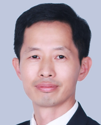 Honggang Li (李红钢)合作论文数Institute of Reproductive Health, Tongji Medical College, Huazhong University of Science and Technology7Tianli Chang合作论文数6Mengdi Zhang合作论文数6
 Lian Hu合作论文数6
 Aihua Liao (廖爱华)合作论文数Institute of Reproductive Health, Tongji Medical College, Huazhong University of Science and Technology;Center for Reproductive Medicine, Huazhong University of Science6
 Ling Zeng (曾灵)合作论文数6Jiahao Yang合作论文数5Qiuzi Shen合作论文数5Jean Damascene Niringiyumukiza合作论文数5
 Yamin Li合作论文数5Min Zou合作论文数5Jing Li合作论文数5YuZhen Guo (郭玉珍)合作论文数5
 Rongzhen Jiang (蒋荣珍)合作论文数Shanghai Sixth People’s Hospital5Lijun Shui (水丽君)合作论文数5
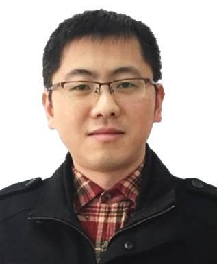 Liquan Zhou (周立全)合作论文数Institute of Reproductive Health, Tongji Medical College, Huazhong University of Science and Technology4
 Xianjin Xiao (肖先金)合作论文数Institute of Reproductive Health, Tongji Medical College, Huazhong University of Science and Technology4
 Yong Zhang (张勇)合作论文数Union Hospital, Tongji Medical College, Huazhong University of Science and Technology4Yinzhao Jia合作论文数3Su Song合作论文数3Li Chen合作论文数3Zili Li合作论文数3
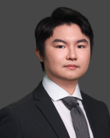 Yanwei Zhang (张晏维)合作论文数International Business School, Hainan University3
 Jinzhao Zhou合作论文数3Gu Xiuli合作论文数3Na Zhao (赵娜)合作论文数3
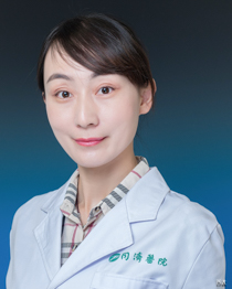 Kezhen Li (李科珍)合作论文数Tongji Hospital, Tongji Medical College, Huazhong University of Science and Technology3Liang Hu合作论文数2Yinhua Song合作论文数2




