Objective: To determine whether treat-to-target urate-lowering therapy (ULT) can prevent joint damage, particularly bone erosion progression, in patients with gout.,Methods: Patients who had experienced recent gout episodes were consecutively enrolled in this prospective, single-center study and treated with ULT. Baseline and 12-month assessments included laboratory testing and ultrasound (US) of bilateral knee, ankle, and first metatarsophalangeal joint (MTPJ), with semi-quantitative scoring for gout-related US lesions and synovial changes. Multivariable logistic regression identified factors linked to bone erosion.,Results:116 patients (mean age 44.0 years, disease duration 7.2 years, 92.2% male) were evaluated. After 12 months, 68% achieved the target serum urate (SUA) level (<360 μmol/L; normal SUA group), and 32% did not (high SUA group). The total score decreased significantly in the normal SUA group but increased slightly in the high SUA group (7.27 vs. 7.68, P = 0.803). The between-group difference in total score changes was significant (-2.05 vs. 0.32, P = 0.032). The difference in bone erosion score showed an upward trend in the high SUA group, while that in the normal SUA group slightly decreased (-0.09 vs. 0.41, P = 0.045). Age, disease duration, and tophus presence were independent risk factors for bone erosion (ORs: 1.020, 1.048, 1.891; all P<0.05).,Conclusion: In patients with gout, ULT that achieves target SUA levels is associated with stabilization or mild reduction of bone erosion, whereas uncontrolled hyperuricemia predicts progression of bone erosion. These findings support that effective treat-to-target ULT protects against structural joint damage in gout.
Purpose: This study aims to develop a computer-aided diagnosis (CAD) model for aiding ultrasonographers in classification of benign or malignant breast lesions in BI-RADS categories 4-5 using ultrasound videos. Materials and Methods: This prospective multicenter study enrolled 532 breast lesions categorized as breast imaging reporting and data system (BI-RADS) 4-5 by ultrasound. Of them, 334 breast lesions were included as training and validation sets. The remaining 198 lesions was assigned to a test set. We acquired both static and dynamic video ultrasound images of lesions for each patient and utilized them to develop a deep learning model. Temporal-guided multi-scale multi-instance learning network (TGMIL) with the video images was also developed. In this study, four metrics were calculated: area under the curve (AUC), accuracy, precision and sensitivity. In addition, pairwise DeLong and McNemar were used as statistical tests. Results: A total of 120 patients with 198 videos (benign, 45.8 ± 11.5 years; malignant, 50.7 ± 11.0 years) were evaluated. On the video-based test set, TGMIL achieved an AUC of 0.824 (95% CI, 0.790–0.857), significantly outperforming junior ultrasonographers (AUC, 0.698; 95% CI, 0.615–0.781; p < 0.05) and attending ultrasonographers (AUC:0.808; 95% CI, 0.735–0.880; p < 0.05). With TGMIL assistance, diagnostic accuracy improved from 69.2% to 85.9% for juniors (p < 0.05) and from 81.8% to 87.3% for attendings (p < 0.05); sensitivity increased from 68.4% to 82.7% for juniors (p < 0.05) and from 80.8% to 86.5% for attendings (p < 0.05). Conclusions: The TGMIL model significantly improved the diagnostic performance of junior and attending ultrasonographers for BI-RADS 4-5 breast lesions.
Background and Clinical Significance: Cutaneous Rosai-Dorfman disease (CRDD) is a rare, benign histiocytic proliferative disorder, accounting for approximately 3% of all Rosai-Dorfman disease (RDD) cases. Currently, the diagnosis of CRDD relies on invasive pathological examination due to the absence of reliable non-invasive alternatives. This case series evaluates the potential utility of high-frequency ultrasound (HFUS) as an adjunctive diagnostic tool for CRDD. Case Presentation: We present three CRDD cases, correlating HFUS features with histopathology. All cases showed hypoechoic lesions with varying infiltration depths and morphologies, though no specific diagnostic features were identified. HFUS clearly delineated involvement of the dermal and subcutaneous layers, assessed morphological characteristics like contour regularity and border definition, and evaluated vascularity. This information is crucial for clinical decision-making. HFUS also demonstrated value in therapeutic follow-up. In Case 1, it objectively showed a reduction in lesion size and decreased internal vascularity, providing clear evidence of treatment response. Conclusions: Although HFUS cannot independently diagnose CRDD and histopathology remains the gold standard, it serves as a valuable complementary tool. HFUS allows evaluation of deeper tissue structures, infiltration depth, and vascularity. As a non-invasive modality, it is useful for treatment monitoring, therapy guidance, and prognosis assessment. Integrating HFUS into the CRDD workflow enables more comprehensive and precise management.
RATIONALE AND OBJECTIVES:Superficial soft tissue masses (STMs) represent a diagnostic dilemma in clinical practice, with ultrasound (US) being the front-line imaging modality available globally. However, the high complexity of STMs in imaging makes subjective evaluation highly dependent on experience, frequently causing inconsistent malignancy assessments. This inconsistency triggers unnecessary benign biopsies and delays treatment for malignant STMs. We developed ST-USNet, a multitask convolutional neural network framework to classify superficial STMs based on manually drawn regions of interest. MATERIALS AND METHODS:This retrospective study included US images of 3168 patients (median age, 58 years; IQR, 46-68 years) with STMs from four institutions between March 2015 and October 2024. The ST-USNet was developed and validated on multi-center data. Its performance was then evaluated on a separate, independent test cohort. The diagnostic performance of ST-USNet was compared with that of radiologists using McNemar tests. RESULTS:The ST-USNet was composed of four sub-models (SM-1, SM-2, SM-a, and SM-b). In the validation cohort, SM-1 was trained to distinguish malignant from benign STMs (AUC: 0.984); SM-2 was to classify the malignant STM subtypes including sarcoma, lymphoma, and metastatic carcinoma (AUC: 0.932, 0.909, 0.922); SM-a was to discriminate between aggressive and indolent lymphoma (AUC: 0.951). SM-b was designed to explore the identification of metastatic carcinoma origin (thyroid, breast, respiratory, digestive, and reproductive systems) as a preliminary analysis; however, due to limited sample sizes, these results should be interpreted as exploratory. ST-USNet achieved high AUCs on the validation cohort and remained effective, albeit with slightly lower performance, on an independent test cohort. In a preliminary reader study (4 radiologists, 85 cases), ST-USNet either significantly outperformed senior radiologists (p = 0.012) or performed comparably to them (p = 0.267, 0.092, 0.332) in all classification tasks and effectively enhanced diagnostic accuracy for both junior and senior radiologists when used as an assistive tool. CONCLUSION:ST-USNet serves as an effective and practical decision support system for superficial STMs classification in clinical oncology, though multi-center prospective validation and continuous model updating are required before clinical deployment.
Introduction: The Breslow thickness of cutaneous melanoma (CM) is related to the surgical approach and is usually measured using a preoperative biopsy. However, tumor thickness is often underestimated by partial biopsies. Objectives: To identify whether high-frequency ultrasound (HFUS) can improve the accuracy of preoperative detection of Breslow thickness. Methods: Partial biopsies and HFUS measurements of Breslow thickness were analyzed in 17 patients with CM. Postoperative histopathologic examination is considered the gold standard. In different thicknesses, HFUS and partial biopsy were compared with postoperative pathology and their effects on tumor T staging and surgical margins. Results: The mean (±SD) Breslow thicknesses measured using HFUS, partial biopsy, and postoperative histopathology were 3.5 ± 2.2 mm, 1.8 ± 1.1 mm, and 3.2 ± 2.1 mm, respectively. The correlation coefficients were 0.76 (partial biopsy and postoperative histopathology) and 0.96 (HFUS and postoperative histopathology), respectively (all p < 0.01). Partial biopsy underestimated the Breslow thickness, and with a gradual increase in CM thickness, the underestimation became increasingly obvious. Partial biopsy led to an underestimation of T staging of the tumor in 8 (8/17 [47.1%]) patients, 4 (4/8 [50.0%]) of whom may have had insufficient surgical peripheral margins. Conversely, HFUS exhibited a significant downward trend of underestimation (0/17 [0%], p = 0.003). However, only 2 (2/17 [11.8%]) patients had slight overestimation for T staging (p = 0.485), and none of the overestimations changed the surgical margins. Conclusions: HFUS can provide an accurate preoperative assessment of Breslow thickness, and correlates better with postoperative histopathology.
The B-mode ultrasound (BUS) based computer-aided diagnosis (CAD) has indicated its effectiveness for liver cancer diagnosis. However, the diagnostic information captured by BUS is confined to lesion structure and internal echogenicity, which inherently limits its diagnostic accuracy to some extent. Contrast-enhanced ultrasound (CEUS) can offer additional hemodynamic information, thereby improving classification performance, but its clinical application is less widespread than BUS. Previous studies have shown that learning using privileged information (LUPI) can facilitate the performance of BUS-based CAD by utilizing the transferable knowledge from CEUS. However, the LUPI framework is constrained to knowledge transfer between the paired data. In clinical practice, many patients receive only the single-modality BUS examination, which cannot be used as extensible data in the LUPI framework for superior transfer learning. To this end, we propose a Multi-View Doubly Supervised Knowledge Distillation (MDSKD) algorithm to address the transfer task of imbalanced ultrasound modalities in liver cancer. Specifically, a multi-view feature selection and fusion module is developed, which designs a multi-view channel ranking mechanism to select important features from three CEUS phase images, and then introduces Bi-Mamba to effectively integrate these discriminative features. Moreover, a doubly supervised knowledge distillation module is proposed to facilitate the knowledge transfer between imbalanced modalities. This module introduces generalized distillation and label consistency distillation to perform supervised transfer task on paired and unpaired ultrasound data, guided by shared and unshared labels. MDSKD is evaluated on a liver cancer dataset that comprises 206 subjects with paired BUS and CEUS data and 151 subjects with single-modal BUS data. It achieves best performance with an accuracy of 92.09 +/- 1.75%, a sensitivity of 91.26 +/- 2.06%, and a specificity of 92.98 +/- 2.34%. These results suggest that the MDSKD can assist sonologists in improving diagnostic accuracy.
PurposeTo explore the predictive value of radiomics features, ultrasound (US), and gene mutation status based on interpretable random forest (RF) models for predicting high Ki-67 expression in papillary thyroid carcinoma (PTC).MethodsThis retrospective analysis included 627 patients with surgically confirmed PTC who underwent testing for BRAF V600E and TERT promoter mutations, as well as immunohistochemical assessment of Ki-67 expression from December 2015 to June 2023. The eligible patients were randomly divided into a training set and a testing set at a ratio of 7:3 according to their binary Ki-67 expression status. A random forest model was constructed using both individual and combined ultrasound radiomics features, conventional ultrasound features, and genetic mutation status to predict high Ki-67 expression in PTC. Using AUC, Brier score and decision curve analysis to verify the clinical utility of the model.ResultThe Rad+US+Gene model demonstrated superior predictive accuracy for high Ki-67 expression, achieving the highest accuracy and AUC, along with the lowest Brier score. In the testing cohort, the Rad+US+Gene model attained an accuracy of 0.883, outperforming Rad+US, US and Rad (0.851). Its AUC reached 0.904, markedly exceeding those of Rad+US (0.854), US (0.823), and Rad (0.851). Regarding calibration, the Rad+US+Gene model also yielded the lowest Brier score (0.0822), compared with Rad+US (0.1073), US (0.1061), and Rad (0.1232), indicating superior predictive accuracy and stability.ConclusionThe integrated model combining radiomics, US features and genetic mutations achieves favorable predictive performance, presenting a new method for the preoperative assessment of Ki-67 expression level in PTC.
The accurate diagnosis of skin diseases relies on combining cortical lesion morphological features from clinical and dermoscopic data with deep subcutaneous lesion characteristics from ultrasound data. However, current multi-modal diagnostic methods mainly emphasize clinical and dermoscopic data analysis, lacking a thorough exploration of subcutaneous tissue features provided by skin ultrasound. Additionally, existing research often overlooks the analysis of relationships between skin labels and categories across different diagnostic tasks, constraining model performance as the number of target tasks grows. This paper proposes a label-guided graph learning network (LGL_Net) based on two-stage cross-modal fusion to achieve accurate diagnosis of skin diseases. Specifically, this paper first establishes a two-stage cross-modal fusion unit (TC_Fusion) to enable the fusion and transmission of morphological features from clinical data and deep lesion features from ultrasound data. Subsequently, a label-guided graph learning unit (LGL_Unit) is constructed to explore the correlations between multilabel data of skin lesions by building a graph convolutional network (GCN) at the label and category levels, thereby improving the accuracy of various skin disease diagnostic tasks. Extensive experiments were conducted on a public dataset (including clinical and dermoscopic data) and a private dataset (including clinical and ultrasound data). The experimental results demonstrate that the proposed method achieves optimal performance on tasks like pathological diagnosis (PG), benign or malignant diagnosis (BM), and 7-Point Checklist scoring (7 PC), offering a new approach for skin disease diagnosis. Our code is available at: https://github.com/Zhaocheng 1/LGL-Net.
ObjectiveTo investigate the feasibility of remotely providing routine ultrasound (US) examinations to patients using a fifth‐generation‐based robot‐assisted tele‐ultrasonography (RATU) system in a real‐world setting.MethodsBetween September 2020 and May 2021, we conducted a prospective and large‐scale study using the RATU system to provide US examinations for patients on a limited‐source island locate. An on‐site radiologist on the island performed US examinations, which served as the reference diagnosis. Five tele‐radiologists then remotely conducted RATU examinations from Central Shanghai. We compared the diagnostic performance of the RATU examinations to that of the on‐site US examinations. Additionally, we assessed the learning curves of the various tele‐radiologists. We also distributed 2 questionnaires to evaluate the usefulness of the clinical application.ResultsIn total, 770 patients were enrolled in the study with a mean age of 55.46 ± 15.02 years (ranging from 19 to 80 years). Out of the total, 501 patients were men, and 269 were women. Across all examination types, the diagnosis in 84.3% (649/770) of RATU examinations was consistent with the on‐site US examination. The learning curve was not significantly different between tele‐radiologists of different seniority. Furthermore, 86.6% (667/770) of participants accepted the RATU examination, and 97.5% (751/770) of patients were willing to pay a fair price.ConclusionRATU's diagnostic performance is still helpful for patients in remote areas, even though it may be slightly inferior to on‐site US examination.
BackgroundEarly detection is clinically crucial for the strategic handling of sarcopenia, yet the screening process, which includes assessments of muscle mass, strength, and function, remains complex and difficult to access. ObjectiveThis study aims to develop a convolutional neural network model based on ultrasound images to simplify the diagnostic process and promote its accessibility. MethodsThis study prospectively evaluated 357 participants (101 with sarcopenia and 256 without sarcopenia) for training, encompassing three types of data: muscle ultrasound images, clinical information, and laboratory information. Three monomodal models based on each data type were developed in the training cohort. The data type with the best diagnostic performance was selected to develop the bimodal and multimodal model by adding another one or two data types. Subsequently, the diagnostic performance of the above models was compared. The contribution ratios of different data types were further analyzed for the multimodal model. A sensitivity analysis was performed by excluding 86 cases with missing values and retaining 271 complete cases for robustness validation. By comprehensive comparison, we finally identified the optimal model (SARCO model) as the convenient solution. Moreover, the SARCO model underwent an external validation with 145 participants (68 with sarcopenia and 77 without sarcopenia) and a proof-of-concept validation with 82 participants (19 with sarcopenia and 63 without sarcopenia) from two other hospitals. ResultsThe monomodal model based on ultrasound images achieved the highest area under the receiver operator characteristic curve (AUC) of 0.827 and F1-score of 0.738 among the three monomodal models. Sensitivity analysis on complete data further confirmed the superiority of the ultrasound images model (AUC: 0.851; F1-score: 0.698). The performance of the multimodal model demonstrated statistical differences compared to the best monomodal model (AUC: 0.845 vs 0.827; P=.02) as well as the two bimodal models based on ultrasound images+clinical information (AUC: 0.845 vs 0.826; P=.03) and ultrasound images+laboratory information (AUC: 0.845 vs 0.832, P=0.035). On the other hand, ultrasound images contributed the most evidence for diagnosing sarcopenia (0.787) and nonsarcopenia (0.823) in the multimodal models. Sensitivity analysis showed consistent performance trends, with ultrasound images remaining the dominant contributor (Shapley additive explanation values: 0.810 for sarcopenia and 0.795 for nonsarcopenia). After comprehensive clinical analysis, the monomodal model based on ultrasound images was identified as the SARCO model. Subsequently, the SARCO model achieved satisfactory prediction performance in the external validation and proof-of-concept validation, with AUCs of 0.801 and 0.757 and F1-scores of 0.727 and 0.666, respectively. ConclusionsAll three types of data contributed to sarcopenia diagnosis, while ultrasound images played a dominant role in model decision-making. The SARCO model based on ultrasound images is potentially the most convenient solution for diagnosing sarcopenia. Trial RegistrationChinese Clinical Trial Registry ChiCTR2300073651; https://www.chictr.org.cn/showproj.html?proj=199199
INTRODUCTION:The aim of this study was to identify whether high-frequency ultrasound (HFUS) could correct the misdiagnosis, confirm equivocal skin lesions, and improve the management after clinical examination. METHODS:In this study, a total of 574 skin lesions from 552 patients were prospectively enrolled. The specific diagnosis and management decisions (treatment/excision, observation) determined by HFUS after clinical examination were recorded during the clinical practice. The area under the receiver operating characteristic curve, accuracy, sensitivity, specificity, positive predictive value, negative predictive value, and the number needed to excise (NNE) before and after HFUS were also evaluated. The pathological results were conducted as golden standards to compare the performance. RESULTS:Among the 574 skin lesions, 290 (50.5%) were malignancies and 284 (49.5%) were benign. The diagnostic accuracy was improved from 77.5% to 90.8% after the HFUS examination. There were 44 lesions wrongfully diagnosed by the initial clinical diagnosis, whereas 28 of 44 (63.6%) lesions were correctly identified by HFUS examination. Of 85 lesions categorized as equivocal skin lesions by clinical examination, 65 (76.5%) were diagnosed correctly after HFUS. Lesion management changed in 72 of 574 (12.5%) after HFUS. Among these lesions, HFUS saved 22 unnecessary excisions and prompted the treatment of 30 malignancies that would be observed based on clinical examination alone. Additionally, the NNE was reduced by 15.4% (NNE, 0.828) after HFUS and 4.6% (NNE, 0.933) before HFUS. CONCLUSIONS:HFUS could be a valuable tool in diagnosing equivocal skin lesions, identifying skin cancers missed by clinical examination, and reducing unnecessary excision of benign lesions while improving NNE.
Objectives: Unlike other body parts, unclarified lesions at the end of extremities have unique challenges due to their small size and interference. Traditional imaging methods struggle with low resolution. HFUS enhances resolution, offering a potential diagnostic value. Methods: From January 2019 to October 2023, the clinical and HFUS data of patients with unclarified lesions at the end of extremities were retrospectively analyzed. Independently, the diagnosis was made using two diagnostic modes (Mode A: only clinical information; Mode B: clinical and HFUS information). The diagnostic performance of the two modes was evaluated across different classification methods. Results: For all lesions, the correct rate of Mode B was higher than that of Mode A (52.8% vs. 18.4%, p < 0.001), and the indeterminate rate decreased by 43.0%. For benign lesions (51.0% vs. 18.2%), subungual lesions (40.8% vs. 21.1%), non-subungual lesions (55.6% vs. 17.8%), and common cases (60.9% vs. 20.3%), the diagnostic correct rate of Mode B was also higher than that of Mode A (all p < 0.05). However, there was no significant difference in rare lesions (9.8% vs. 4.9%) and malignant lesions (62.9% vs. 19.4%) between the two modes (both p > 0.05). Moreover, the indeterminate rate for all categories of lesions significantly diminished. Otherwise, Mode B demonstrated strong performance for malignant lesions (85.7% vs. 42.9%, p < 0.001). Conclusions: Adding HFUS can significantly improve the accuracy of diagnosing unclarified lesions at the end of extremities and reduce uncertainty, especially for benign and common lesions. HFUS has also demonstrated better performance in screening for malignant lesions.
The abnormal tumor mechanical microenvironment due to specific cancer-associated fibroblasts (CAFs) subset and low tumor immunogenicity caused by inefficient conversion of active chemotherapeutic agents are two key obstacles that impede patients with desmoplastic tumors from achieving stable and complete immune responses. Herein, it is demonstrated that FAP-α+CAFs-induced stromal stiffness accelerated tumor progression by precluding cytotoxic T lymphocytes. Subsequently, a cascade-responsive nanoprodrug capable of re-educating FAP-α+CAFs and amplifying tumor immunogenicity for potentiated cancer mechanoimmunotherapy is ingeniously designed. Benefiting from the active targeted release of angiotensin II receptor antagonist (losartan) guided by FAP-α cleavable peptide and the efficient conversion of topoisomerase I inhibitor (7-Ethyl-10-hydroxycamptothecin) prodrug under high glutathione/esterase within tumor cells, this regimen created an immune-activated landscape that retarded primary tumor growth and counteracted resistance to immune checkpoint inhibitor in mice with triple-negative breast cancer. This nanoprodrug-assisted mechanoimmunotherapy can serve as a universal strategy for conferring efficient tumoricidal immunity in "immune excluded" desmoplastic tumor interventions.
Background:To investigate the clinical efficacy of narrow-margin modified Mohs microsurgery (mMMS) in the treatment of extramammary Paget's disease (EMPD). Methods:A retrospective cohort review was conducted on 52 patients with EMPD who were treated at the Skin Disease Hospital of Tongji University in Shanghai between 2017 and 2023. The primary objectives of this study were to assess the long-term local recurrence rates of tumors treated with narrow-margin mMMS and to explore the final margin width as well as the factors that may influence postoperative recurrence. Results:A total of 52 patients were included in this retrospective study. Most patients were male (n = 48, 92.3%) with a mean age of 69.5 years (SD:9.08, range:44-91). The follow-up rate was 78.7% (41/52), and the mean follow-up time was 36.17 months (SD:18.25, range 5.8-62.5). The recurrence rate was 9.7% (4/41) and the 5-year tumor-free rate was 85.9%. Approximate 95% of tumors with 1 cm of non-scrotal skin extension or 1.5 cm of scrotal skin extension could be completely cleared. Univariable analysis revealed that hypopigmented patches (HR=14.0, 95% CI=1.269,154.395, p = 0.031) correlated with tumors recurrence. Conclusion:Narrow-margin mMMS is the ideal therapy combine a disease control rate with more satisfying functional results. Determination of tumor boundaries requires attention to skin lesions with hypopigmented macules. The initial resection margins width of the extension cut can be reduced in non-scrotal skin lesions at the time of surgery to minimize pointless margin expansion.
OBJECTIVES:To evaluate the value of ultrasound (US) and shear wave velocity (SWV) to assess muscle in postmenopausal women with osteosarcopenia (OSP). METHODS:This study included 145 postmenopausal women, comprising 115 osteopenia/osteoporosis participants without sarcopenia (OP alone) and 30 OSP participants. All received the evaluation of bone mineral density (BMD), appendicular skeletal muscle mass index (ASMI), handgrip strength, calf circumference, 6-meter walking speed, and 5-time chair stand test. The cross-sectional area (RFcsa), thickness (RFthickness), and mean SWV of rectus femoris (RF) were measured by US and shear wave elastography. The clinical characteristics, RFcsa, RFthickness, and SWV, were compared between OP alone and OSP to determine the independent predictors of OSP. Receiver operating characteristic (ROC) curve analysis was used to evaluate the value of US and SWV for assessment of OSP. RESULTS:The RFcsa, RFthickness, SWV, and BMD of OSP were lower than those of OP alone (all P < 0.05). Through multivariate analysis, the diagnostic performance of the prediction model (area under the ROC curve, AUC, 0.917) composed of RFcsa and SWV was superior to RFcsa (AUC, 0.847), RFthickness (AUC, 0.797), and SWV (AUC, 0.740) alone. Moreover, the prediction model achieved 70.0% sensitivity, 93.0% specificity, and 88.3% accuracy. CONCLUSIONS:The RFcsa, RFthickness, and SWV have potential value in assessing muscle in OSP and can be applied to routine clinical management of postmenopausal women. ADVANCES IN KNOWLEDGE:US-measured RF thickness, CSA, and SWV could assist detection and clinical management of OSP in postmenopausal women.
Heterogeneous morphological features and data imbalance pose significant challenges in rare thyroid carcinoma classification using ultrasound imaging. To address this issue, we propose a novel multitask learning framework, Channel-Spatial Attention Synergy Network (CSASN), which integrates a dual-branch feature extractor - combining EfficientNet for local spatial encoding and ViT for global semantic modeling, with a cascaded channel-spatial attention refinement module. A residual multiscale classifier and dynamically weighted loss function further enhance classification stability and accuracy. Trained on a multicenter dataset comprising more than 2000 patients from four clinical institutions, our framework leverages a residual multiscale classifier and dynamically weighted loss function to enhance classification stability and accuracy. Extensive ablation studies demonstrate that each module contributes significantly to model performance, particularly in recognizing rare subtypes such as FTC and MTC carcinomas. Experimental results show that CSASN outperforms existing single-stream CNN or Transformer-based models, achieving a superior balance between precision and recall under class-imbalanced conditions. This framework provides a promising strategy for AI-assisted thyroid cancer diagnosis.
 Huixiong Xu (徐辉雄)合作论文数School of Forensic Medicine and Science, Fudan University154
 Liping Sun (孙丽萍)合作论文数School of Medicine, Tongji University;Tenth People's Hospital of Tongji University73
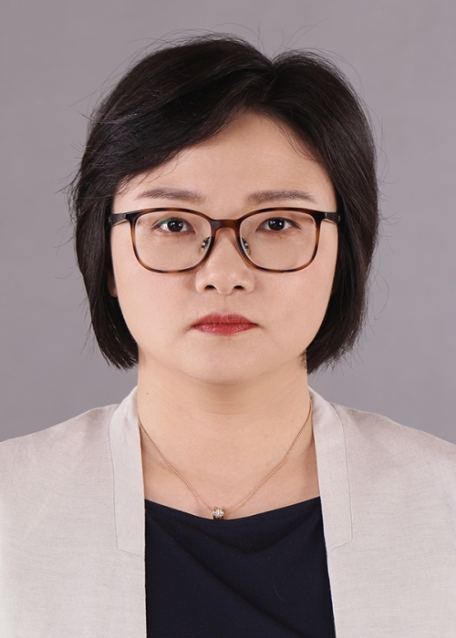 Yifeng Zhang (张一峰)合作论文数Shanghai Tenth People's Hospital61
 Xiaolong Li (李晓龙)合作论文数57
 Linna Liu (刘琳娜)合作论文数48Jun-Mei Xu (徐军妹)合作论文数46Qiao Wang合作论文数41Chang Liu合作论文数41Bo Xiao-Wan合作论文数31
 Chong-Ke Zhao合作论文数28Liu Bo-Ji合作论文数26Wei-Wei Ren合作论文数Tongji Univ, Shanghai Peoples Hosp 10, Ultrasound Res & Educ Inst, Dept Med Ultrasound,Sch Med, Shanghai 200072, Peoples R China20An-Qi Zhu合作论文数18Dan Wang合作论文数Tongji Univ, Shanghai Peoples Hosp 10, Ultrasound Res & Educ Inst, Dept Med Ultrasound,Sch Med, Shanghai 200072, Peoples R China18
 Dan-Dan Li合作论文数18
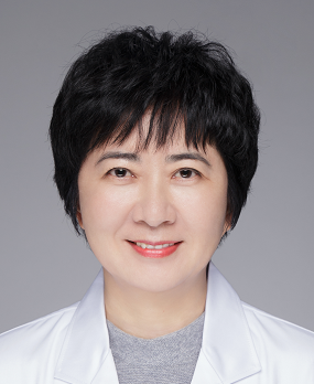 Rong Wu (吴蓉)合作论文数Shanghai General Hospital17Xu Xiao-Hong合作论文数16Lu Feng合作论文数16Ya-Ping He合作论文数Tongji Univ, Shanghai Peoples Hosp 10, Ultrasound Res & Educ Inst, Dept Med Ultrasound,Sch Med, Shanghai 200072, Peoples R China14Li-Fan Wang合作论文数13Dan-Dan Shan合作论文数13Zi-Tong Chen合作论文数12Hui-Jun Fu合作论文数12
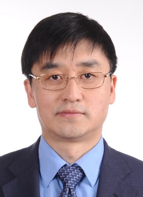 Shen Qu (曲伸)合作论文数School of Medicine, Tongji University12
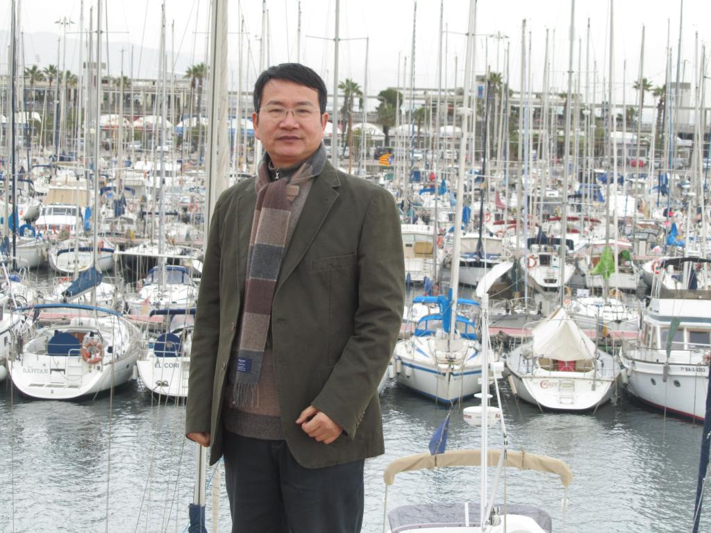 Shuguang Zheng (郑曙光)合作论文数School of Law,Ningbo University,Ningbo ,China12
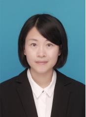 Wenwen Yue (岳雯雯)合作论文数School of Medicine, Tongji University;Tenth People's Hospital of Tongji University10Shuai Wang (王帅)合作论文数10Guang Xu合作论文数9Yong He (何勇)合作论文数9
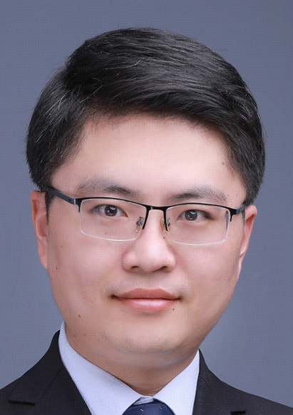 Kun Zhang (张坤)合作论文数School of Medicine, University of Electronic Science and Technology of China9Minghua Yao合作论文数8
 Jun Shi (施俊)合作论文数School of Communication and Information Engineering, Shanghai University8Ya-Qin Zhang合作论文数7Yujing ZHAO (赵宇靖)合作论文数7
 Jian Wu (吴剑)合作论文数Zhongshan Hospital, Fudan University7Yuanyuan Liu (刘媛媛)合作论文数7
 Fang Ma (马方)合作论文数Shanghai Sixth People's Hospital7Hui Shi合作论文数6
 Ai Peng (彭艾)合作论文数School of Medicine, Tongji University6Feng-Shan Jin合作论文数5
 Hui Zhao合作论文数5Ming-Xu Li合作论文数5Li Liang合作论文数5
 Jing Zhang (张敬)合作论文数School of Life Science and Technology, Tongji University5Na Ni合作论文数4Yi-Kang Sun合作论文数4Chuan Qin合作论文数4Jian-Na Yan合作论文数4




