BACKGROUND AND OBJECTIVES:While neurosurgical posterior fossa decompression with duraplasty (PFDD) may provide clinical and radiological improvement for Chiari malformation-I (CM-I)-syringomyelia, the comparative efficacy and safety of PFDD without vs with intradural tonsillar manipulation (posterior fossa decompression with tonsil resection) has remained controversial for over 5 decades. METHODS:We conducted a retrospective cohort study of 1231 CM-I-syringomyelia patients treated at our institution from 2003 to 2024, comparing 2 techniques: standard PFDD and foramen magnum and foramen of magendie dredging (FMMD, a modified posterior fossa decompression with tonsil resection procedure). Propensity score matching was used to balance baseline characteristics between the 2 groups. The primary outcomes were syrinx regression >50%, while secondary outcomes encompassed symptom-related parameters, syrinx regression, complication-related parameters, and reoperation rate. RESULTS:A total of 1231 patients with CM-I were included, of whom 310 (25.2%) were in the PFDD group, and 921 (74.8%) were in the FMMD group. Per treatment analysis demonstrated no increase in odds of complications for FMMD ( P > .05). PFDD was noninferior to FMMD in clinical improvement and syrinx regression ( P = .147, P = .169, respectively). Syrinx regression (>50% reduction) was superior following FMMD (78% vs 60%, P < .001). PFDD had a higher rate of revision surgery than FMMD (15.5% vs 4.1%, log-rank P < .001). CONCLUSION:At our center, FMMD demonstrated greater effectiveness in managing syringomyelia, with higher rates of syrinx regression (>50% reduction), a lower rate of revision surgery, and no increase in complications compared with PFDD. Nonetheless, PFDD was similar to FMMD regarding clinical improvement and syrinx regression.
INTRODUCTION:Chronic spinal cord injury (SCI) is a severe neurological disorder resulting from trauma or disease, characterized by persistent motor, sensory, and autonomic deficits below the level of the lesion. As the condition becomes long-standing, the management of multisystem complications - particularly those affecting motor, respiratory, cardiovascular, gastrointestinal, and urinary functions - emerges as a major clinical challenge. These complications not only impair quality of life but also contribute to increased rates of rehospitalization and impose significant economic burdens on healthcare systems. DISCUSSION:Although no curative therapy is currently available, treatment strategies have evolved into a comprehensive, multilayered framework incorporating rehabilitation, pharmacological interventions, neuromodulation techniques, and surgical procedures. Nevertheless, existing management approaches remain predominantly reactive and symptom-oriented, with notable limitations in long-term effectiveness, accessibility, and sustained patient engagement. This review synthesizes recent advances in understanding the pathophysiology of chronic SCI complications and critically evaluates current therapeutic limitations. A fundamental paradigm shift is urgently required - one that moves beyond traditional reactive models toward proactive, patient-centered care grounded in multidisciplinary collaboration and individualized treatment plans. CONCLUSION:Given the numerous challenges and the limitations of the current management strategies, further research is urgently needed for the management of chronic spinal cord injury complications. Future progress is expected to be driven by innovations in neural regeneration and precision medicine, aiming to transform clinical management from passive symptom mitigation to active functional restoration, thereby improving long-term outcomes for individuals living with chronic SCI.
BACKGROUND AND OBJECTIVE:Whether extent of resection (EOR) improves overall survival (OS) in World Health Organization (WHO) grade 4 primary spinal cord astrocytoma (SCA) remains controversial owing to limited and conflicting evidence. This study aimed to evaluate the impact of EOR on the prognosis of patients with WHO grade 4 primary SCA. METHODS:We retrospectively analyzed 59 cases of WHO grade 4 primary SCA (2014-2024) to assess the impact of EOR on survival with subgroup analysis based on tumor location and Ki-67 expression. RESULTS:In the whole cohort, Kaplan-Meier analysis showed no significant OS difference between the EOR ≥50% and EOR <50% groups (17 vs 10 months, P = .171). However, subgroup analyses found that EOR ≥50% significantly improved OS in noncervical SCA (36 vs 10 months, P = .034) and high Ki-67 SCA (16 vs 6 months, P = .016). CONCLUSION:This study demonstrates that the surgical benefit in WHO grade 4 primary SCA may be influenced by tumor location and Ki-67. We propose a hypothesis-generating conceptual framework to guide future research directions.
OBJECTIVE:Tethered cord syndrome (TCS) results in neurological dysfunction due to spinal cord tethering. Because MRI sensitivity, particularly for occult TCS (OTCS), is limited by variable image quality and anatomy, the authors aimed to develop and externally validate a simple MRI-based diagnostic index derived from dural sac morphology to improve diagnosis across multicenter datasets. METHODS:This retrospective multicenter study included 280 participants from 5 hospitals, encompassing patients with simple TCS, complex TCS, and OTCS along with healthy controls. Participants were allocated into training, internal validation, external test, and external OTCS test datasets. Dural sac morphological features were manually measured on axial T2-weighted MRI scans from levels L1-2 through L5-S1. Logistic regression identified key morphological features at L1-2 and L2-3 levels, which were integrated into a diagnostic index incorporating demographic adjustments for age and sex. Model performance was assessed using receiver operating characteristic curve analysis across all datasets. RESULTS:The final diagnostic model demonstrated strong and consistent diagnostic efficacy. In the training dataset, the corrected diagnostic index achieved a sensitivity of 0.921 and specificity of 0.779 (area under the curve 0.930). High sensitivity (≥ 0.9) and satisfactory specificity (≥ 0.7) were reliably maintained across the internal validation, external test, and OTCS datasets. CONCLUSIONS:This study establishes a practical MRI-based diagnostic index that leverages dural sac morphological parameters at levels L1-2 and L2-3 to accurately diagnose TCS, including challenging occult cases. The model's reliance on straightforward anatomical measurements and demographic corrections eliminates dependence on advanced imaging protocols or subjective interpretations, presenting a robust imaging biomarker that can significantly improve early detection and clinical management of TCS.
The pathways of clearance of cerebrospinal fluid (CSF) in the spine remains incompletely understood. While our minimally invasive subarachnoid-subarachnoid bypass technique has demonstrated efficacy in Syringomyelia related to spinal subarachnoid obstruction, its therapeutic utility is significantly limited in extensive spinal adhesive arachnoiditis (ESAA; >5 spinal levels involvement) or Lumbosacral Outlet Obstruction with Syringomyelia. This study evaluates thecal-peritoneal shunting as a novel intervention for ESAA or Lumbosacral Outlet Obstruction with Syringomyelia. We prospectively evaluated 20 consecutive Extensive Spinal Adhesive Arachnoiditis or Lumbosacral Outlet Obstruction with Syringomyelia patients undergoing thecal-peritoneal shunting, with a mean follow-up of 14 months (range 12–20 months). Intraspinal pressure was measured intraoperatively at sites cranial to arachnoid adhesions and preoperatively caudal to adhesions during myelography. Twenty baseline-matched small spinal meningioma or schwannoma patients served as controls with intraoperative pressure measurements. The procedure yielded clinical improvement in 16 patients (80
Introduction:Coronal imbalance in degenerative spinal deformity is difficult to correct, particularly when the fractional curve is rigid and the lumbosacral foundation is oblique. Research question:Is staged lateral lumbar interbody fusion with targeted mini-open posterior correction associated with stepwise coronal and sagittal realignment and maintenance of coronal alignment at follow-up? Material and methods:We retrospectively reviewed 51 consecutive patients treated with multi-level lateral lumbar interbody fusion followed by posterior percutaneous fixation with or without targeted mini-open osteotomy. Standing full-length radiographs were assessed preoperatively, after stage I, after stage II, and at latest follow-up. Coronal endpoints were coronal Cobb angle, coronal vertical axis, and fractional-curve take-off angle. Sagittal endpoints included lumbar lordosis, pelvic incidence minus lumbar lordosis mismatch, T1 pelvic angle, and sagittal vertical axis. Clinical outcomes were assessed preoperatively and at follow-up. Results:Coronal Cobb angle improved from 35.62° preoperatively to 21.93° after stage I, 10.20° after stage II, and 10.91° at follow-up. Coronal vertical axis improved from 28.50 mm to 14.95 mm, and fractional-curve take-off angle from 17.28° to 5.88°. Lumbar lordosis increased from 22.40° to 35.66°, while pelvic incidence minus lumbar lordosis mismatch, T1 pelvic angle, and sagittal vertical axis also improved. Back-pain visual analog scale and Oswestry Disability Index improved significantly at follow-up. Discussion and conclusion:Staged lateral lumbar interbody fusion with targeted mini-open posterior correction was associated with stepwise coronal and sagittal realignment, and the improvement in mean coronal alignment was maintained at follow-up.
Recent discoveries focused on the role of intraspinal pressure (ISP) in metabolite clearance after spinal cord injury (SCI) have initiated intense research on CSF inflow and outflow pathways. This study aimed to investigate whether the pulsatile ISP wave serves as the primary driver of posttraumatic syringomyelia (PTS) formation and progression, and to determine if a novel subarachnoid‑subarachnoid (S‑S) bypass procedure can effectively attenuate this abnormal pulsatile ISP wave in affected PTS patients. In this prospective cohort study of patients with PTS, ISP across the injury site was monitored intraoperatively both before and after placement of the S-S bypass tube. Neurological impairment was assessed using standardized scales at baseline, one year postoperatively, and at the final follow-up. A logistic regression model was used to analyze prognostic factors associated with surgical outcomes. All 64 enrolled patients underwent S-S bypass surgery; 54 (84.4
Adhesive arachnoiditis (AA) is a rare form of chronic degenerative pathology associated with persistent inflammation in the arachnoid matter of the spinal cord. Despite the existing knowledge, the detailed pathological mechanisms underlying AA are not fully understood. This study aimed to elucidate through comprehensive single nuclei RNA sequencing (snRNA-seq) to delineate the transcriptomic landscape of AA. From six arachnoid membrane samples, a total of 52,886 cells met the quality control standards for analysis. The main cell populations identified with specific gene markers were as follows: fibroblasts, glial cells, microglial cells, endothelial cells, mural cells, plasma cells, and T cells. Downstream analysis of fibroblasts, glial cells, and microglial cells was performed. Notably, fibroblast subsets 1 and 3 demonstrated a strong association with AA. Among them, subcluster 3 demonstrated elevated expression of genes COL1A1, COL3A1, and FN1, indicative of enhanced Wnt/β-catenin and extracellular matrix (ECM) synthesis pathways. Subcluster 3 was predicted to progressively transform into subcluster 1. In subcluster 1, there was a significant upregulation of genes such as BMP and ALPL, signaling enhanced activation of calcification-related pathways. This was highly relevant to end-stage arachnoid ossification formation. After being activated, microglial cells transformed into inflammatory disease-associated microglial cells and continued to express high levels of chemokines CCL2, CCL4, IL-1β, and other inflammatory factors NAMPT, INPP5D and NLRP3. This might be the main reason why AA recurrence is frequently observed in patients. These insights enhance our understanding of the pathological progression of AA and may contribute to the identification of novel therapeutic targets.
(1) Background: Patients with mild cervical spondylotic myelopathy (CSM) who delay surgery risk progression. While PET evaluates spinal cord function, its cost and radiation limit its use. (2) Methods: In this prospective study, patients with mild cervical spondylosis underwent preoperative 18F-FDG PET-MRI. Narrowed spinal levels were classified based on whether SUVmax was decreased. Follow-up assessments were conducted. Two machine learning models using MRI T2-based radiomics were developed to identify stenotic levels and decreased SUVmax. (3) Results: Patients with normal SUVmax showed greater symptom improvement. The radiomics models performed well, with AUCs of 0.981/0.962 (training/testing) for stenosis detection and 0.830/0.812 for predicting SUVmax decline. The model outperformed clinicians in predicting SUVmax decline, improving the AUC by 10%. (4) Conclusion: Patients with preserved SUVmax have better outcomes. MRI-based radiomics shows potential for identifying stenosis and predicting spinal cord function changes for preoperative assessment, though larger studies are needed to validate its clinical utility.
BACKGROUND: Revision surgery for patients with persistent, recurrent, or progressive syringomyelia following Foramen Magnum Decompression for Chiari malformation with syringomyelia (CM-SM) is not uncom- mon and presents both strategic and technical challenges. METHODS: We conducted a retrospective study including all patients who underwent revision Cerebellar Tonsillectomy (CTL) for CM-SM between 2003 and 2023. Additionally, we performed univariate and multivariate analyses to identify possible factors contributing to failed CTL outcomes. RESULTS: Sixty-three consecutive patients (13 males; average age 45.86 +/- 11.18 years) underwent surgical treat- ment for persistent (n = 29), progressive (n = 21), or recurrent (n = 13) syringomyelia, with an average interval of 65.57 +/- 73.33 months (range: 3-480 months) between the two surgeries. Factors significantly associated with the effec- tiveness of the revision CTL included dural incision and tonsil manipulation during the first surgery, severe intradural adhesions during the revision CTL, and spinal cord atrophy before the revision CTL. Multivariate logistic regression revealed that dural incision (P = 0.031, odds ratio [OR] = 6.779, 95% confidence interval {CI} [1.187 38.719]), tonsillar manipulation (P = 0.037, OR = 7.432, 95% CI [1.131 48.835]), and severe intradural adhesions (P = 0.030, OR = 11.465, 95% CI [1.264 103.967]) constituted risk fac- tors significantly statistical associated with prognosis out- comes. Long-term follow-up (average 18.75 +/- 6.86 months, range: 12-72 months) of revision CTL demonstrated clinical stabilization in 55.6% of cases for at least 1.5 years. The complication rate for revision CTL was 14.3% (n = 9). CONCLUSIONS: Dural incision and tonsillar manipula- tion during the first surgery, spinal cord atrophy prior to revision CTL, and severe intradural adhesions during revi- sion CTL are significant risk factors associated with poor prognosis in revision surgery for CM-SM.
OBJECTIVE Current treatment options for posttraumatic syringomyelia (PTS) lack clear standardization and often result in common complications. This study aims to introduce a novel minimally invasive technique for a modified subarachnoid-subarachnoid (S-S) bypass procedure for PTS. METHODS The study included 20 consecutive patients with symptomatic PTS who underwent the modified S-S bypass. The surgical technique of modified S-S bypass involved two-laminae fenestration based on preoperative MRI and myelography results showing normal subarachnoid space above and below the injury site. A passer was used to create a subcutaneous tunnel, and two medical-grade silicone tubes were inserted into the normal subarachnoid space at the cranial and caudal ends. Bypass tubes were placed in the subcutaneous tunnel, and dural closure was achieved using 6-0 nonabsorbable sutures. The mean follow-up period was 14.9 (range 12-18) months, with neurological function assessed using a standard grading system and MRI used to evaluate the change in syrinx size. RESULTS Seventeen patients demonstrated clinical improvement, while 3 remained stable. The mean preoperative syrinx length on MRI was 16.9 spinal levels, with a mean Syringomyelia Tension Index (STI) of 58.1%. The postoperative mean STI was 28.4%, significantly lower than preoperative values (p < 0.05). CONCLUSIONS Modified S-S bypass, which can be performed without myelotomy and without the bypass tubes interfering with the adhesion, was not only a safe and effective surgical technique, but may also be a physiologically better way of treating PTS.
Current surgical options for treating non-traumatic adhesive syringomyelia, particularly in patients with a history of previous spinal canal surgery, lack clear standardization and frequently lead to common complications such as recurrent adhesions, tube disconnections, and neurological injuries related to myelotomy, resulting in the need for multiple revision surgeries. This study aims to introduce a novel, minimally invasive technique of modified subarachnoid-subarachnoid (S − S) bypass procedure for syringomyelia associated with postoperative arachnoid adhesions and to prospectively evaluate its surgical outcomes. This prospective study included 11 consecutive patients with syringomyelia related to postoperative arachnoid adhesions who underwent modified S-S bypass surgery, with a mean follow-up period of 18.3 months (range: 16–20 months). Neurological function was assessed using standardized grading systems, and changes in syrinx size were evaluated using MRI. Ten patients demonstrated clinical improvement, while one patient remained stable. Notably, the preoperative and postoperative ASIA motor and sensory scores showed significant improvement (88.45 ± 11.01 vs. 89.64 ± 9.99, p < 0.05; 200.73 ± 15.52 vs. 203.09 ± 15.29, p < 0.001). The mean syrinx tension index postoperatively was significantly lower than the preoperative value (74.28 ± 14.34
The sedimentation sign offers an efficient method for evaluating lumbar spinal stenosis. However, limited research exists regarding its applicability to post-operative MRI scans. This study aims to assess the viability of utilizing the nerve root sedimentation sign (NRSS) and Schizas classification (SC) in the evaluation of post-operative lumbar stenosis. Patients were classified into seven groups using SC: A1, A2, A3, A4, B, C and D. The dural sac cross-sectional area (DSCA), anterior-posterior dural sac diameter (AP), and the Oswestry disability index (ODI) of each group were compared. The difference in DSCA between direct and indirect decompression surgery was also compared to confirm whether the deformation of the spinal canal will affect the results. 232 postoperative patients were evaluated. The variance of analysis showed that DCSA, AP and ODI had significant differences among the SC groups and NRSS groups postoperatively (P < 0.01). Comparison of DSCA results between direct and indirect decompression postoperative patients showed significant differences between groups A1 and A2, A2 and A3. AP comparison results showed significant differences between groups A1 and A2. ODI comparison results showed significant differences between groups A2 and A3. Among OLIF patients, groups A1 and A2 had mean DSCA values greater than 100mm2, while in the T/PLIF group, groups A1-4 had mean DSCA values greater than 100mm2. Both sedimentation signs are applicable for the evaluation of postoperative lumbar spinal stenosis. We recommend updating the definition of negative nerve root sedimentation sign to nerve root sedimentation on the dorsal side of the dural sac, with an occupying area less than half, and greater than half considered positive sign. Sedimentation sign is mainly formed by the interaction of gravity, extradural pressure, and nerve root tension.
BACKGROUND AND OBJECTIVES:The exact pathways of fluid outflow within the central canal (CC) of the spinal cord are not completely understood. The best management approach for patients with post-traumatic syringomyelia that progresses to the cranial end of the CC, also known as post-traumatic syringobulbia (PT-syringobulbia), is still a topic of debate. This study aims to introduce a new surgical indication for the foramen magnum and foramen of Magendie dredging (FMMD) procedure in patients with PT-syringobulbia and prospectively assess its surgical outcomes. METHODS:The study included 15 consecutive patients with symptomatic PT-syringobulbia who underwent the FMMD procedure and 20 patients who underwent traditional arachnolysis. The surgical procedure included decompression of the foramen magnum, removal of all potential intradural factors, and clearance of any possible obstructions at the foramen magnum and the foramen of Magendie. The major presenting symptoms or signs were assessed in terms of symptom improvement, stabilization, or deterioration. Preoperative MRI and postoperative MRI were used to assess syringobulbia resolution. The mean follow-up period was 24 months (range 12-60 months). RESULTS:Twelve patients showed clinical improvement, and 3 were stable. The mean length of the syrinx observed on preoperative MRI was 17.3 spinal levels, and the mean syringobulbia/medulla oblongata index was 69%. The mean syringobulbia/medulla oblongata index observed on postoperative MRI was 29%. The values were significantly lower than the preoperative values ( P < .01). Statistical analysis revealed no significant differences in age, sex, or initial clinical/radiological presentation between the FMMD and arachnolysis groups ( P > .05). However, the FMMD group showed superior outcomes compared with the arachnolysis group, including better clinical improvement, syringomyelia regression, lower complication rates, and reduced revision surgery rates ( P < .01). CONCLUSION:FMMD emerges as a potentially safe and effective surgical strategy for post-traumatic syringobulbia by restoring physiological fluid outflow through the cranial end of CC in midterm follow-up.
BACKGROUND:The natural history of clinically stable patients with Chiari I malformation (CM-I)-syringomyelia is uncertain. To understand their outcomes, we examined conservatively managed CM-I-syringomyelia patients' long-term clinical and radiological courses. METHODS:We enrolled 156 mild CM-I-syringomyelia cases (Japanese Orthopaedic Association (JOA) score ≥13) managed non-surgically between 1994 and 2014 and followed them periodically until December 2024 for significant progressive myelopathy that we termed 'obvious deterioration'. Obvious deterioration was defined as a ≥2-point decline in JOA score to less than 13. Spontaneous syrinx resolution was radiologically defined as >50% reduction in syrinx length or maximal axial diameter on T1-weighted MRI. RESULTS:The entire cohort had over 1401 patient-years of follow-up. 55 patients exhibited clinical deterioration, yielding an annual progression rate of 3.9%. Obstructive sleep apnoea-hypopnoea syndrome (OSAHS) (HR=1.841, 95% CI 0.999 to 3.392; p=0.049), positive Babinski sign (HR=2.252, 95% CI 1.229 to 4.125; p=0.009) and without spontaneous resolution (HR=20.308, 95% CI 4.804 to 85.849; p<0.001) independently predicted later clinical obvious deterioration. Spontaneous resolution of CM-I-syringomyelia was more frequent with cervical syringes (HR=2.12, 95% CI 1.224 to 3.674; p=0.007) and absence of OSAHS (HR=3.83, 95% CI 1.376 to 10.640; p=0.01). CONCLUSION:This study showed that the natural course of myelopathy in CM-I-syringomyelia varies according to the OASHS status, Babinski sign and spontaneous syrinx resolution. Additionally, baseline characteristics, including the spinal region of the syrinx and the absence of OSAHS, correlated with spontaneous syrinx resolution.
Lumbar spondylolysis of a single lumbar vertebra with a fracture of the pedicle on the opposite side, as well as fractures of both pedicles and bilateral spondylolysis, have been extensively reported in the literature. These cases are commonly linked to factors such as trauma, sports activities, and spinal surgeries. We report a unique case of a unilateral lumbar spondylolysis with a fracture on the opposite side including the pedicle and lamina. To the best of our knowledge, this specific case has not been previously reported. Additionally, we provide a comprehensive assessment of the existing literature on this subject.
Purpose:Perioperative neurocognitive disorder (PND) is common in elderly surgical patients and severely affects postoperative recovery. However, effective prevention is still lacking. Potential perioperative cerebral stressors (including inappropriate sedative/analgesic depth and imbalanced cerebral oxygen supply/demand) may be important contributing factors. We developed an anesthesia management protocol based on multimodal brain monitoring to achieve standardized, individualized, and real-time regulation of sedative/analgesic depth and cerebral oxygen saturation and investigated whether it could reduce the incidence of PND and its underlying mechanisms. Patients and Methods:Patients (aged ≥65 years) were randomized into Groups C (n=88) and E (n=93). Patients in Group E received multimodal brain monitoring-guided anesthesia management, and those in Group C received BIS-guided anesthesia management. The Montreal Cognitive Assessment (MoCA) was performed both before and seven days after surgery. The postoperative pain scores were recorded. Resting-state functional MRI data were analyzed to examine functional connectivity (FC). Results:Group E demonstrated a numerically lower incidence of PND (15.50% vs 21.59% in Group C), but this difference was not statistically significant. Patients in Group E had increased FC within the right pulvinar, right sub-gyral region, and right inferior parietal lobule (P < 0.05). Significantly lower pain scores were observed in Group E at rest (1h: P=0.04; 24h: P=0.04) and during movement (1h: P=0.03). Conclusion:These results suggest that multimodal brain monitoring-guided anesthesia management may protect neurocognition by enhancing FC within cognition-associated brain regions and attenuating postoperative acute pain. And multimodal brain monitoring-guided anesthesia management may confer a clinically relevant reduction in PND incidence compared to BIS-guided management in elderly surgical patients.
 Fengzeng Jian (菅凤增)合作论文数.Department of Neurosurgery,Xuanwu Hospital,Capital Medical University,Beijing ,China74
 Zan Chen (陈赞)合作论文数Xuanwu Hospital Capital Medical University51
 Kai Wang合作论文数47
 Zhenlei Liu (刘振磊)合作论文数Xuanwu Hospital, Capital Medical University35Xingwen Wang (王兴文)合作论文数33Can Zhang合作论文数27Zuowei Wang (王作伟)合作论文数Beijing Hospital27
 Wanru Duan (段婉茹)合作论文数Department of Anesthesiology and Critical Care Medicine, Johns Hopkins University School of Medicine26
 Xunming Ji (吉训明)合作论文数Department of Neurosurgery, Xuanwu Hospital, Capital Medical University;Graduate School, Capital Medical University23
 Feng Ling (凌锋)合作论文数Department of Neurosurgery, Xuanwu Hospital, Capital Medical University21Lei Zhang合作论文数Sch & Hosp Stomatol, Peking Univ14
 Jian Guan (关健)合作论文数Chinese Academy of Medical Sciences & Peking Union Medical College14Yue-Qi Du合作论文数11Wenjuan Gao (高文娟)合作论文数11Qingyu Yao合作论文数10
 Chenghua Yuan合作论文数10CaiYing Ge (葛彩英)合作论文数10WanDong Su (苏万东)合作论文数9
 Junhui Liu合作论文数9
 Fengdong Zhao (赵凤东)合作论文数School of Medicine, Zhejiang University9Lei Cheng合作论文数8XiQing Zhao (赵喜庆)合作论文数8Longbing Ma合作论文数7Xuejuan Wei合作论文数7Shanhang Jia合作论文数6
 Yuxiang Zhang合作论文数6
 Xuyang Zhang合作论文数6XinYing Liu (刘新颖)合作论文数6LiDan Jiang (蒋丽丹)合作论文数6
 Wang Kai合作论文数Beijing Tian Tan Hospital, Capital Medical University6
 Shan Zhi合作论文数School of Medicine, Zhejiang University6
 Ling Chen (陈凌)合作论文数Chinese PLA General Hospital6
 Bao Huang合作论文数5Miao Guo合作论文数5Min Kong (孔慜)合作论文数5TianBao Luo (罗天宝)合作论文数5
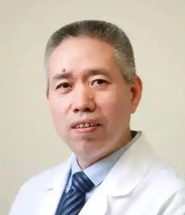 Shunwu Fan (范顺武)合作论文数Sir Run Run Shaw Hospital, Zhejiang University School of Medicine5Shao-Cheng Liu合作论文数4Liyong Sun合作论文数4Cheng Ye合作论文数4
 Gao Zeng合作论文数4
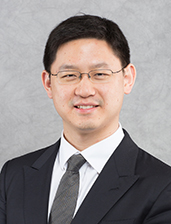 Pui Yin Jason Cheung合作论文数Department of Orthopaedics and Traumatology, Li Ka Shing Faculty of Medicine, The University of Hong Kong4JianXin Du (杜建新)合作论文数4
 He Wang (王鹤)合作论文数广西医科大学附属肿瘤医院妇瘤科4
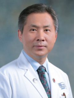 Hongqi Zhang (张鸿祺)合作论文数Xuanwu Hospital, Capital Medical University4
 Tianlong Wang (王天龙)合作论文数Xuanwu Hospital Capital Medical University4
 Jian Chen合作论文数Department of Electrical & Computer Engineering;Clemson University;Department of Electrical & Computer Engineering, Clemson University4
 Guowei Lv (吕国蔚)合作论文数Institute for Hypoxia Medicine, Capital Medical University;Institute of Basic Medical Sciences, Chinese Academy of Medical Sciences4Pingchuan Xia合作论文数3




