BACKGROUND/AIMS:Lenvatinib resistance and immune exclusion limit outcomes in hepatocellular carcinoma (HCC). We hypothesized that metabolic rewiring orchestrates resistance to lenvatinib and programmed cell death protein 1 (PD-1) blockade. METHODS:We established lenvatinib-sensitive/lenvatinib-resistant (LS/LR) HCC models and employed multi-omics (proteomics/RNA-seq), chromatin immunoprecipitation, luciferase, and RNA immunoprecipitation assays to map hexokinase domain containing protein 1 (HKDC1) regulation. Tumor immunity was profiled by scRNA-seq, multiplex fluorescent immunohistochemistry, and flow cytometry. Spermidine (SPD)+lenvatinib efficacy was tested in cell lines and patient-derived organoids/xenografts. Therapeutic effects were tested in an immunocompetent hydrodynamic HCC model with hepatocyte-specific Hkdc1 deletion and were analyzed a postoperative cohort (n=40) treated with lenvatinib+PD-1. RESULTS:HKDC1, upregulated in LR HCC, was transcriptionally activated by upstream stimulatory factor 1 (USF1) and promoted spermine synthase (SMS)-mediated polyamine rewiring. This impaired CD8+ T-cell metabolism, reversible by HKDC1 knockdown or SPD. SPD synergized with lenvatinib, triggering autophagy and suppressing tumor growth in vitro and in vivo. High HKDC1 predicted poor response and survival in patients receiving lenvatinib+aPD-1. CONCLUSIONS:A USF1/HKDC1/SMS axis couples polyamine metabolism to immune dysfunction and lenvatinib resistance. HKDC1 is a predictive biomarker and therapeutic node and supports polyamine-axis modulation to sensitize HCC to lenvatinib plus PD-1 therapy.
Resistance to anti-PD-1/PD-L1 immune checkpoint blockade continues to be a critical challenge undermining its therapeutic efficacy in clinical applications. Most of the resistance mechanisms characterized to date have predominantly involved external factors beyond PD-L1. Here, we unexpectedly discovered that PD-L1 itself possesses E3 ubiquitin ligase activity to induce β2m ubiquitylation and subsequent degradation, which notably reduces MHC-I levels on the surface of tumor cells and antigen-presenting cells, thereby contributing to tumor cell evasion of recognition by CD8+ T cells and ultimately resulting in resistance to anti-PD-1/PD-L1 immunotherapy, particularly in tumors with low basal β2m expression. Disrupting the E3 ubiquitin ligase activity of PD-L1 or interfering with the PD-L1–β2m interaction dramatically enhanced the sensitivity of tumor cells to PD-L1 blockade therapy. Our study reveals a previously unknown function of PD-L1 in the immune evasion of tumor cells, expanding our understanding of intrinsic resistance mechanisms to immune checkpoint blockade therapy.
Abstract Comprising 90% of all liver cancers, hepatocellular carcinoma (HCC) is the second leading cause of cancer-related death worldwide. Despite decades of research, prognosis remains poor: approximately 800,000 people are diagnosed with HCC each year, and ∼80% die within five years. Therapeutically, there is a critical unmet need for more effective treatments. Resistance to sorafenib, the current first-line multi-target tyrosine kinase inhibitor, has become increasingly common. Moreover, more than 150 clinical trials evaluating targeted agents, immunotherapies, and combination regimens have failed due to insufficient efficacy, underscoring the urgent need for novel, mechanism-based therapeutic strategies. Emerging evidence highlights the central role of metabolic reprogramming in carcinogenesis, treatment resistance, and recurrence across multiple cancers, including HCC. Ornithine aminotransferase (OAT) is a key enzyme in this process, linking amino acid metabolism, particularly glutamine and proline, to polyamine biosynthesis and cancer cell proliferation. OAT is overexpressed in HCC and validated as a therapeutic target in preclinical models. We previously demonstrated that the OAT inactivator LHJ-2-79 effectively inhibits tumor growth in HepG2 and Hep3B xenografts and significantly suppresses alpha-fetoprotein (AFP) secretion, a key HCC biomarker, in Hep3B and HepG2 cells. However, no OAT inactivators have yet advanced to clinical trials. To address this gap, our laboratory recently synthesized SS-1-148, a novel OAT inactivator. Here, we report the effects of SS-1-148 on HCC using IC50 profiling, enzyme expression assays, xenograft survival studies, RNA-seq analysis, and metabolomic data. Our findings reveal cell line-dependent differences in SS-1-148 potency, OAT activity inhibition, tumor growth suppression, metabolic gene expression, and alterations in proline and polyamine levels. In vivo, SS-1-148 inhibited Huh-7 xenograft growth but not Huh-6 tumors. Metabolomic analyses of LHJ-2-79 and SS-1-148 in Huh-6 and Huh-7 cells further demonstrated distinct effects on proline and putrescine levels, suggesting different mechanisms of action between the two inactivators. Collectively, these results support OAT as a promising therapeutic target in HCC and lay the foundation for further development of OAT-based treatment strategies. Citation Format: Wenan Qiang, Yi Yang, Tommy Ouyang, Vivian Chen, Alice Qiu, Richard B. Silverman. Targeting ornithine aminotransferase as a promising therapeutic strategy for hepatocellular carcinoma [abstract]. In: Proceedings of the American Association for Cancer Research Annual Meeting 2026; Part 1 (Regular Abstracts); 2026 Apr 17-22; San Diego, CA. Philadelphia (PA): AACR; Cancer Res 2026;86(7 Suppl):Abstract nr 3068.
Objective: Conventional RECIST has limitations in predicting neoadjuvant therapy's pathological response. This study explored the clinical application of RecistTM in predicting neoadjuvant therapy efficacy.Methods: The retrospective study included 124 NSCLC patients with baseline tumor marker levels ≥3 times the normal upper limit, who completed ≥2 cycles of neoadjuvant therapy before surgery. Efficacy was evaluated using RecistTM, RECIST, and pathological standards. The study compared the efficacy and EFS evaluated by the three criteria and the predictive ability of RecistTM and RECIST for pCR.Results: The concordance between RecistTM and postoperative pathological results (Weighted Kappa = 0.571) was significantly higher than that of RECIST (Weighted Kappa = 0.096). The pCR rate of tmCR patients using RecistTM was significantly higher than that of PR patients using RECIST (71.87% vs. 46.07%, p < 0.01). RecistTM showed better performance in predicting pCR than RECIST (AUC: 0.828 vs. 0.573, p < 0.001). tmCR and pCR were independent predictors of EFS.Conclusion: In patients with baseline tumor marker levels >3 times the cutoff, RecistTM had better predictive performance for pathological response than conventional RECIST. tmCR defined by RecistTM may be a potential surrogate endpoint for evaluating short - term neoadjuvant therapy efficacy in this subgroup.
To investigate the mechanism by which heterogeneous nuclear ribonucleoprotein A2/B1 (hnRNPA2B1) regulates the maturation of LncRNA MM2P to promote M2 polarization of macrophages in gastric cancer. In vitro experiments showed that hnRNPA2B1 could promote M2 polarization of iBMDM cells, enhance the maturation and expression of MM2P, and increase STAT6 phosphorylation levels. When MM2P was knocked out, hnRNPA2B1 could no longer further promote cell M2 polarization. iBMDM cells with hnRNPA2B1-induced M2 polarization could enhance the metastasis and invasion of MFC cells, improve clonogenic ability, and increase cell viability. In vivo experiments demonstrated that hnRNPA2B1 could promote the infiltration and polarization of CD206 cells and facilitate tumor growth. hnRNPA2B1 can regulate macrophage M2 polarization by promoting the maturation and expression of MM2P, thereby enhancing the malignant behavior of gastric cancer cells and tumor growth.
China faces the world’s largest diabetes burden, with a 12.4
Acute kidney injury (AKI) is a prevalent and critical clinical condition characterized by high morbidity and mortality. Recently, numerous studies have implicated ferroptosis, an iron-dependent programmed cell death process, in the pathophysiology of AKI. Despite this, the mechanism underlying the widespread occurrence of ferroptosis in AKI remains elusive. To address this, our study analyzed snRNA-seq data from AKI and healthy renal tissues. The analysis revealed notable differences in ferroptosis activity within proximal tubule (PT) cells of AKI patients, specifically highlighting a strong correlation between ferroptosis and the expression of genes GPX4, FTH1, and FTL. Spatial transcriptomics confirmed that the genes GPX4, FTH1, and FTL play a crucial role in driving ferroptosis propagation in AKI. Furthermore, utilizing a mouse model of bilateral renal ischemia-reperfusion injury, we validated the emergence of ferroptosis mediated by these key genes following AKI. The findings from our in vivo experiments were consistent with the spatial transcriptomics data. Chromatin accessibility and transcription factor analysis identified KLF6 as a repressor of ferroptosis-related genes. An in-depth analysis of PT revealed a subpopulation closely associated with ferroptosis. The cellular microenvironment within this subpopulation may regulate ferroptosis through the SPP1 signaling pathway, ultimately influencing the outcome of PT following AKI. In conclusion, this study elucidates the crucial role of GPX4, FTH1, and FTL in ferroptosis propagation during AKI and underscores the potential therapeutic benefits of targeting ferroptosis in the management of AKI.
Triple-negative breast cancer (TNBC) remains a challenging malignancy to treat, underscoring the urgent need to explore novel and effective therapeutic targets. In this study, we found that carnitine palmitoyltransferase 1A (CPT1A), the central and rate-limiting enzyme for fatty acid oxidation (FAO) in lipid metabolism, is significantly correlates with poor survival outcomes in TNBC patients and is highly expressed in TNBC patient samples. Inhibition of CPT1A greatly suppresses TNBC tumor growth. Mechanistically, we discovered that beyond disruption of the canonical metabolic functions for tumor cell survival, CPT1A depletion markedly triggers cGAS/ STING activation due to lipid accumulation-induced elevation of mitochondrial reactive oxygen species (ROS), leading to mitochondrial damage and subsequent mtDNA cytosolic release, which ultimately promotes neutrophil intratumoral infiltration and acquisition of a tumor-killing phenotype, thereby effectively inhibiting tumor growth. Our current findings suggest that inhibition of CPT1A potently activates the cGAS/STING pathway, significantly enhancing the engagement of neutrophils for tumor abrogation.
BackgroundTo investigate the cost-effectiveness of toripalimab plus chemotherapy versus chemotherapy alone for advanced non-small cell lung cancer (NSCLC) patients from a societal perspective.MethodsA partitioned-survival model estimated the costs and cost-effectiveness of toripalimab plus chemotherapy versus standard chemotherapy for advanced NSCLC over 20 years. Clinical data were derived from the CHOICE-01 trial, and cost and utility inputs were gathered from Yaozh.com, expert interviews, and a nationwide hospital-based survey. Costs were reported in 2022 US dollars, and outcomes included quality-adjusted life-years (QALYs) and incremental cost-effectiveness ratios (ICERs), with a 5% discount rate was applied. Sensitivity, subgroup, and scenario analyses verified the robustness of results.ResultsToripalimab plus chemotherapy resulted in 3.048 QALYs and a total cost of $60,813, with an ICER of $19,066 per QALY gained, below China's 3 times GDP per capita threshold ($38,223). Robustness is confirmed through sensitivity, subgroup and scenario analyses.ConclusionsToripalimab plus chemotherapy is a cost-effective option for treatment-naive advanced NSCLC compared to chemotherapy alone, providing valuable evidence for clinical and reimbursement decision-making.
OBJECTIVE:Thrombocytopenia is the main limiting toxicity of chemotherapy, which has not been adequately addressed until now. However, no exact studies have been conducted on the secondary prevention of chemotherapy-induced thrombocytopenia.The aim of this study is to explore the effect of Chinese herbal medicine on the secondary prevention of chemotherapy-induced thrombocytopenia (CIT) in malignant solid tumors. METHODS:This study was a prospective randomized controlled trial. The participants were divided into two groups: treatment group and control group. In the treatment group, chemotherapy was combined with Danggui Buxue Decoction (DBD) with extra flavor. The control group received chemotherapy alone. From May 2019 to May 2022, ninety patients with previous history of ≥grade 1 CIT were recruited.The CIT severity after chemotherapy, effect of CIT on chemotherapy, and economic impact of CIT were compared by statistics. RESULTS:CIT severity differed significantly between the treatment and control groups (χ2=29.514, p<0.001). Grade 3-4 CIT was significantly reduced in the treatment group compared with the control group (6.67 % vs. 57.78 %, χ2 =26.914, p<0.001)). With the self-controlled study method, no significant decrease in platelets was seen in the treatment group compared with platelets before enrollment (z=0.308, p=0.758). Meanwhile, platelets in the control group appeared to be significantly decreased(z=3.514, p<0.001). There was no significant difference between the two groups regarding the number of chemotherapy cycles (F=0.294, p=0.589). However, due to CIT, dose downregulation, chemotherapy termination, and regimen changes were more prevalent in the control group(42.2 % vs. 68.9 %; 15.6 % vs. 62.2 %; 15.6 % vs. 40.0 %; both p<0.05). Thus the overall cost was significantly lower than that of the control group (F=12.825, p=0.001). Moreover, there was no significant difference between the treatment and the control group in any other respect adverse side effect. CONCLUSION:Chinese herbal medicines (DBD) may prevent further CIT and minimize its adverse effects on chemotherapy, including dose downregulation, chemotherapy termination, and regimen changes . It could be considered as an effective and economical treatment option for secondary prevention of CIT. (ChiCTR1900022996).
Metabolic rewiring is a defining feature of malignant cells, enabling them to dynamically exploit nutrient resources to meet bioenergetic problems at different growth stages. Beyond the classical Warburg effect, recent studies have shown that neoplasms demonstrate a marked dependency on lipid metabolism, using free fatty acids to support cellular proliferation and regeneration via fatty acid oxidation (FAO). As a central component of lipid metabolism, FAO exerts dual immunomodulatory functions within tumors. Although numerous studies have described the enzymatic reactions of the FAO pathway in different malignancies, relatively few have investigated the pharmacological disruption of these enzymatic checkpoints and the resulting immunological consequences. Moreover, existing therapeutic strategies have failed to achieve a risk–benefit balance, limiting the clinical translation of FAO-directed approaches. To better understand the therapeutic implications of FAO, we investigated the mechanistic pathways mediated by mitochondrial rate-limiting enzymes, with a particular focus on the carnitine palmitoyltransferase 1 enzyme family—the critical gatekeeper controlling the entry of fatty acids into mitochondrial oxidation instead of CPT2. We comprehensively evaluated its role in tumor biology and also highlight future research directions to inform rational intervention strategies.
It is important to systematically identify tumor suppressor genes (TSGs) to improve our understanding of tumorigenesis and develop strategies for early diagnosis and mitigating disease progression. In the present study, we used an in vivo genome-wide clustered regularly interspaced short palindromic repeats (CRISPR)-CRISPR-associated protein 9 (Cas9) screen and identified FPS/FES-related (FER) as a TSG. Single-cell RNA sequencing (scRNA-seq) revealed that normal cells with low FER expression exhibited elevated malignant transformation potential and stemness properties. FER knockout promoted the tumorigenic transformation, characterized by high colony-forming efficiency and suspension growth ability, acquired tumorigenicity in vivo, increased metabolic activity, dedifferentiation properties, and immune evasion. Moreover, analysis revealed that low FER expression tumors share molecular phenotypes with FER knockout cells, suggesting the consistent role of FER in tumor initiation and progression. Taken together, our findings not only provide insights into the essential role of FER as a tumor suppressor in tumor initiation and progression but also highlight its potential as a target for future clinical diagnosis. (c) 2024 The Pathological Society of Great Britain and Ireland.
BackgroundTyrosine kinase inhibitors (TKIs) are standard first-line treatments for advanced non-small-cell lung cancer (NSCLC) with driver gene mutations. The Response Evaluation Criteria in Solid Tumors (RECIST) are limited in predicting long-term patient benefits. A tumour marker-based evaluation criteria, RecistTM, was used to investigate the potential for assessing targeted-therapy efficacy in lung cancer treatment.MethodsWe retrospectively analysed patients with stage IIIA–IV NSCLC and driver gene mutations, whose baseline tumour marker levels exceeded the pre-treatment cut-off value three-fold and who received TKI-targeted therapy as a first-line treatment. We compared efficacy, progression-free survival (PFS), and overall survival (OS) between RecistTM and RECIST.FindingsThe median PFS and OS differed significantly among treatment-response subgroups based on RecistTM but not RECIST. The predicted 1-, 2-, and 3-year disease-progression risk, according to area under the receiver operating characteristic curve, as well as the 1-, 3-, and 5-year mortality risk, differed significantly between RecistTM and RECIST. The median PFS and OS of tmCR according to RecistTM, was significantly longer than (CR+PR) according to RECIST. Imaging analysis revealed that the ΔPFS was 11.27 and 6.17 months in the intervention and non-intervention groups, respectively, suggesting that earlier intervention could extend patients' PFS.InterpretationRecistTM can assess targeted-therapy efficacy in patients with advanced NSCLC and driver gene mutations, along with tumour marker abnormalities. RecistTM surpasses RECIST in predicting short- and long-term patient benefits, and allows the early identification of patients resistant to targeted drugs, enabling prompt intervention and extending the imaging-demonstrated time to progression.
Objective To elucidate the underlying mechanism by which the proliferation and migration abilities of human umbilical cord mesenchymal stem cells (hUC-MSCs) determine their therapeutic efficacy in rheumatoid arthritis treatment.Methods The DBA/1J mice were utilized to establish a collagen-induced RA (CIA) mouse model and to validate the therapeutic efficacy of hUC-MSCs transfected with CD151 siRNA. RNA-seq, QT-PCR and western blotting were utilized to evaluate the mRNA and protein levels of the PI3K/AKT pathway, respectively.Results IFN-γ significantly enhanced the proliferation and migration abilities of hUC-MSCs, up-regulating the expression of CD151, a gene related to cell proliferation and migration. Effective inhibition of this effect was achieved through CD151 siRNA treatment. However, IFN-γ did not affect hUC-MSCs differentiation or changes in cell surface markers. Additionally, transplantation of CD151-interfered hUC-MSCs (siRNA-CD151-hUC-MSCs) resulted in decreased colonization in the toes of CIA mice and worse therapeutic effects compared to empty vector treatment (siRNA-NC-hUC-MSCs).Conclusion IFN-γ facilitates the proliferation and migration of hUC-MSCs through the CD151/PI3K/AKT pathway. The therapeutic efficacy of siRNA-CD151-hUC-MSCs was found to be inferior to that of siRNA-NC-hUC-MSCs.
Although neoadjuvant chemoradiotherapy treatment followed by surgical resection is the recommended treatment for locally advanced rectal cancer (LARC), response rates remain poor. In proficient mismatch repair (pMMR) rectal cancer, combination (vs. monotherapy) immunotherapy has begun to show promise. This study involved 87 LARC patients undergoing short-course radiotherapy (SCRT), followed by CAPOX (capecitabine and oxaliplatin), in combination with the immune checkpoint inhibitor tislelizumab. Following neoadjuvant therapy, 81 patients underwent surgery, achieving an R0 resection rate of 98.7%. Pathological complete response (pCR) was observed in 41 patients (50.6%), with responders (patients with tumor regression grade TRG 0/TRG 1 or complete clinic response) constituting 69% (60/87). Grade 3 adverse events occurred in 11.5% of participants, and there was one case of grade 4 myasthenia gravis. Imaging Mass Cytometry (IMC) analysis demonstrated higher infiltration of M1 macrophages were in responders. Spatial analysis further identified significant aggregation of PD-L1+ myofibroblastic cancer-associated fibroblasts (MyoCAFs), a unique cell population, within a 10 µm radius to tumor cells, in non-responders; and dynamic analysis showed that post-treatment PD-L1+ MyoCAFs continued to increase in the non-responder group, who also had more exhausted CD8+T cells, possibly explaining their worse response. Our study affirms the efficacy and safety of neoadjuvant SCRT combined with immunochemotherapy in LARC, highlighting the importance of assessing the spatial distribution of immune cells in the tumor microenvironment (TME) for predicting treatment responses. ClinicalTrials.gov registration: NCT05515796.
Despite the notable efficacy of anti-PD1 therapy in the management of hepatocellular carcinoma (HCC) patients, resistance in most individuals necessitates additional investigation. For this study, we collected tumor tissues from nine HCC patients receiving anti-PD1 monotherapy and conducted RNA sequencing. These findings revealed significant upregulation of GSDME, which is predominantly expressed by tumor-associated macrophages (TAMs), in anti-PD1-resistant patients. Furthermore, patients with elevated levels of GSDME+ macrophages in HCC tissues presented a poorer prognosis. The analysis of single-cell sequencing data and flow cytometry revealed that the suppression of GSDME expression in nontumor cells resulted in a decrease in the proportion of M2-like macrophages within the tumor microenvironment (TIME) of HCC while concurrently augmenting the cytotoxicity of CD8 + T cells. The non-N-terminal fragment of GSDME within macrophages combines with PDPK1, thereby activating the PI3K-AKT pathway and facilitating M2-like polarization. The small-molecule Eliprodil inhibited the increase in PDPK1 phosphorylation mediated by GSDME site 1. The combination of Eliprodil and anti-PD1 was effective in the treatment of both spontaneous HCC in c-Myc + /+;Alb-Cre + /+ mice and in a hydrodynamic tail vein injection model, which provides a promising strategy for novel combined immunotherapy.
Mesenchymal stem cells (MSCs) are widely distributed pluripotent stem cells with powerful immunomodulatory capacity. MSCs transplantation therapy (MSCT) is widely used in the fields of tissue regeneration and repair, and treatment of inflammatory diseases. Apoptosis is an important way for tissues to maintain cell renewal, but it also plays an important role in various diseases. And many studies have shown that MSCs improves the diseases by regulating cell apoptosis. The regulation of MSCs on apoptosis is double-sided. On the one hand, MSCs significantly inhibit the apoptosis of diseased cells. On the other hand, MSCs also promote the apoptosis of tumor cells and excessive immune cells. Furthermore, MSCs regulate apoptosis through multiple molecules and pathways, including three classical apoptotic signaling pathways and other pathways. In this review, we summarize the current evidence on the regulation of apoptosis by MSCs.
BackgroundHepatocellular carcinoma (HCC) remains a significant global health challenge with limited treatment options. Lenvatinib, a tyrosine kinase inhibitor, has shown promise but is often undermined by the development of drug resistance.MethodsUtilizing high-throughput sequencing, we investigated the molecular mechanisms underlying lenvatinib resistance in HCC cells, with a focus on metabolic pathways. Key genes, including GALNT6, were validated through quantitative real-time PCR. The effects of GALNT6 knockdown on lenvatinib sensitivity were examined in vitro and in vivo. O-GalNAc glycosylation was assessed using Vicia Villosa Lectin. Immune cell infiltration and interactions were analyzed in the TCGA-LIHC cohort, with further validation by Western blotting and immunohistochemistry.ResultsOur findings indicate that lenvatinib resistance in HCC is driven by the mucin-type O-glycosylation pathway, with GALNT6 playing a critical role. Knockdown of GALNT6 led to reduced O-GalNAc glycosylation, including the modification of LAPTM5, resulting in decreased LAPTM5 activity and autophagy inhibition. Additionally, GALNT6 silencing disrupted the PDGFA-PDGFRB axis, impairing the activation of cancer-associated fibroblasts (CAFs) and reducing their secretion of SPP1, which collectively diminished lenvatinib resistance.ConclusionsGALNT6 is integral to the resistance mechanisms against lenvatinib in HCC by modulating autophagy and CAF activation. Targeting GALNT6 offers a promising strategy to enhance lenvatinib efficacy and improve therapeutic outcomes in HCC.
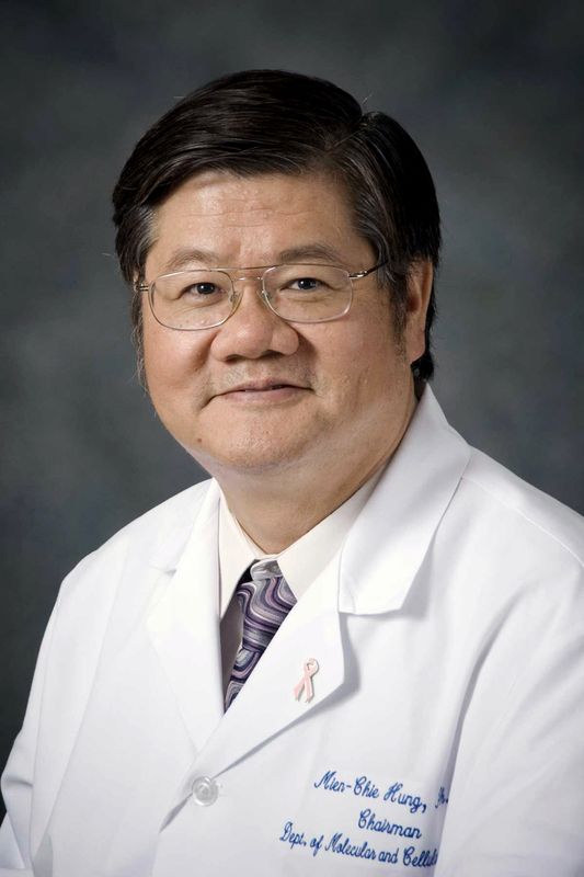 Mien-Chie Hung (洪明奇)合作论文数China Medical University;China Medical University Hospital14
 Jung-Mao Hsu合作论文数Division of Molecular and Genomic Medicine;National Health Research Institutes;Division of Molecular and Genomic Medicine, National Health Research Institutes13Jennifer L Hsu合作论文数Univ Texas MD Anderson Canc Ctr, Dept Mol & Cellular Oncol, Houston, TX 77030 USA12Chia-Wei Li合作论文数Univ Texas MD Anderson Canc Ctr, Dept Mol & Cellular Oncol, Houston, TX 77030 USA11
 Weiya Xia合作论文数Department of Molecular and Cellular Oncology, The University of Texas MD Anderson Cancer Center10
 Li-Chuan Chan合作论文数Boehringer Ingelheim Pharmaceuticals, Inc.9Linlin Sun合作论文数Univ Texas MD Anderson Canc Ctr, Dept Mol & Cellular Oncol, Houston, TX 77030 USA8
 Junwei Hou合作论文数Tongji University, Tongji Univ.8Xueqin Yang (杨雪琴)合作论文数8
 Yongkun Wei合作论文数Department of Molecular and Cellular Oncology, The University of Texas MD Anderson Cancer Center7Lei Nie合作论文数Univ Texas MD Anderson Canc Ctr, Dept Mol & Cellular Oncol, Houston, TX 77030 USA6Zhi Dai (代智)合作论文数Zhongshan Hospital, Fudan University6
 Gui-Qi Zhu合作论文数5
 Jong-Ho Cha合作论文数Univ Texas MD Anderson Canc Ctr, Dept Mol & Cellular Oncol, Houston, TX 77030 USA5Baozhen Ke合作论文数Univ Texas MD Anderson Canc Ctr, Dept Mol & Cellular Oncol, Houston, TX 77030 USA5Xu Mingfang合作论文数5Biao Wang合作论文数5Chunxiao Liu (刘春晓)合作论文数5Xunjie Kuang合作论文数4Yanhui Xu (徐彦辉)合作论文数4
 Dong Wang (王东)合作论文数4Yun Wu合作论文数4Rongce Zhao (赵荣策)合作论文数3Yeh Chen合作论文数3Jia-Liang Cai合作论文数3Jun-Xian Du合作论文数3Jing-Lei Wan合作论文数3Zheng-Jun Zhou合作论文数3Yintao Li合作论文数3
 Xianghou Xia (夏想厚)合作论文数Zhejiang Cancer Hospital, the University of Chinese Academy of Science3Zu Ye合作论文数Univ Texas MD Anderson Canc Ctr, Dept Mol & Cellular Oncol, Houston, TX 77030 USA3Yufan Qiu合作论文数Univ Texas MD Anderson Canc Ctr, Dept Mol & Cellular Oncol, Houston, TX 77030 USA3Huan Huang合作论文数3Yu-Chuan Wang合作论文数Institute of New Drug Development, China Medical University3Yun You合作论文数3Shi-Ping Chen合作论文数3Li-Na Song合作论文数3
 Ri-Yao Yang合作论文数Univ Texas MD Anderson Canc Ctr, Dept Mol & Cellular Oncol, Houston, TX 77030 USA3
 Xiuwu Bian (卞修武)合作论文数School of Medicine, University of Science and Technology of China;The First Hospital Affiliated to Army Medical University3
 Lai Yun-Ju合作论文数3Yamaguchi Hirohito合作论文数Dept Mol & Cellular Oncol, Univ Texas MD Anderson Canc Ctr3
 Chiung-wen June Chang合作论文数3
 Jia Fan (樊嘉)合作论文数Zhongshan Hospital, Fudan University;Liver Cancer Institute, Fudan University;Shanghai Medical College, Fudan University3
 John A. Tainer合作论文数Department of Molecular and Cellular Oncology, Division of Basic Science Research, The University of Texas MD Anderson Cancer Center;Department of Structural Biology, The University of Texas MD Anderson Cancer Center;Department of Integrated Diffraction Analysis Technologies Program, The University of California Lawrence Berkeley National Laboratory;Skaggs Institute for Chemical Biology, Scripps Research Institute3
 Jian Zhou (周健)合作论文数Shanghai Sixth People’s Hospital Affiliated to Shanghai Jiao Tong University School of Medicine3
 Gabriel N. Hortobagyi合作论文数Department of Breast Medical Oncology, Division of Cancer Medicine, The University of Texas MD Anderson Cancer Center3
 Kebin Huang (黄克斌)合作论文数School of Chemistry and Pharmaceutical Sciences, Guangxi Normal University3Yanli Xiong (熊艳丽)合作论文数3
 Huirong Yang (杨慧蓉)合作论文数Department of Systems Biology for Medicine, School of Basic Medical Sciences, Fudan University;Institutes of Biomedical Sciences, Fudan University3




