Peripherally inserted central catheters (PICCs) are essential for long-term infusion in vulnerable pediatric patients. Optimal tip placement in the lower third of the superior vena cava or at the cavoatrial junction is critical to prevent serious complications. Verifying correct tip position in infants and toddlers is challenging because of very small anatomic target zones, non-standard radiograph acquisition, interference from other devices, low contrast, and high risk of catheter migration. Existing automated segmentation methods, mostly developed for adults, perform poorly on pediatric images. We retrospectively collected 1184 PICC patients from three medical centers, including 280 pediatric cases (210 neonates, 46 infants, 24 toddlers), with appropriate ethical approval. We introduce TopNet, a topology-preserving embedded network designed for automated PICC segmentation in pediatric patients. TopNet maintains catheter continuity and enables precise tip localization under difficult conditions. Quantitative and qualitative evaluations show superior segmentation and tip localization on both internal and external validation.
Background:Accurate and early differentiation of focal pancreatic solid lesions (FPSLs) in the outpatient setting remains a major clinical challenge. Benign inflammatory conditions, such as focal autoimmune pancreatitis (fAIP) and mass-forming chronic pancreatitis (MFCP), often appear similar to pancreatic ductal adenocarcinoma (PDAC) in clinical features and conventional imaging findings, leading to diagnostic uncertainty and potential unnecessary pancreaticoduodenectomy. Current serum biomarkers lack accuracy, and invasive diagnostic procedures are limited by sampling variability, highlighting the need for a reliable, non-invasive triage tool suitable for outpatient care. Venous phase contrast-enhanced computed tomography (CECT) best captures pancreatic parenchymal and lesional enhancement patterns, and radiomics from this phase can quantify subtle, visually imperceptible differences in enhancement homogeneity, tissue heterogeneity, and periductal parenchymal remodelling. Therefore, this study aimed to develop and temporally validate an integrated model that combines venous phase CECT radiomic features with key clinical and laboratory variables to better differentiate FPSLs in an outpatient population. Methods:In this retrospective study, outpatients with FPSLs who underwent venous-phase CECT from May 2013 to May 2024 were consecutively enrolled, and diagnoses were based on international consensus criteria (fAIP), or cytology/surgery (MFCP and PDAC). The cohort was randomly divided into training and internal validation sets at a 7:3 ratio. Additionally, 11 fAIP patients and 19 PDAC patients were included in the independent temporal validation analysis. Clinical variables, including demographics, symptoms and laboratory parameters, were collected concurrently with imaging. Quantitative radiomics features were extracted from manually segmented lesions on CECT images. Model discrimination was assessed using receiver operating characteristic (ROC) analysis and decision curve analysis (DCA). Results:The mean age of the three groups of FPSLs patients was 57.21±10.76 (fAIP), 48.25±12.14 (MFCP), and 60.55±9.66 (PDAC) years, respectively. The majority of patients were male, and the pancreatic head was the most common lesion location across all groups (P<0.01). For differentiating fAIP from PDAC, the combined clinical-radiomics nomogram demonstrated strong diagnostic performance, achieving an area under the curve (AUC) of 0.95, 0.91 and 0.88 in the training, internal validation, and temporal validation cohorts, respectively. Similar results were seen in distinguishing MFCP from PDAC. However, although the radiomics model showed initial promise in differentiating fAIP from MFCP in the training set, its performance declined in the validation set. Conclusions:Integrating CECT-based radiomic features with clinical data results in a compelling, non-invasive tool for characterizing FPSLs. Future investigations should prioritize the integration of multi-modal data streams to enhance diagnostic precision.
Purpose: To quantify the agreement of prostate cancer radiomic features within a reader, between readers, and across scanners for T2-weighted imaging (T2WI), diffusion-weighted imaging (DWI; b = 1500 s/mm2), and apparent diffusion coefficient (ADC). Materials and Methods: Seventeen men with biopsy-proven prostate cancer underwent 3.0 T magnetic resonance imaging on two platforms. Two radiologists contoured the dominant lesion. Reader 1 repeated the segmentation approximately 4 weeks later on the same examination. For each sequence and prespecified comparison, feature-wise agreement across patients was quantified using the concordance correlation coefficient (CCC). CCCs were summarized as median (interquartile range) and categorized as poor (< 0.40), moderate (0.40-0.69), good (0.70-0.89), or excellent (>= 0.90). Results: Within a given scanner, prostate cancer radiomic features showed consistently high agreement for both Reader 1 repeat segmentation and Reader 1 versus Reader 2, with most features in the good and excellent ranges across T2WI, DWI, and ADC. In contrast, the cross-scanner agreement was low, even for the same reader and session, and the majority of features were categorized as poor. Sequence-wise, as shown in Figure 2, adjusted T2WI outperformed DWI and ADC for cross-scanner comparisons, yet still fell short of within-scanner performance. Conclusion: Prostate cancer radiomic features demonstrated good reproducibility on a single scanner but poor cross-scanner reproducibility. For future radiomic research, researchers should incorporate scanner type into model analyses and perform data harmonization before integrating data from different manufacturers.
PURPOSE:To address the issues of interpretability and learning efficiency in traditional single-stream frameworks, as well as the limitations in capturing long-range dependencies and semantic relationships caused by the dependence of two-stream architectures. METHODS:This paper proposes GLACF, a triple-stream global-to-local attention framework for deformable MR image registration, integrating CNNs, Transformers, and coarse-to-fine strategies to enhance alignment accuracy. Its key innovations include: a triple-stream feature extraction based on Transformers for enriched feature representation; multi-scale decoupling blocks (MSDBs) that leverage low-resolution features to generate coarse displacement fields; and a voxel-wise local attention module (VLFM) that refines high-resolution features to produce precise displacement fields. RESULTS:Experimental results on three public 3D brain MRI datasets (e.g., LONI LBPA40, IXI, and OASIS) demonstrate that GLACF consistently outperforms state-of-the-art methods in registration similarity, smoothness, and invertibility. Specifically, on LONI LBPA40, GLACF achieves a Dice similarity coefficient (DSC) of 72.7%, structural similarity index measure (SSIM) of 0.973, percentage of negative Jacobian determinants (% of |Jϕ| ≤ 0) of 0.15, and 95th percentile Hausdorff distance (HD95) of 6.088. On IXI, it attains 76.8% DSC, 0.952 SSIM, 0.48% of |Jϕ| ≤ 0, and 3.284 HD95. On OASIS, it reaches 89.1% DSC, 0.973 SSIM, 0.55% of |Jϕ| ≤ 0, and 1.328 HD95. CONCLUSION:These results confirm the robustness and accuracy of GLACF across diverse datasets, highlighting its strong potential for clinical translation in high-fidelity image registration scenarios.
OBJECTIVE:The growing number of studies directly comparing artificial intelligence (AI) to physicians in diagnostic tasks often focuses on performance outcomes, overlooking fundamental methodological rigor. This scoping review aims to critically appraise the methodological quality of this body of literature, identifying key challenges and proposing a framework to enhance the fairness, standardization, and clinical relevance of future comparisons. MATERIALS AND METHODS:We conducted a systematic search of PubMed, Scopus, and Web of Science for studies published between January 1, 2020, and October 31, 2025, following the PRISMA-ScR guidelines. From 8,851 screened records, 120 studies met the inclusion criteria for direct AI-physician comparison. Data on study characteristics, dataset quality, task design, physician configuration, and reporting transparency were extracted and synthesized narratively. RESULTS:Our analysis of 120 studies revealed a field characterized by significant methodological heterogeneity. Key issues include a predominant focus on retrospective studies (75.8%), frequent information asymmetry between AI and physicians (20.8%), limited clinical relevance in task design despite superficial fidelity, and insufficient physician sample sizes (60.8% had ≤ 10 readers). Furthermore, we found a widespread neglect of time constraints (absent in 50.8% of studies) and a critical lack of transparency regarding code and data availability. CONCLUSION:Current research on AI-physician diagnostic comparisons is often hampered by methodological weaknesses that undermine the validity and generalizability of its findings. To ensure the generation of reliable and clinically meaningful evidence, future studies must prioritize prospective designs, ensure fairness in experimental conditions, and adhere to higher standards of transparency. We propose the AI vs. Physician Study Checklist (AIPSC) as a practical tool to guide the design and reporting of more robust and systematic evaluations, ultimately fostering the responsible integration of AI into clinical practice.
Accurate preoperative T and TNM staging of clear cell renal cell carcinoma (ccRCC) is crucial for diagnosis and treatment, but these assessments often depend on subjective radiologist judgment, leading to interobserver variability. This study aims to design and validate two CT-based deep learning models and evaluate their clinical utility for the preoperative T and TNM staging of ccRCC. Data from 1,148 ccRCC patients across five medical centers were retrospectively collected. Specifically, data from two centers were merged and randomly divided into a training set (80
BACKGROUND:Distinguishing between benign and malignant testicular lesions on clinical magnetic resonance imaging (MRI) is crucial for guiding treatment planning. However, conventional MRI-based radiomics to identify testicular cancer requires expert machine learning knowledge. This study aims to investigate the potential of utilizing automatic machine learning (AutoML) based on MRI to diagnose testicular lesions without the need for expert algorithm optimization. METHODS:Retrospective preoperative MRI scans from 115 patients diagnosed with testicular disease through pathology were obtained. A total of 1781 radiomics features were extracted from each lesion on the T2-weighted images. Intraclass and interclass correlation coefficients were used to evaluate the intra-observer and interobserver agreements for each radiomics feature. We developed an AutoML method based on the tree-based pipeline optimization tool (TPOT) algorithm to construct a discriminant model. The best pipeline was determined through 100 repeated operations using a 5-fold cross-validation algorithm in TPOT. The model was evaluated for accuracy, sensitivity, and specificity using the area under the curve (AUC) value of the receiver operating characteristic (ROC) curve. Shapley Additive exPlanations were used to illustrate the optimization results. RESULTS:Utilizing the TPOT method, 100 diagnostic models were developed to identify testicular lesions. The best model was determined based on the highest AUC in the training cohort. The prediction model yielded AUC values of 0.989 (95% confidence interval [CI]: 0.985-0.993) and 0.909 (95% CI: 0.893-0.923) in the training and testing cohorts, respectively. CONCLUSIONS:AutoML, based on the TPOT algorithm, holds potential as a noninvasive method for effectively discriminating between benign and malignant testicular lesions.
To investigate the efficacy of [68Ga]Ga-FAPI-04 PET/CT for assessing viable tumours (VTs) after local regional treatment (LRT) in hepatocellular carcinoma (HCC) patients. The related imaging features of HCC after LRT are preliminarily discussed. A cohort of 37 LRT patients with HCC (encompassing 51 lesions) was retrospectively included from a prospective parent study (ChiCTR2000039099), and sequential PET/CT using [18F]FDG and [68Ga]Ga-FAPI-04 was performed. The diagnostic accuracies of [68Ga]Ga-FAPI-04 and [18F]FDG PET/CT and multiphasic CT/MRI for detecting VTs after LRT were calculated and analysed. Pathological examination was considered the gold standard for VT diagnosis, and clinical follow-up was used as the reference standard. The SUVmax and tumour-to-background ratio (TBR) derived from [18F]FDG and [68Ga]Ga-FAPI-04 PET/CT were calculated and compared. Moreover, the SUVmax, target-to-normal liver ratio (TNR) of VT, tumour necrosis (TN), benign rim (BR), and normal liver (NL) from different imaging modalities after LRT for HCC were compared. Both the sensitivity (96.0
Oesophageal cancer is a serious threat to human health and life due to its high incidence levels and mortality rates. Early detection and diagnosis are crucial. However, existing oesophageal cancer detection models are plagued with missed detections and false-positives, especially with small and irregular lesions. To address these challenges, a novel approach called JS-DETR has been proposed, which combines Joint position channel attention and Shape adaptation improvement loss with DEtection TRansformer (DETR). In JS-DETR, several key improvements are made to enhance the accuracy of automated oesophageal cancer detection and localization. First, the DETR backbone is reinforced by incorporating joint position channel attention, enhancing the model's ability to learn and utilize crucial features effectively. Second, shape adaptation improvement loss is employed to refine the model's regression loss function, resulting in more accurate predictions of the precise locations of oesophageal cancer targets. Finally, transfer learning is utilized to fine-tune the enhanced model by transitioning it from the COCO dataset to the oesophageal cancer barium swallow imaging dataset. The JSDETR model achieves a precision of 76.9%, a recall of 79.4%, an average precision of 87.0%, and an F1-score of 78.1% in the experimental results. Compared to other currently popular object detection models. The JSDETR model enables more precise detection and localization of oesophageal cancer, offering clinicians amore accurate means of oesophageal cancer detection. Our code is available at https://github.com/learningmuch/JSDETR.
BACKGROUND:Most attention-based networks fall short in effectively integrating spatial and channel-wise information across different scales, which results in suboptimal performance for segmenting coronary vessels in x-ray digital subtraction angiography (DSA) images. This limitation becomes particularly evident when attempting to identify tiny sub-branches. PURPOSE:To address this limitation, a multi-scale dual attention embedded network (named MDA-Net) is proposed to consolidate contextual spatial and channel information across contiguous levels and scales. METHODS:MDA-Net employs five cascaded double-convolution blocks within its encoder to adeptly extract multi-scale features. It incorporates skip connections that facilitate the retention of low-level feature details throughout the decoding phase, thereby enhancing the reconstruction of detailed image information. Furthermore, MDA modules, which take in features from neighboring scales and hierarchical levels, are tasked with discerning subtle distinctions between foreground elements, such as coronary vessels of diverse morphologies and dimensions, and the complex background, which includes structures like catheters or other tissues with analogous intensities. To sharpen the segmentation accuracy, the network utilizes a composite loss function that integrates intersection over union (IoU) loss with binary cross-entropy loss, ensuring the precision of the segmentation outcomes and maintaining an equilibrium between positive and negative classifications. RESULTS:Experimental results demonstrate that MDA-Net not only performs more robustly and effectively on DSA images under various image conditions, but also achieves significant advantages over state-of-the-art methods, achieving the optimal scores in terms of IoU, Dice, accuracy, and Hausdorff distance 95%. CONCLUSIONS:MDA-Net has high robustness for coronary vessels segmentation, providing an active strategy for early diagnosis of cardiovascular diseases. The code is publicly available at https://github.com/30410B/MDA-Net.git.
Most attention-embedded networks fall short in effectively integrating spatial / channel-wise information across diverse scales, leading to suboptimal performance for coronary vessels segmentation in X-ray digital subtraction angiography images. To address this limitation, a multi-scale across attention incorporated network (named MS2A-Net) is introduced. MS2A-Net accepts original and enhanced images as inputs, leveraging complementary information provided by the different contrasts within the images. Furthermore, MS2A is designed to integrate features across multiple levels, scales and sources, for effectively extracting deep semantic information. After the incorporation of features with adaptive weightings, segmentation accuracy is refined. Qualitative and quantitative experiment results prove that MS2A-Net not only outperforms state-of-the-art tactics but also shows superior performance, e.g., higher intersection over union, Dice coefficient, broader areas under receiver operating characteristic curves.
Objectives This study aims to evaluate the feasibility and effectiveness of deep learning-based super-resolution techniques to reduce scan time while preserving image quality in high-resolution prostate diffusion-weighted imaging (DWI) with readout-segmented echo-planar imaging (rs-EPI). Methods We retrospectively and prospectively analyzed prostate rs-EPI DWI data, employing deep learning super-resolution models, particularly the Multi-Scale Self-Similarity Network (MSSNet), to reconstruct low-resolution images into high-resolution images. Performance metrics such as structural similarity index (SSIM), Peak signal-to-noise ratio (PSNR), and normalized root mean squared error (NRMSE) were used to compare reconstructed images against the high-resolution ground truth (HRGT). Additionally, we evaluated the apparent diffusion coefficient (ADC) values and signal-to-noise ratio (SNR) across different models. Results The MSSNet model demonstrated superior performance in image reconstruction, achieving maximum SSIM values of 0.9798, and significant improvements in PSNR and NRMSE compared to other models. The deep learning approach reduced the rs-EPI DWI scan time by 54.4 % while maintaining image quality comparable to HRGT. Pearson correlation analysis revealed a strong correlation between ADC values from deep learning-reconstructed images and the ground truth, with differences remaining within 5 %. Furthermore, all models showed significant SNR enhancement, with MSSNet performing best across most cases. Conclusions Deep learning-based super-resolution techniques, particularly MSSNet, effectively reduce scan time and enhance image quality in prostate rs-EPI DWI, making them promising tools for clinical applications.
531 Background: Primary liver cancer (PLC), comprising hepatocellular carcinoma (HCC) and cholangiocarcinoma (CCA), is a leading cause of cancer mortality globally. The combined hepatocellular cholangiocarcinoma (cHCC-CC) subtype may be less common but is relevant to treatment efficacy. We therefore evaluated the diagnostic accuracy of various approaches in distinguishing these liver cancers. Methods: Patients diagnosed with HCC, CCA, and cHCC-CC at Beijing University Cancer Hospital and Institute, China were included. Radiologists of varying expertise independently assessed MRI scans, and we measured their diagnostic consistency. Radiomic features were extracted from MRI scans, and machine learning was applied to differentiate the cancer types. Results: Standard imaging was insufficient to reliably characterize cHCC-CC. Abdominal imaging experts (AIEs) had a higher mean sensitivity for HCC and CCA, 88% and 84% respectively, while non-experts (NIEs) had a lower sensitivity of 50% for HCC and 38% for CCA (HCC: p=0.03, CCA: p=0.008). Radiomic analysis found ‘Sphericity’ and ‘ClusterShade’ as the most relevant features. However, radiomics algorithms were also not sufficient to distinguish cHCC-CC from either HCC or CCA. Regarding sensitivity, the radiomic-based model was not better than radiologists for any of the three classes (p=0.065 for HCC, p=0.426 for CCA, and p=1.0 for cHCC-CC). The random forest algorithm yielded an accuracy of 76% in the test set, since it correctly classified most HCC and CCA, while only one quarter of cHCC-CC tumors. Conclusions: Until improved diagnostic tools are available, biopsy of liver cancer remains critical to the detection, diagnosis, and effective treatment of these cancers.
Introduction and Objectives Primary liver cancer (PLC), comprising hepatocellular carcinoma (HCC) and cholangiocarcinoma (CCA), is a leading cause of cancer mortality globally. The combined hepatocellular-cholangiocarcinoma (cHCC-CC) subtype may be less common but is relevant to treatment efficacy. We therefore evaluated the diagnostic accuracy of various approaches in distinguishing these liver cancers. Materials and Methods Patients diagnosed with HCC, CCA, and cHCC-CC at Beijing University Cancer Hospital and Institute, China were included. Radiologists of varying expertise independently assessed MRI scans, and we measured their diagnostic consistency. Radiomic features were extracted from MRI scans, and machine learning was applied to differentiate the cancer types. Results Standard imaging was insufficient to reliably characterize cHCC-CC. Abdominal imaging experts (AIEs) had a higher mean sensitivity for HCC and CCA, 88% and 84% respectively, while non-experts (NIEs) had a lower sensitivity of 50% for HCC and 38% for CCA (HCC: p=0.03, CCA: p=0.008). Radiomic analysis found ‘Sphericity’ and ‘ClusterShade’ as the most relevant features. However, radiomics algorithms were also not sufficient to distinguish cHCC-CC from either HCC or CCA. Regarding sensitivity, the radiomic-based model was not better than radiologists for any of the three classes (p=0.065 for HCC, p=0.426 for CCA, and p=1.0 for cHCC-CC). The random forest algorithm yielded an accuracy of 76% in the test set, since it correctly classified most HCC and CCA, while only one quarter of cHCC-CC tumors. Conclusions Histopathological analysis, complemented by imaging as indicated, remains essential for accurate detection, diagnosis, and treatment of liver cancers.
To investigate the prognostic significance of histone acetylation (HAc) regulators in esophageal cancer (EC) and develop a transcriptome-based HAc_score reflecting epigenetic and immunogenomic states. Expression and mutation from EC were analyzed to identify prognostic HAc regulators via univariable Cox models. Consensus clustering defined HAc-related expression patterns. Differentially expressed genes (DEGs) among clusters were functionally enriched. A principal component-based HAc_score was constructed from prognostic DEGs and tested for associations with overall survival, tumor mutational burden (TMB), immunophenoscore (IPS), and immune cell infiltration. Three HAc-related expression patterns showed distinct biological and immune features. From shared DEGs, 19 prognostic genes defined two molecular subtypes and served as the basis for the HAc_score. Higher HAc_score was associated with better overall survival, particularly in early-stage disease. HAc_score correlated inversely with TMB and positively with IPS components, suggesting a transcriptionally active, immunogenic phenotype despite lower mutation burden. Combining HAc_score with TMB improved risk stratification. HAc_score quantifies HAc–linked transcriptional states in EC and reflects tumor–immune interactions. It stratifies survival risk and complements TMB, supporting its potential use as a prognostic biomarker and integrative epigenetic–immune signature.
Abstract Background Anoikis resistance is a hallmark characteristic of oncogenic transformation, which is crucial for tumor progression and metastasis. The aim of this study was to identify and validate a novel anoikis‐related prognostic model for prostate cancer (PCa). Methods We collected a gene expression profile, single nucleotide polymorphism mutation and copy number variation (CNV) data of 495 PCa patients from the TCGA database and 140 PCa samples from the MSKCC dataset. We extracted 434 anoikis‐related genes and unsupervised consensus cluster analysis was used to identify molecular subtypes. The immune infiltration, molecular function, and genome alteration of subtypes were evaluated. A risk signature was developed using Cox regression analysis and validated with the MSKCC dataset. We also identify potential drugs for high‐risk group patients. Results Two subtypes were identified. C1 exhibited a higher level of CNV amplification, immune score, stromal score, aneuploidy score, homologous recombination deficiency, intratumor heterogeneity, single‐nucleotide variant neoantigens, and tumor mutational burden compared to C2. C2 showed a better survival outcome and had a high level of gamma delta T cell and activated B cell infiltration. The risk signature consisting of four genes (HELLS, ZWINT, ABCC5, and TPSB2) was developed (area under the curve = 0.780) and was found to be an independent prognostic factor for overall survival in PCa patients. Four CTRP‐derived and four PRISM‐derived compounds were identified for high‐risk patients. Conclusions The anoikis‐related prognostic model developed in this study could be a useful tool for clinical decision‐making. This study may provide a new perspective for the treatment of anoikis‐related PCa.
Objectives: Dysregulation of RNA modifications has emerged as a contributor to cancer, but the clinical implication of RNA modification-related genes remains largely unclear. The study focused on well-studied RNA modification modalities (m(6)A, m(1)A, m(5)C and m(7)G) in bladder cancer, and proposed a machine learning-based integrative approach for establishing a consensus RNA modification-based signature.Methods: Multiple publicly available bladder cancer cohorts were enrolled. A novel RNA modification-based classification was proposed via consensus clustering analysis. RNA modification-related genes were subsequently selected through WGCNA. A machine learning-based integrative framework was implemented for constructing a consensus RNA modification-based signature.Results: Most RNA modifiers were dysregulated in bladder tumours at the multi-omics levels. Two RNA modification clusters were identified, with diverse prognostic outcomes. A consensus RNA modification-based signature was established, which displayed stable and powerful efficacy in prognosis estimation. Notably, the signature was superior to conventional clinical indicators. High-risk tumours presented the activation of tumourigenic pathways, with the activation of metabolism pathways in low-risk tumours. The low-risk group was more sensitive to immune-checkpoint blockade, with the higher sensitivity of the high-risk group to cisplatin and paclitaxel. Genes in the signature: AKR1B1, ANXA1, CCNL2, OAS1, PTPN6, SPINK1 and TNFRSF14 were specially expressed in distinct T lymphocytes of bladder tumours at the single-cell level, potentially participating in T cell-mediated antitumour immunity. They were transcriptionally and post-transcriptionally modulated, and might become potentially actionable therapeutic targets.Conclusions: Altogether, the consensus RNA modification-based signature may act as a reliable and hopeful tool for improving clinical decision-making for individual bladder cancer patients.
441 Background: Current guidelines emphasize the use of CT or MRI for hepatocellular carcinoma (HCC) diagnosis, with strict regulation of biopsies. However, Hepatocellular-Cholangiocarcinoma (cHCC-CC), a liver cancer variant displaying histological elements of both HCC and cholangiocellular carcinoma (CCC), typically exhibits radiological features of both HCC and CCC on imaging studies. Additionally, platinum drugs represent the most promising initial treatment option for patients with unresectable or advanced cHCC-CC, deviating significantly from the treatment approach for HCC. Consequently, our study aims to assess the efficacy of imaging techniques in distinguishing between HCC, CCC, and cHCC-CC, highlighting its clinical significance. Methods: We conducted a database search to identify patients diagnosed with HCC, CCC, and cHCC-CC from June 2010 to September 2020. After implementing quality control measures, our study comprised 68 MRI scans from patients, including 30 with HCC, 23 with CCC, and 15 with cHCC-CC. Histological evidence confirming the diagnoses was obtained within a maximum of four weeks before or after the MRI scans. Subsequently, seven radiologists from Asia, Europe, North America, and South America, including abdominal imaging experts (AIEs) and non-abdominal imaging experts (NIEs) or trainees, independently and blindly assessed the MRI scans. They evaluated various MRI features and established a differential diagnosis encompassing HCC, CCC, and cHCC-CC. Results: The AIEs demonstrated high proficiency in utilizing MRI images exclusively for diagnosing HCC (70%-100%) and CCC (73.9%-91.3%). They significantly outperformed NIEs/trainees (all p values < 0.01), achieving accuracy rates of 26.7%-66.7% for HCC and 21.7%-60.9% for CCC. However, their ability to accurately distinguish cHCC-CC (6.7%-53.3%) was limited and comparable to NIEs/trainees (26.7%-46.7%). Additionally, there was greater consistency in MRI feature assessment among AIEs for HCC and CCC when compared to cHCC-CC. Notably, no significant differences were observed in the impact of a cirrhotic background on the diagnosis of HCC and cHCC-CC among AIEs. Furthermore, there was no significant intercontinental variability in overall liver cancer diagnosis and the diagnosis rates of the three types of liver cancer by AIEs. Conclusions: MRI imaging demonstrated effective differentiation between HCC and CCC, especially when interpreted by experts in abdominal imaging. Nevertheless, the ability to accurately detect cHCC-CC was notably constrained across all participating radiologists. Consequently, liver biopsy continues to play a pivotal role in ensuring diagnostic precision and facilitating the selection of appropriate medical treatment strategies.
Background Current guidelines emphasize the use of CT or MRI for hepatocellular carcinoma (HCC) diagnosis and restricting biopsies. However, hepatocellular-cholangiocarcinoma (cHCC-CC), a variant displaying features of both HCC and cholangiocellular carcinoma (CCC), presents mixed radiological traits. Unlike HCC, platinum drugs are the most promising primary treatment for unresectable cHCC-CC. Our study assesses imaging techniques' efficacy in distinguishing HCC, CCC, and cHCC-CC, emphasizing its clinical importance.
Methamphetamine (MA) is a neurological drug, which is harmful to the overall brain cognitive function when abused. Based on this property of MA, people can be divided into those with MA abuse and healthy people. However, few studies to date have investigated automatic detection of MA abusers based on the neural activity. For this reason, the purpose of this research was to investigate the difference in the neural activity between MA abusers and healthy persons and accordingly discriminate MA abusers. First, we performed event-related potential (ERP) analysis to determine the time range of P300. Then, the wavelet coefficients of the P300 component were extracted as the main features, along with the time and frequency domain features within the selected P300 range to classify. To optimize the feature set, F_score was used to remove features below the average score. Finally, a Bidirectional Long Short-term Memory (BiLSTM) network was performed for classification. The experimental result showed that the detection accuracy of BiLSTM could reach 83.85%. In conclusion, the P300 component of EEG signals of MA abusers is different from that in normal persons. Based on this difference, this study proposes a novel way for the prevention and diagnosis of MA abuse.
 Peipei Zhang合作论文数36
 Zhaoyan Feng (冯朝燕)合作论文数34Basen Li合作论文数32ChaoYan Feng (冯朝燕)合作论文数29Zan Ke合作论文数26Jie Cai合作论文数24Ming Deng (邓明)合作论文数24
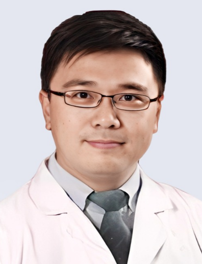 Liang Wang (汪良)合作论文数Union Hospital, Tongji Medical College, Huazhong University of Science and Technology18Chanyuan Fan合作论文数17Huijuan You (游慧娟)合作论文数School of Pharmacy, Tongji Medical College, Huazhong University of Science and Technology16Wei Cai (蔡卫)合作论文数Tongji Hospital, Tongji Medical College, Huazhong University of Science and Technology12Yin Xi合作论文数8Wenzhi Lv合作论文数8
 Zhen Kang合作论文数8feng qixing合作论文数7
 Junfeng Gao (高军峰)合作论文数‘School of Biomedical Engineering, South-Central Minzu University7
 Daoyu Hu (胡道予)合作论文数Tongji Hospital, Tongji Medical College, Huazhong University of Science and Technology7Qiao-Yuan Lu合作论文数5Isaac Rodriguez合作论文数5Yuquan Qian合作论文数5Jinke Xie合作论文数5
 Xu Yan合作论文数5
 Zhiquan Hu (胡志全)合作论文数Tongji Hospital, Tongji Medical College of Huazhong University of Science & Technology5Castrillon German A合作论文数5
 Jianping Dai (戴建平)合作论文数Beijing Tiantan Hospital, Capital Medical University5
 Andreas Teufel合作论文数Department of Medicine I, Johannes Gutenberg University5Stefan O. Schoenberg合作论文数5
 Christel Weiss合作论文数Medizinische Fakultät Mannheim, Universität Heidelberg5
 Yingshi Sun (孙应实)合作论文数Beijing Cancer Hospital5Muzaffer Reha Ümütlü合作论文数4Kailun Sun (孙凯伦)合作论文数4
 Shengzhou Xu合作论文数4Qiubai Li合作论文数4Sah Vivek Kumar合作论文数4
 Liming Xia (夏黎明)合作论文数Tongji Hospital, Tongji Medical College, Huazhong University of Science and Technology4
 Jihong Liu (刘继红)合作论文数Tongji Hospital, Tongji Medical College, Huazhong University of Science and Technology4Juan Alberto Garay Mora合作论文数3Tan Qinghai合作论文数3
 Matthias Frolich合作论文数3DingYi Feng (冯定义)合作论文数3
 Matthias Ebert合作论文数School of Translational Medicine, Medical Faculty Mannheim, Heidelberg University3Zheng Hua合作论文数3
 Chenxu Zhu合作论文数State Key Lab Prot & Plant Gene Res, Peking Univ3
 Guoping Wang (王国平)合作论文数School of Basic Medicine, Tongji Medical College, Huazhong University of Science and Technology;Tongji Hospital, Tongji Medical College, Huazhong University of Science and Technology3
 Liang Li (李亮)合作论文数Wuhan National High Magnetic Field Center, School of Physics, Huazhong University of Science and Technology3




