BACKGROUND:Striatal dopamine transporter (DAT) availability is a reliable marker of dopaminergic degeneration and has been shown to predict both motor worsening and cognitive decline in Parkinson's disease (PD). Although cross-sectional studies suggest that lower DAT is associated with autonomic dysfunction, its prognostic value for longitudinal autonomic progression remains unknown. METHODS:This study investigated the longitudinal association between striatal DAT availability and autonomic dysfunction in 1,219 individuals with PD. Single-photon emission computed tomography of DAT (DAT-SPECT) and Scales for Outcomes in Parkinson's Disease-Autonomic (SCOPA-AUT) scores were assessed at baseline and annually over a 5-year follow-up period. A linear mixed-effects model was employed to evaluate the interaction between DAT uptake (baseline or time-varying) and time. RESULTS:The mean baseline age and disease duration of the 1,219 participants were 63.0 years and 1.2 years, respectively. SCOPA-AUT scores increased by 0.8 points per year (P < 0.001). Linear mixed-effects models demonstrated that the interaction between baseline DAT availability and time was significant in the overall striatum (β = -0.54, P = 0.012), after adjustment for age, sex, baseline disease duration, DAT asymmetry index, MDS-UPDRS-III scores, and levodopa equivalent daily dose. The interaction between time-varying striatal DAT and time produced consistent results (β = -0.63, P = 0.018). CONCLUSION:Higher striatal DAT uptake was associated with slower progression of autonomic dysfunction, extending its established prognostic value beyond motor and cognitive decline.
Dropped head syndrome, caused by neck muscle weakness from neurological diseases, severely impairs an individuals ability to support and move their head, causing pain and making everyday tasks challenging. Our long-term goal is to develop an assistive powered neck exoskeleton that restores natural movement. However, predicting a users intended head movement remains a key challenge. We leverage virtual reality (VR) to collect coupled eye and head movement data from healthy individuals to train models capable of predicting head movement based solely on eye gaze. We also propose a novel multi-layer controller selection framework, where head control strategies are evaluated across decreasing levels of abstractionfrom simulation and VR to a physical neck exoskeleton. This pipeline effectively rejects poor-performing controllers early, identifying two novel gaze-driven models that achieve strong performance when deployed on the physical exoskeleton. Our results reveal that no single controller is universally preferred, highlighting the necessity for personalization in gaze-driven assistive control. Our work demonstrates the utility of VR-based evaluation for accelerating the development of intuitive, safe, and personalized assistive robots.
Neck muscle weakness causes the inability to raise and move the head, leading to fatigue, neck pain, and a “head-on-chest” posture (dropped head syndrome) in severe cases, which significantly affects quality of life. Static neck collars are the current standard of care. However, these collars are passive, which cannot restore the head-neck movement necessary for daily tasks. Emerging robotic devices like powered neck exoskeletons were developed to enable head-neck movements. Previous laboratory tests showed improved patients’ ability to follow prescribed trajectories; however, the ability to assist with daily tasks of such a robotic device remains unknown. In this paper, the functional range of motion allowed by a state-of-the-art powered neck exoskeleton was compared to a clinic-standard static neck collar in healthy adults performing simulated daily tasks wearing these devices. Results showed a greater head range of motion and consequently less compensatory torso movements while wearing the neck exoskeleton in its transparent mode. Participants rated the neck exoskeleton more favorably than the static collar in terms of comfort and ability to perform the tasks. Results also revealed the range of motion limits of the current neck exoskeleton for these daily tasks. These results provided justifications for using neck exoskeletons to restore daily functions and offered critical insights into future refinement of this technology to enable head range of motion for critical daily activities.
Neck weakness limits head control and quality of life for individuals with Amyotrophic Lateral Sclerosis (ALS). The Utah Neck Exoskeleton can restore neck motion, but current control methods—joystick and gaze tracking—have limited accessibility and reliability. These preliminary offline analyses investigate neck electromyography (EMG) as an alternative control modality from ALS patients. EMG signals were recorded from four male participants with ALS while performing neck flexion/extension, axial rotation, and lateral deviation. The resulting dataset was used to train convolutional neural networks (CNNs) per patient to classify either head position or movement direction from EMG features offline. Position classification significantly outperformed direction classification, with a mean accuracy of 82.5% $\pm ~0.010$ across participants. Performance was consistent when controlling one, two, or all three neck degrees of freedom. A subset of participants with sufficient residual motor function also completed neck movements while talking or chewing. Classification accuracy decreased during talking and chewing, although these effects were not statistically significant. Importantly however, training CNNs with diverse data that included periods of talking and chewing improved algorithm robustness across all conditions. These findings suggest that neck EMG signals can reliably predict intended head movements in ALS, even in the presence of weak and often confounding muscle activity. Offline accuracy and real-time computational speed suggest the approach is feasible for future online user-in-the-loop studies. Altogether, this pilot work advances EMG-based assistive technology for individuals with severe motor impairments, laying the groundwork for clinically viable, intuitive control systems.
Wearable exosuits assist human movement in tasks ranging from rehabilitation to daily activities; specifically, head-neck support is necessary for patients with certain neurological disorders. Rigid-link exoskeletons have shown to enable head-neck mobility compared to static braces, but their bulkiness and restrictive structure inspire designs using ``soft" actuation methods. In this paper, we propose a fabric pneumatic artificial muscle-based exosuit design for head-neck support. We describe the design of our prototype and physics-based model, enabling us to derive actuator pressures required to compensate for gravitational load. Our modeled range of motion and workspace analysis indicate that the limited actuator lengths impose slight limitations (83% workspace coverage), and gravity compensation imposes a more significant limitation (43% workspace coverage). We introduce compression force along the neck as a novel, potentially comfort-related metric. We further apply our model to compare the torque output of various actuator placement configurations, allowing us to select a design with stability in lateral deviation and high axial rotation torques. The model correctly predicts trends in measured data where wrapping the actuators around the neck is not a significant factor. Our test dummy and human user demonstration confirm that the exosuit can provide functional head support and trajectory tracking, underscoring the potential of artificial musclebased soft actuation for headneck mobility assistance.
This paper presents a novel cable-driven exosuit intended for head-neck support and movement assistance. Mobility limitations in the head-neck, such as dropped head syndrome, can result from various neurological disorders. Current solutions, ranging from static neck collars to rigid-link robotic neck exoskeletons, are unsatisfactory. Neck collars are the most used clinically but fail to restore head-neck motion. Rigid-link neck exoskeletons can enable head movement but are bulky and restrictive. In this paper, we present the design of this exosuit, an analysis of its ability to balance the gravitational moment of the head in simulation, and the results of a user study comparing its kinematic performance to a state-of-the-art rigid-link neck exoskeleton. The exosuit is able to support the head across its full range of motion according to simulation results. It fits users of different sizes and participants exhibited more natural head-neck movement wearing the exosuit as compared to wearing the rigid-link exoskeleton. The exosuit allowed more head rotations than the rigid-link neck exoskeleton and required less compensatory torso movement for three daily tasks (looking for traffic, drinking from a bottle, and picking up an object from the floor). Its absolute range of motion was also much larger than the one allowed by the rigid-link neck exoskeleton. These results demonstrate the kinematic benefits of a cable-driven neck exosuit and provide justification for studying the use of such an exosuit for head-neck movement assistance in patient groups.
Background DNase2a, a key enzyme responsible for clearing cytoplasmic double-stranded DNA, prevents cytosolic DNA accumulation. Accumulating evidence suggests that aberrant cytosolic DNA accumulation contributes to Parkinson’s disease (PD) pathogenesis, yet the role of DNase2a in PD remains unclear. Methods We examined the effects of neuronal DNase2a and cytosolic damaged DNA on α-synuclein (α-Syn) accumulation in cultured neurons and male A53T transgenic mice, and investigated the underlying mechanism by which α-Syn modulates DNase2a expression. Results The levels of DNase2a were markedly reduced in the brain of A53T α-Syn transgenic mice, accompanied by increased cytoplasmic DNA accumulation. Decreased neuronal DNase2a led to persistent cytosolic DNA accumulation and suppressed NEDD4-mediated α-Syn ubiquitination and degradation, exacerbating α-Syn accumulation and PD pathology in vitro and in vivo. Moreover, A53T α-Syn further aggravated cytosolic DNA accumulation and then repressed MEF2C-mediated DNase2a transcription via activating the cGAS-STING-IFN pathway, forming a deleterious loop between DNase2a and α-Syn. Consistently, neuronal DNase2a deficiency in WT mice drove α-Syn pathology and dopaminergic neuronal degeneration, leading to motor deficits characteristic of PD, while neuronal DNase2a overexpression in A53T transgenic mice significantly ameliorated motor deficits by reducing α-Syn accumulation and preserving dopaminergic neuron integrity. Conclusions Our findings reveal that DNase2a deficiency disrupts α-Syn degradation and accelerates PD pathogenesis, suggesting that DNase2a is a potential therapeutic target for PD.
Basic science. To characterize robotically controlled cervical traction applied to intact and C4–C5 facet injury cadaveric models and compare the radiographic results with those of weight-pulley traction. Manual application of weight-pulley cervical traction for dislocated facet reduction or cervical deformity correction has many limitations. Robotic cervical traction has demonstrated mechanical proof of concept but has not yet demonstrated reduction of dislocated facets in multiple cadaveric specimens. Nine cephalus to T4 adult human cadaveric specimens were obtained. Intact specimens (n=4) were placed first into weight-pulley traction with Gardner-Wells tongs and then into robotic traction, and radiographs were obtained at each weight interval. Intervertebral disc heights were measured at all visualized levels. Posterior cervical dissection was performed in 5 cadaveric specimens for the iatrogenic creation of bilateral C4–C5 facet dislocation injury. These specimens were placed into weight-pulley traction, and force was applied in 5-lb increments until reduction was achieved. The injuries were then re-created, and the injured specimens were then placed into robotic traction, and force was applied in 5 lb increments until reduction was obtained. Radiographs were obtained at each force increment in the injured specimens in both traction systems. In the intact cadaveric specimens, there was no radiographic difference in intervertebral disc space height at 90 lbs of force between weight-pulley and robotic traction. In the 3 successful C4–C5 facet dislocation injury specimens, radiographic reduction was achieved at similar traction forces between robotic (mean force 31.7±11.9 lbs) and weight-pulley (mean force: 30.0±15.0 lbs) traction. Robotic traction slip-detection functions prevented unnecessary overdistraction in one injured cadaver. Radiographic outcomes for robotic cervical traction and weight-pulley traction were similar at identical traction forces in intact cadaveric specimens. C4–C5 facet dislocation injuries were reduced at a similar traction force when compared with weight-pulley traction and may prevent overdistraction.
The long-term goal of this work is to restore dexterous and intuitive head-neck motion to patients with Amyotrophic Lateral Sclerosis (ALS). ALS is an idiopathic disease characterized by progressive paralysis. Some patients experience neck weakness such that their heads permanently drop to their chests, causing pain and extreme difficulty eating, navigating, and socializing. We previously developed the Utah Neck Exoskeleton, a powered neck brace that supports the head and uses electric motors to move the head in a large range of motion, counteracting head drop. However, the exoskeleton has been controlled either with a joystick or gaze tracking, both of which are difficult to use for parts of the ALS population. Here, we show that the residual neck muscles of ALS patients with neck weakness can be used to determine intended neck position and motion. Electromyographic (EMG) signals were recorded from the neck muscles of two individuals with ALS, low clinical functional scores, and self-reported neck weakness. EMG was then mapped to either steady-state head position or the direction of head motion using convolutional neural networks. Despite the patients having neck weakness and limited range of motion, EMG signals were sufficient to accurately classify both steady-state head position and the direction of head motion (97.1% and 83.12% median accuracy, respectively). As such, this work demonstrates that EMG may serve as a dexterous and intuitive control modality for real-time head-neck movement, and in conjunction with the Utah Neck Exoskeleton, may ultimately improve quality of life for individuals with head drop.Clinical RelevanceResidual neck muscle activity in ALS patients can be recorded via surface EMG and potentially used to reliably predict intended head position and motion.
The predominant use of electromyography (EMG) with the extremities has led to specific form factors conducive to the arms and legs. Here, we describe the design and validation of a new wearable for recording EMG from the neck. EMG from the neck is useful for intraoperative neuromonitoring, outpatient monitoring of disease progression, and control of assistive technology. The current approach of using adhesive electrodes is time consuming and not practical for extended at-home use. Here, we introduce a low-profile, high-density EMG neckband that supports the unique requirements of the neck. The EMG neckband ensures broad muscle coverage across a range of neck sizes without hindering neck mobility or function (e.g., breathing, eating, speaking). Relative to the clinical and research standard of adhesive electrodes, the EMG neckband provides significantly faster donning and doffing times (seconds instead of minutes) and comparable signal quality and myoelectric control. This work constitutes an important step towards the translation of neck EMG as an assistive and diagnostic wearable, which in turn may improve quality of life for individuals with neuromuscular impairments.
-Attaching a wearable device to the user's body for comfort and function while accommodating the differences and changes in body shapes often represents a challenge. In this letter, we propose an approach that addresses this problem through granular jamming, where a granule-filled membrane stiffens by rapidly decreasing the internal air pressure (e.g., vacuum), causing the granule material to be jammed together due to friction. This structure was used to conform to complex shapes of the human body when it is in the soft state while switching to the rigid state for proper robot functions by jamming the granules via vacuum. We performed an experiment to systematically investigate the effect of multiple design parameters on the ability of such jamming- based interfaces to hold against a lateral force. Specifically, we developed a bench prototype where modular granular-jamming structures are attached to objects of different sizes and shapes via a downward suspension force. Our data showed that the use of jamming is necessary to increase the overall structure stability by 1.73 to 2.16 N. Furthermore, using three modules, high suspension force, and a low membrane infill (similar to 25%) also contribute to high resistance to lateral force. Our results lay a foundation for future implementation of wearable attachments using granular-jamming structures.
STUDY DESIGN:Basic science. OBJECTIVE:To characterize robotically controlled cervical traction applied to intact and C4-C5 facet injury cadaveric models and compare the radiographic results with those of weight-pulley traction. SUMMARY OF BACKGROUND DATA:Manual application of weight-pulley cervical traction for dislocated facet reduction or cervical deformity correction has many limitations. Robotic cervical traction has demonstrated mechanical proof of concept but has not yet demonstrated reduction of dislocated facets in multiple cadaveric specimens. METHODS:Nine cephalus to T4 adult human cadaveric specimens were obtained. Intact specimens (n=4) were placed first into weight-pulley traction with Gardner-Wells tongs and then into robotic traction, and radiographs were obtained at each weight interval. Intervertebral disc heights were measured at all visualized levels. Posterior cervical dissection was performed in 5 cadaveric specimens for the iatrogenic creation of bilateral C4-C5 facet dislocation injury. These specimens were placed into weight-pulley traction, and force was applied in 5-lb increments until reduction was achieved. The injuries were then re-created, and the injured specimens were then placed into robotic traction, and force was applied in 5 lb increments until reduction was obtained. Radiographs were obtained at each force increment in the injured specimens in both traction systems. RESULTS:In the intact cadaveric specimens, there was no radiographic difference in intervertebral disc space height at 90 lbs of force between weight-pulley and robotic traction. In the 3 successful C4-C5 facet dislocation injury specimens, radiographic reduction was achieved at similar traction forces between robotic (mean force 31.7±11.9 lbs) and weight-pulley (mean force: 30.0±15.0 lbs) traction. Robotic traction slip-detection functions prevented unnecessary overdistraction in one injured cadaver. CONCLUSIONS:Radiographic outcomes for robotic cervical traction and weight-pulley traction were similar at identical traction forces in intact cadaveric specimens. C4-C5 facet dislocation injuries were reduced at a similar traction force when compared with weight-pulley traction and may prevent overdistraction.
Neck muscle weakness due to amyotrophic lateral sclerosis (ALS) can result in dropped head syndrome, adversely impacting the quality of life of those affected. Static neck collars are currently prescribed to hold the head in a fixed upright position. However, these braces are uncomfortable and do not allow any voluntary head-neck movements. By contrast, powered neck exoskeletons have the potential to enable head-neck movements. Our group has recently improved the mechanical structure of a state-of-the-art neck exoskeleton through a weighted optimization. To evaluate the effect of the structural changes, we conducted an experiment in which patients with ALS were asked to perform head-neck tracking tasks while using the two versions of the neck exoskeleton. We found that the neck muscle activation was significantly reduced when assisted by the structurally enhanced design compared to no assistance provided. The improved structure also improved kinematics tracking performance, allowing users to better achieve the desired head poses. In comparison, the previous design did not help reduce the muscle effort required to perform these tasks and even slightly worsened the kinematic tracking performance. It was also found that biomechanical benefits gained from using the structurally improved design were consistent across participants with both mild and severe neck weakness. Furthermore, we observed that participants preferred to use the powered neck exoskeletons to voluntarily move their heads and make eye contact during a conversation task rather than remain in a fixed upright position. Each of these findings highlights the importance of the structural design of neck exoskeletons in achieving desired biomechanical benefits and suggests that neck exoskeletons can be a viable method to improve the daily life of patients with ALS.
Abstract Objective This article introduces a dynamic neck brace to measure the full range of motion (RoM) of the head–neck. This easy-to-wear brace was used, along with surface electromyography (EMG), to study changes in movement characteristics after neck dissection (ND) in a clinical setting. Methods The brace was inspired by the head–neck anatomy and was designed based on the head–neck movement of 10 healthy individuals. A 6 degrees-of-freedom open-chain structure was adopted to allow full RoM of the head–neck with respect to the shoulders. The physical model was realized by 3D printed materials and inexpensive sensors. Five subjects, who underwent unilateral selective ND, were assessed preoperative and postoperative using this prototype during the head–neck motions. Concurrent EMG measurements of their sternocleidomastoid, splenius capitis, and trapezius muscles were made. Results Reduced RoM during lateral bending on both sides of the neck was observed after surgery, with a mean angle change of 8.03° on the dissected side (95% confidence intervals [CI], 3.11–12.94) and 9.29° on the nondissected side (95% CI, 4.88–13.69), where CI denotes the confidence interval. Axial rotation showed a reduction in the RoM by 5.37° (95% CI, 2.34–8.39) on the nondissection side. Neck extension showed a slight increase in the RoM by 3.15° (95% CI, 0.81–5.49) postoperatively. Conclusions This brace may serve as a simple but useful tool in the clinic to document head–neck RoM changes in patients undergoing ND. Such a characterization may help clinicians evaluate the surgical procedure and guide the recovery of patients.
Objectives: Falls in hospitals pose a significant safety risk, leading to injuries, prolonged hospitalization, and lasting complications. This study explores the potential of augmented reality (AR) technology in healthcare facility design to mitigate fall risk. Background: Few studies have investigated the impact of hospital room layouts on falls due to the high cost of building physical prototypes. This study introduces an innovative approach using AR technology to advance methods for healthcare facility design efficiently. Methods: Ten healthy participants enrolled in this study to examine different hospital room designs in AR. Factors of interest included room configuration, door type, exit side of the bed, toilet placement, and the presence of IV equipment. AR trackers captured trajectories of the body as participants navigated through these AR hospital layouts, providing insights into user behavior and preferences. Results: Door type influenced the degree of backward and sideways movement, with the presence of an IV pole intensifying the interaction between door and room type, leading to increased sideways and backward motion. Participants displayed varying patterns of backward and sideways travel depending on the specific room configurations they encountered. Conclusions: AR can be an efficient and cost-effective method to modify room configurations to identify important design factors before conducting physical testing. The results of this study provide valuable insights into the effect of environmental factors on movement patterns in simulated hospital rooms. These results highlight the importance of considering environmental factors, such as the type of door and bathroom location, when designing healthcare facilities.
We describe a multimodal dataset of paired head and eye movements acquired in controlled virtual reality environments. Our dataset includes head and eye movement for n = 25 participants who interacted with four different virtual reality environments that required coordinated head and eye behaviors. Our data collection involved two visual tracking tasks and two visual searching tasks. Each participant performed each task three times, resulting in approximately 1080 seconds of paired head and eye movement and 129,611 data samples of paired head and eye rotations per participant. This dataset enables research into predictive models of intended head movement conditioned on gaze for augmented and virtual reality experiences, as well as assistive devices like powered exoskeletons for individuals with head-neck mobility limitations. This dataset also allows biobehavioral and mechanism studies of the variability in head and eye movement across different participants and tasks. The virtual environment developed for this data collection is open sourced and thus available for others to perform their own data collection and modify the environment.
Blood-brain barrier (BBB) dysfunction plays a pivotal role in the pathology of chronic cerebral hypoperfusion (CCH)-related neurodegenerative diseases. Continuous endothelial cells (EC) that line the blood vessels of the brain are important components of the BBB to strictly control the flow of substances and maintain the homeostatic environment of the brain. However, the molecular mechanisms from the perspective of EC-induced BBB dysfunction after CCH are largely unknown. In this study, the BBB function was assessed using immunostaining and transmission electron microscopy. The EC dysfunction profile was screened by using EC enrichment followed by RNA sequencing. After identified the key EC dysfunction factor, C-kit, we used the C-kit inhibition drug (imatinib) and C-kit down-regulation method (AAV-BR1-C-kit shRNA) to verify the role of C-kit on BBB integrity and EC transcytosis after CCH. Furthermore, we also activated C-kit with stem cell factor (SCF) to observe the effects of C-kit on BBB following CCH. We explored that macromolecular proteins entered the brain mainly through EC transcytosis after CCH and caused neuronal loss. Additionally, we identified receptor tyrosine kinase C-kit as a key EC dysfunction molecule. Furthermore, the pharmacological inhibition of C-kit with imatinib counteracted BBB leakage by reducing caveolae-mediated transcytosis. Moreover, treatment with AAV-BR1-C-kit shRNA, which targets brain EC to inhibit C-kit expression, also ameliorated BBB leakage by reducing caveolae-mediated transcytosis. Furthermore, the SCF increased the permeability of the BBB by actively increasing caveolae-mediated transcytosis. This study provides evidence that C-kit is a key BBB permeability regulator through caveolae-mediated transcytosis in EC after CCH.
BackgroundCerebral autosomal dominant arteriopathy with subcortical infarcts and leukoencephalopathy (CADASIL) is one of the most common inherited cerebral small vessel diseases caused by the NOTCH3 gene mutation. This mutation leads to the accumulation of NOTCH3 extracellular domain protein (NOTCH3ECD) into the cerebral arterioles, causing recurrent stroke, white matter lesions, and cognitive impairment. With the development of gene sequencing technology, cysteine-sparing mutations can also cause CADASIL disease, however, the pathogenicity and pathogenic mechanisms of cysteine-sparing mutations remain controversial.ObjectiveTo analyze the pathogenicity and pathological features of cysteine-sparing mutations in both in vitro and in vivo mouse models.MethodsA cysteine-sparing mutant of NOTCH3ECD R75Q was constructed by lentiviral transfection in vitro, and the NOTCH3 R75Q knock-in mouse model was constructed by CRISPR/Cas-mediated genome engineering in vivo. A cycloheximide pulse-chase experiment was used to analyze the degradation of NOTCH3 extracellular domain proteins, and the deposition characteristics of NOTCH3ECD were quantitatively analyzed by immunohistochemical staining. The characteristics of the smooth muscle cells and granular osmiophilic materials were observed using electron microscopy.ResultsWe elucidated that the NOTCH3 R75Q mutation is pathogenic. NOTCH3ECD R75Q was found to be resistant to protein degradation and more likely to cause abnormal aggregation of NOTCH3ECD, resulting in reduced cell activity in vitro. The NOTCH3 R75Q mouse model showed pathological characteristics of CADASIL, with age-dependent NOTCH3ECD, granular osmiophilic material, and degenerated smooth muscle cells detected in the brain.ConclusionTo our knowledge, this is the first study to analyze the pathogenicity of NOTCH3 R75Q cysteine-sparing mutations in both in vitro and in vivo models. We demonstrate that NOTCH3ECD induced by NOTCH3 R75Q mutation has toxic effects on cells and reveal the deposition characteristics of NOTCH3ECD in the brain. This provides a feasible model and lays the foundation for further studies on the pathogenesis and therapeutic strategies of NOTCH3 cysteine-sparing mutations.
In 2023, the National Science Foundation (NSF) and the National Institute of Health (NIH) brought together engineers, scientists, and clinicians by sponsoring a conference on computational modelling in neurorehabiilitation. To facilitate multidisciplinary collaborations and improve patient care, in this perspective piece we identify where and how computational modelling can support neurorehabilitation. To address the where, we developed a patient-in-the-loop framework that uses multiple and/or continual measurements to update diagnostic and treatment model parameters, treatment type, and treatment prescription, with the goal of maximizing clinically-relevant functional outcomes. This patient-in-the-loop framework has several key features: (i) it includes diagnostic and treatment models, (ii) it is clinically-grounded with the International Classification of Functioning, Disability and Health (ICF) and patient involvement, (iii) it uses multiple or continual data measurements over time, and (iv) it is applicable to a range of neurological and neurodevelopmental conditions. To address the how, we identify state-of-the-art and highlight promising avenues of future research across the realms of sensorimotor adaptation, neuroplasticity, musculoskeletal, and sensory & pain computational modelling. We also discuss both the importance of and how to perform model validation, as well as challenges to overcome when implementing computational models within a clinical setting. The patient-in-the-loop approach offers a unifying framework to guide multidisciplinary collaboration between computational and clinical stakeholders in the field of neurorehabilitation.
Background: Cerebral autosomal-dominant arteriopathy with subcortical infarction and leukoencephalopathy (CADASIL) is an inherited small-vessel disease that affects the white matter of the brain. Recent studies have confirmed that the deposition of NOTCH3ECD is the main pathological basis of CADASIL; however, whether different mutations present the same pathological characteristics remains to be further studied. Some studies have found that mitochondrial dysfunction is related to CADASIL; however, the specific effects of NOTCH3ECD on mitochondrial remain to be determined. Objective: We aimed to explore the role of mitochondrial dysfunction in CADASIL. Methods: We established transgenic human embryonic kidney-293T cell models (involving alterations in cysteine and non-cysteine residues) via lentiviral transfection. Mitochondrial function and structure were assessed using flow cytometry and transmission electron microscopy, respectively. Mitophagy was assessed using western blotting and immunofluorescence. Results: We demonstrated that NOTCH3ECD deposition affects mitochondrial morphology and function, and that its protein levels are significantly correlated with mitochondrial quality and can directly bind to mitochondria. Moreover, NOTCH3ECD deposition promoted the induction of autophagy and mitophagy. However, these processes were impaired, leading to abnormal mitochondrial accumulation. Conclusions: This study revealed a common pathological feature of NOTCH3ECD deposition caused by different NOTCH3 mutations and provided new insights into the role of NOTCH3ECD in mitochondrial dysfunction and mitophagy.
 Sunil K. Agrawal合作论文数Department of Mechanical Engineering, The Fu Foundation School of Engineering and Applied Science, Columbia University15
 Jiewen Zhang (张杰文)合作论文数Neurology Department, Henan Provincial Peoples Hospital10
 Biing-Chwen Chang合作论文数Columbia Univ, Dept Mech Engn, New York, NY 10027 USA8Ruihua Sun合作论文数7Junkui Shang合作论文数5
 Chunlei Tang合作论文数Fudan University, Fudan University5Fengyu Wang (王凤羽)合作论文数5
 Yun Xiong (熊赟)合作论文数College of Computer Science and Artificial Intelligence, Fudan University;Shanghai Key Laboratory of Data Science, Fudan University5Wan Wang合作论文数4
 Plasek Joseph M合作论文数Division of General Medicine and Primary Care, Brigham and Women's Hospital, Boston, MA.4
 David Westfall Bates合作论文数Department of Health Policy and Management, Harvard T.H. Chan School of Public Health, Harvard University;Division of General Internal Medicine and Primary Care, Department of Medicine, Brigham and Women’s Hospital4
 Li Zhou合作论文数Brigham and Women's Hospital4
 Yingying Shi (施迎迎)合作论文数Xiamen Maternity and Child Health Hospital4
 Brandon A Sherrod合作论文数2Department of Neurological Surgery, University of Utah3Chenhao Gao合作论文数3Huayuan WANG (王华园)合作论文数3Gai Li合作论文数3
 Marcus Mazur合作论文数Primary Children's Hospital, University ofUtah;and.;Primary Children's Hospital, University ofUtah3
 Edoardo Battaglia合作论文数Department of Mechanical Engineering, University of Utah3Xi Yan合作论文数2Shuai CHEN (陈帅)合作论文数2
 Min Jeoung Kang合作论文数Division of General Internal Medicine and Primary Care, Brigham & Women’s Hospital, Boston, USA2Weiwei Qin合作论文数2
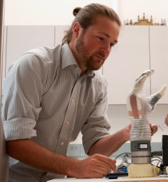 Jacob A. George合作论文数Department of Mechanical Engineering, University of Utah2Sallie M Long合作论文数2FengMin Shao (邵凤民)合作论文数2
 Fuben He (何福本)合作论文数2XiaoMing Qin (秦晓明)合作论文数2
 Quan Yuan合作论文数2
 Daniel S. Brown合作论文数Robotics Center, University of Utah;School of Computing, University of Utah2
 Andrews Jinsy合作论文数2
 Yande Liang (梁延德)合作论文数School of Mechanical Engineering, Dalian University Of Technology2Andrew T. Dailey合作论文数2
 Jinglin Li (李静林)合作论文数The State Key Laboratory of Networking and Switching Technology, School of Computer Science, Beijing University of Posts and Telecommunications2Wei Li (李玮)合作论文数2
 Jing Ma合作论文数Harvard Medical School, Harvard University2Mark B. Bromberg合作论文数2
 Enrico Pagello合作论文数University of Padua2
 Joseph Brignone合作论文数Wearable Robotics Laboratory, Department of Mechanical Engineering, University of Utah;Robotics Center, University of Utah1
 Ian Bales合作论文数Solitude Mountain Resort;University of Utah1Priya Kulkarni合作论文数1Wenjing Wang合作论文数1Scott H Troob合作论文数1Lillian Yang合作论文数1Ade Obayemi合作论文数1Sevan Dulgarian合作论文数1
 Adetokunbo Obayemi合作论文数1Runrun Wang合作论文数1
 Meihan Wan合作论文数1




