How intrinsically disordered regions (IDRs) shape chromatin binding and nuclear organization of transcription factors (TFs) remains unclear. We used proximity-assisted photoactivation (PAPA), a single-molecule protein-protein interaction sensor, to investigate how IDRs might influence TF interactions with each other and with chromatin in live cells. We found that the Sp1 DNA binding domain (DBD) interacted poorly with chromatin and did not colocalize with Sp1. Weak interaction of the isolated IDR with full-length Sp1 was enhanced by fusion to various unrelated DBDs. Live imaging of Drosophila polytene chromosomes confirmed that an IDR could confer sharp locus specificity on an otherwise nonspecific DBD. These findings suggest that TF specificity emerges on chromatin when ensembles of diverse, unstructured interactions are scaffolded by transient DNA contacts.
The classical view of gene regulation complexes as stable, modular machines needs amending based on emerging insights into their dynamic nature. Whereas recent advances in structural biology have provided high-resolution snapshots of these complex machines, single-molecule and live-cell imaging techniques reveal a more fluid picture: biological function emerges not from static architectures but from transient, dynamic assemblies that continually exchange their components and whose activity is tuned through kinetic control. In this Perspective, we propose dynamic, reversible assembly as a framework for understanding the mechanisms of RNA processing and gene regulation. Drawing on specific case studies from ribosome biogenesis, spliceosomes, small RNAs and transcription factors, we explore how ribonucleoprotein complexes and transcriptional ensembles form and dissolve in time, how protein intrinsically disordered regions collectively enable transcription factors to achieve specificity, and the kinetic principles underlying the fidelity, adaptability and robustness of cellular processes and their related pathologies. In doing so, we show how molecular interactions are governed by rates rather than by equilibrium affinities, providing a foundation for time-integrated structure-function studies.
Transcription factor (TF) upregulation accompanies many cellular state transitions, yet how increased TF abundance impacts gene regulation remains unclear. Two broad models are often invoked, whereby higher TF levels amplify the expression of preexisting target genes, or, by mass-action binding, expand genome engagement and regulation to lower-affinity sites. We sought to elucidate how these two regulatory modes contribute to cell differentiation in a well-characterized myogenic system by upregulating the expression of the myogenic TF MyoD1 in C2C12 myoblasts. Unexpectedly, elevated MyoD1 levels impaired myoblast fusion (a hallmark of myogenic differentiation), yet enabled robust contraction in myotubes that did form. Live-cell single-molecule imaging and CUT and RUN profiling revealed that elevated MyoD1 dosage increased total genome-wide chromatin binding and broadened genome occupancy by preferentially engaging lower-affinity sites. Integrating CUT and RUN with RNA sequencing (RNA-seq) experiments linked expanded MyoD1 binding to upregulation of cell adhesion genes. Cell mixing and fractionated RNA-seq experiments supported a two-population model in which an adhesion-gene-upregulated, unfused myoblast population supported contraction of myotubes formed by fusion-competent cells. Ectopic expression of several individual MyoD1-upregulated cell adhesion genes was sufficient to recapitulate the "off script" myotube contraction phenotype. Together, these results support a MyoD1 dose-dependent "spillover" model, in which increased TF abundance broadens cis-regulatory engagement and produces distinct cell differentiation outcomes.
Mutations in the pioneer transcription factor FOXA1 occur in 10%-40% of prostate cancers and broadly alter chromatin accessibility. In a cohort of 874 primary and metastatic tumors, we confirm frequent Wing2 missense mutations and indels, as well as C-terminal truncating frameshifts. To define their functional impact, we performed single-nucleus multiome profiling in mouse prostate organoids expressing representative alleles, including overexpressed wild-type FOXA1. Each subgroup produces distinct chromatin and transcriptional changes, but all perturb epithelial lineage specification. Indel mutants promote basal-like states, whereas C-terminal truncations, Wing2 missense mutations, and elevated wild-type FOXA1 drive secretory L1-like luminal fates. Integrated RNA-seq, ATAC-seq, and ChIP-seq reveal that L1-like specification involves a hybrid androgen receptor/FOXA1 motif and cooperation with POU2F1. In vivo, these same alleles, combined with Trp53/Pten loss, shift tumor histology from basal-like to secretory luminal phenotypes.
The human placenta is built by trophoblast cells that fuse together, secrete hormones, and invade the uterus, and defects in these processes contribute to pregnancy disorders such as preeclampsia. Because cell-cell fusion and hormone secretion are inherently non-cell-autonomous processes, their regulators have remained inaccessible to conventional pooled CRISPR screens. Here, we developed an arrayed CRISPR screen in fusogenic BeWo trophoblasts that simultaneously quantifies fusion and hCG secretion across 412 gene perturbations. The screen revealed that these two hallmark functions of trophoblast differentiation are genetically separable. We characterized the strongest novel hit, ZBTB10, in trophoblast stem cells, organoids, and placental tissue and find that ZBTB10 is an essential regulator of human trophoblast differentiation. ZBTB10 is required for invasive extravillous trophoblast differentiation and supports syncytiotrophoblast maturation, establishing it as a cross-lineage regulator that both activates and represses distinct trophoblast fate programs. Together, these findings provide a genetic platform and phenotypic dissection of how regulatory networks control human placental development.
Recombinant adeno-associated viral vectors (rAAV) are a powerful tool for gene delivery but have a limited DNA carrying capacity. Efforts to expand this genetic payload have focused on engineering the vector components, such as dual trans-splicing vectors which double the delivery size by exploiting the natural concatenation of rAAV genomes in host nuclei. We hypothesized that inefficient dual vector transduction could be improved by modulating host factors which affect concatenation. Since factors mediating concatenation are not well defined, we performed a genome-wide screen to identify host cell regulators. We discover that Homologous Recombination (HR) is inhibitory to dual vector transduction. We demonstrate that depletion or inhibition of HR factors BRCA1 and Rad51 significantly increase reconstitution of a large split transgene by increasing both concatenation and expression from rAAVs. Our results define roles for DNA damage repair in rAAV transduction and highlight the potential for pharmacological intervention to increase genetic payload of rAAV vectors.
Gene expression is essential for life and development, allowing the cell to modulate mRNA production in response to intrinsic and extracellular cues. Initiation of gene transcription requires a highly regulated molecular process to assemble multisubunit complexes into the preinitiation complex (PIC). Attempts to visualize these processes have been driven largely by electron microscopy, with near atomic-level resolution producing static snapshots complemented by low-resolution fluorescence cell imaging. Here, we review how new advances in superresolution single-molecule imaging in live cells can track transcription across vast spatiotemporal scales. We discuss how recent imaging research has fundamentally recast our understanding of PIC assembly from a stable, ordered process to one constantly in flux, dominated by multivalent weak interactions. We also discuss future advancements that will further expand our ability to measure PIC assembly in concert with cellular behavior, predict complex interactions computationally, and target undruggable transcription factors to treat human disease.
Type II nuclear receptors (T2NRs) require heterodimerization with a common partner, the retinoid X receptor (RXR), to bind cognate DNA recognition sites in chromatin. Based on previous biochemical and overexpression studies, binding of T2NRs to chromatin is proposed to be regulated by competition for a limiting pool of the core RXR subunit. However, this mechanism has not yet been tested for endogenous proteins in live cells. Using single-molecule tracking (SMT) and proximity-assisted photoactivation (PAPA), we monitored interactions between endogenously tagged RXR and retinoic acid receptor (RAR) in live cells. Unexpectedly, we find that higher expression of RAR, but not RXR, increases heterodimerization and chromatin binding in U2OS cells. This surprising finding indicates the limiting factor is not RXR but likely its cadre of obligate dimer binding partners. SMT and PAPA thus provide a direct way to probe which components are functionally limiting within a complex TF interaction network providing new insights into mechanisms of gene regulation in vivo with implications for drug development targeting nuclear receptors.
Cells are built from vast networks of interdependent molecular interactions. Here, we combine proximity-assisted photoactivation (PAPA) with automated fast single-molecule tracking (fSMT) to probe subunit interactions within endogenous protein complexes in live human cells. PAPA-fSMT revealed that the inactive positive transcription elongation factor b (P-TEFb):7SK ribonucleoprotein complex is predominantly mobile, not tethered to chromatin, and detected interaction of specific heterogeneous nuclear ribonucleoproteins (hnRNPs) with the 7SK complex. Cyclin-dependent kinase 9 (Cdk9) inhibition liberated hnRNP R from large RNAs, increased hnRNP R binding to 7SK, and evicted P-TEFb from 7SK within minutes-consistent with rapid, homeostatic negative feedback regulation of P-TEFb by competing protein-RNA interactions. Association with the coactivator BRD4 increased P-TEFb chromatin binding, which depended on the BRD4 bromodomains. Finally, PAPA detected the release of P-TEFb from 7SK by the HIV transcriptional activator Tat. Our results illuminate aspects of P-TEFb regulation that were previously inaccessible in live cells and open a route to probe subunit interactions and exchange within endogenous regulatory complexes.
Transcription factor (TF) expression and dosage regulate developmental cell fate decisions. Increased TF dosage has been predicted to enhance expression of high-affinity target genes but also increase the binding of lower-affinity loci. The relative importance of high- versus lower-affinity TF binding in guiding cell fate decisions remains unclear. To test the roles of TF dosage, we examined the effects of increasing the dosage of MyoD1, the “master regulator of myogenesis”, on skeletal muscle differentiation. Unexpectedly, increased MyoD1 dosage inhibited canonical myogenesis and redirected myoblast differentiation towards forming spontaneously contracting myotubes. This novel phenotype was driven by the MyoD1-dose-dependent upregulation of non-myogenic genes, including cell adhesion genes whose ectopic expression also inhibited classical myogenic differentiation and enabled myotube contraction. Live-cell single-molecule imaging showed that elevated MyoD1 dosage increased total chromatin binding and CUT&RUN profiling demonstrated that this increase occurred via preferential binding to lower-affinity loci. Integration of CUT&RUN, ATAC-seq and RNA-seq experiments revealed that increased MyoD1 binding correlated to the upregulation of otherwise lowly expressed genes. These findings suggest that increased MyoD1 dosage induced a selective gene regulatory expansion from high- to lower-affinity cis-regulatory elements, activating a broader ensemble of target genes, revealing a TF dose-dependent mechanism that can trigger distinct developmental programs.
Eukaryotic transcription factors (TFs) contain both structured DNA-binding domains (DBDs) and intrinsically disordered regions (IDRs). While the structures and sequence preferences of DBDs have been extensively characterized, the role of IDR-mediated interactions in chromatin binding and nuclear organization remains poorly understood, in part because these interactions have been difficult to measure in living cells. Here, we use a recently developed single-molecule technique, proximity-assisted photoactivation (PAPA), to investigate how IDRs influence TF associations with each other and with chromatin, focusing on the factors Sp1 and Klf1. We find that the number and patterning of aromatic and basic residues within IDRs govern both TF self-association and chromatin binding. Unexpectedly, the isolated DBD of Sp1 binds chromatin very weakly and non-specifically. The isolated IDR, by contrast, interacts poorly with chromatin-bound wild-type Sp1, yet this interaction is enhanced when even minimal DNA-binding capacity is restored. Strikingly, replacing Sp1's native DBD with those of heterologous TFs recovers both IDR-mediated interactions and chromatin association, despite divergent sequence preferences. PAPA measurements also reveal extensive heterotypic interactions between wild-type Sp1 and other TFs. Together, these results establish PAPA as a powerful method for studying unstructured interactions in their native context and suggest that IDRs participate in widespread cooperative associations scaffolded by transient DBD-DNA contacts, which concentrate disordered regions along chromatin. In contrast to classical models, we propose that TF specificity in vivo emerges not solely from DBD sequence preferences, but from a constellation of weak, dynamic, and diverse interactions mediated by IDRs.
Lineage transcription factors (TFs) provide one regulatory level of differentiation crucial for the generation and maintenance of healthy tissues. To probe TF function by measuring their dynamics during adult intestinal homeostasis, we established HILO-illumination-based live-cell single-molecule tracking (SMT) in mouse small intestinal enteroid monolayers recapitulating tissue differentiation hierarchies in vitro. To increase the throughput, capture cellular features, and correlate morphological characteristics with diffusion parameters, we developed an automated imaging and analysis pipeline, broadly applicable to two-dimensional culture systems. Studying two absorptive lineage-determining TFs, we found an expression level-independent contrasting diffusive behavior: while Hes1, key determinant of absorptive lineage commitment, displays a large cell-to-cell variability and an average fraction of DNA-bound molecules of ∼32%, Hnf4g, conferring enterocyte identity, exhibits more uniform dynamics and a bound fraction of ∼56%. Our results suggest that TF diffusive behavior could indicate the progression of differentiation and modulate early versus late differentiation within a lineage.
During human development, a temporary organ is formed, the placenta, which invades the uterine wall to support nutrient, oxygen, and waste exchange between the mother and fetus until birth. Most of the human placenta is formed by a syncytial villous structure lined by syncytialized trophoblasts, a specialized cell type that forms via cell-cell fusion of underlying progenitor cells. Genetic and functional studies have characterized the membrane protein fusogens Syncytin-1 and Syncytin-2, both of which are necessary and sufficient for human trophoblast cell-cell fusion. However, identification and characterization of upstream transcriptional regulators regulating their expression have been limited. Here, using CRISPR knockout in an in vitro cellular model of syncytiotrophoblast development (BeWo cells), we found that the transcription factor TFEB, mainly known as a regulator of autophagy and lysosomal biogenesis, is required for cell-cell fusion of syncytiotrophoblasts. TFEB translocates to the nucleus, exhibits increased chromatin interactions, and directly binds the Syncytin-1 and Syncytin-2 promoters to control their expression during differentiation. Although TFEB appears to play a critical role in syncytiotrophoblast differentiation, ablation of TFEB largely does not affect lysosomal gene expression or lysosomal biogenesis in differentiating BeWo cells, suggesting a previously uncharacterized role for TFEB in controlling the expression of human syncytins.
During human development, a subset of differentiating fetal cells form a temporary organ, the placenta, which invades the uterine wall to support nutrient, oxygen, and waste exchange between the mother and fetus until birth. Most of the human placenta is formed by a syncytial villous structure which arises via cell-cell fusion of underlying fetal trophoblast stem cells. Genetic and functional studies have characterized the membrane protein fusogens, Syncytin-1 and Syncytin-2, that are both necessary and sufficient for human trophoblast cell-cell fusion. However, identification and characterization of upstream transcriptional regulators regulating their expression has been limited. Here, using CRISPR knockout in an in vitro cellular model of syncytiotrophoblast development (BeWo cells), we find that the transcription factor TFEB, mainly known as a regulator of autophagy and lysosomal biogenesis, is required for cell-cell fusion of syncytiotrophoblasts. TFEB translocates to the nucleus, exhibits increased chromatin interactions, and directly binds the Syncytin-1 and Syncytin-2 promoters to control their expression during differentiation. While TFEB appears to play an important role in syncytiotrophoblast differentiation, ablation of TFEB largely does not affect lysosomal gene expression or lysosomal biogenesis in differentiating BeWo cells, suggesting that TFEB plays an alternative role in placental cells.### Competing Interest StatementRT and XD are co-founders of Eikon Therapeutics, Inc. The other authors have no competing interests.
Recent insights into genome organization have emphasized the importance of A/B chromatin compartments. While our previous research showed that Brd2 depletion weakens compartment boundaries and promotes A/B mixing [1][1], Hinojosa-Gonzalez et al.[2][2] were unable to replicate the findings. In response, we revisited our Micro-C data and successfully replicated the original results using the default parameters in the cooltools software package. We show that, after correcting inconsistencies with the selection and phasing of the compartment profiles, the decrease in B compartment strength persists but the change in compartment identity is to a much lesser extent than originally reported. To further assess the regulatory role of Brd2, we used saddle plots to determine the strength of compartmentalization and observed a consistent decrease of compartment strength especially at B compartments upon Brd2 depletion. This study highlights the importance of selecting appropriate parameters and analytical tools for compartment analysis and carefully interpreting the results.### Competing Interest StatementThe authors have declared no competing interest. [1]: #ref-1 [2]: #ref-2
How molecules interact governs how they move. Single-molecule tracking (SMT) thus provides a unique window into the dynamic interactions of biomolecules within live cells. Using transcription regulation as a case study, we describe how SMT works, what it can tell us about molecular biology, and how it has changed our perspective on the inner workings of the nucleus. We also describe what SMT cannot yet tell us and how new technical advances seek to overcome its limitations. This ongoing progress will be imperative to address outstanding questions about how dynamic molecular machines function in live cells.
 Xavier Darzacq合作论文数Genetics, Genomics & Development Faculty, Department of Molecular and Cell Biology, University of California, Berkeley59
 Claudia Cattoglio合作论文数Dept Mol & Cell Biol, Univ Calif Berkeley24
 Anders Hansen合作论文数University of Cambridge20
 Zhe Liu (刘喆)合作论文数School of Basic Medical Sciences, Tianjin Medical University19
 Gina M Dailey合作论文数17
 James T. Kadonaga合作论文数Department of Molecular Biology, School of the Biological Sciences, University of California, San Diego16
 Luke Lavis合作论文数Howard Hughes Medical Institute14
 Eva Nogales合作论文数Department of Molecular and Cell Biology, College of Letters & Science, University of California, Berkeley;Howard Hughes Medical Institute;Molecular Biophysics and Integrative Bioimaging Division, Lawrence Berkeley National Laboratory14
 Katherine A. Jones合作论文数Salk Institute for Biological Studies13
 Graham Thomas合作论文数12
 Liangqi Xie合作论文数Howard Hughes Med Inst, Janelia Res Campus12Siegfried Ruppert合作论文数12Claire Dugast-Darzacq合作论文数12
 Arie Admon合作论文数12William S. Dynan合作论文数12Carla J. Inouye合作论文数11
 Timothy Hoey合作论文数Tenaya Therapeutics10
 Robert A. Coleman合作论文数Department of Cell Biology, Albert Einstein College of Medicine10
 Anna Maurer合作论文数Dept Mol & Cell Biol, Univ Calif Berkeley9
 Vinson B Fan合作论文数Biol Sci Bldg, Univ Michigan9K Yokomori合作论文数9Meagan N Esbin合作论文数Dept Mol & Cell Biol, Univ Calif Berkeley9
 Anders Näär合作论文数Nutritional Sciences & Toxicology, College of Natural Resources, University of California, Berkeley9Grimm Jonathan B合作论文数9Peng Dong合作论文数9
 Zhengjian Zhang合作论文数Janelia Farm Research Campus, Howard Hughes Medical Institute9
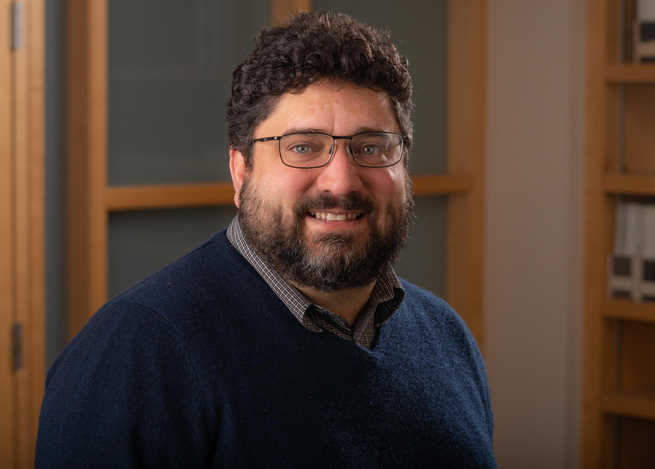 Richard Freiman合作论文数Department of Molecular Biology, Cell Biology, and Biochemistry, Brown University9
 Andrey Revyakin合作论文数QB3 Institute, University of California9
 Michael Marr合作论文数Section of Biochemistry;Molecular and Cell Biology;Cornell University;Molecular and Cell Biology, Cornell University9C. Peter Verrijzer合作论文数9Alec Heckert合作论文数8
 Oscar N Whitney合作论文数University of California8Fong Yick W合作论文数8Brian D. Dynlacht合作论文数Department of Pathology, Grossman School of Medicine, NYU;Perlmutter Cancer Center, Grossman School of Medicine, NYU8
 Richard Myers合作论文数HudsonAlpha Institute for Biotechnology;Department of Genetics, University of Alabama at Birmingham Medical Center;Department of Biological Sciences, University of Alabama in Huntsville8Wright Kevin J合作论文数7
 Ulrike Heberlein合作论文数Howard Hughes Medical Institute;Janelia Farm Research Campus;Department of Anatomy, University of California, San Francisco7James A. Goodrich合作论文数Department of Chemistry and Biochemistry;University of Colorado at Boulder;Department of Chemistry and Biochemistry, University of Colorado at Boulder7
 Mark D. Biggin合作论文数Lawrence Berkeley National Laboratory7
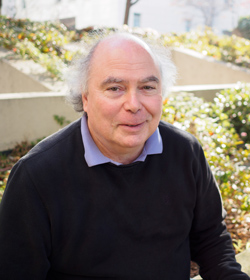 Donald Rio合作论文数Rio Lab, Department of Molecular & Cell Biology, College of Letters & Science, University of California, Berkeley;Division of Biochemistry and Molecular Biology, College of Letters & Science, University of California, Berkeley7Adrian Hinkle合作论文数6JL CHEN合作论文数6
 Liza Dahal合作论文数Molecular and Cell Biology, University of California Berkeley6
 Hannah Greenwald合作论文数Natl Sci Fdn Engn Res Ctr Reinventing Nations Urba6S Zhou合作论文数6
 Basem Al-Shayeb合作论文数Univ Calif Berkeley, Dept Plant & Microbial Biol, Berkeley, CA 94720 USA6
 Tsung-Han Stanley Hsieh合作论文数6
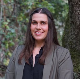 Lauren Kennedy合作论文数Stanford Univ6
 Sheila Teves合作论文数Department of Biochemistry & Molecular Biology, Faculty of Medicine, The University of British Columbia6




