Background and AimsThe immune microenvironment (IME) plays a crucial role in the progression of hepatocellular carcinoma (HCC). In HCC, the IME is often compromised by hepatitis B virus (HBV) infection, chronic inflammation, and fibrosis. Both antiviral therapy (AVT) and the alleviation of inflammation and fibrosis (AIF) have been shown to improve prognosis. However, the relationship among the IME of HCC, AVT, and AIF remains unclear.MethodsA total of 140 and 110 primary HBV-related HCC patients were enrolled as training and validation sets, respectively, to establish a HCC-immune microenvironment score (H-IME score). Immunohistochemistry was performed to assess the number of granzyme B+ (GrB+) and Foxp3+ cells, as well as the expression of CTLA-4, PD-1, LAG3, TIGIT, TIM3, and VISTA. Another cohort consisting of 114 recurrent HBV-related HCC patients with paired primary and recurrent tissues was used to study the relationship among the IME of HCC, AVT, and AIF.ResultsThe H-IME score, including GrB, Foxp3, CTLA-4, PD-1, LAG3, and TIGIT, was established to evaluate the IME. A higher H-IME score indicates stronger immunosuppressive activities. Both AVT and AIF were found to inhibit immunosuppressive activities in the IME. Compared to primary tumors, the H-IME scores of recurrent tumors in the effective AVT group (e-AVT, classified by HBV DNA) with AIF decreased, while the scores increased in the non-AVT group without AIF.ConclusionsThe IME of HCC is closely related to AVT and AIF. e-AVT can enhance anti-tumor activities in the IME by alleviating inflammation and fibrosis.
Three-dimensional cultured organoids recapitulate histopathologic and molecular characteristics of the primary tissues. The rise and development of organoids provide an important platform for clinical medicine research. Patient-derived organoids could be used as models to help clinicians to better understand the pathophysiology of related diseases and formulate corresponding treatment strategies; Organoids derived from tumors could be utilized as a drug screening system for precision medicine, helping clinicians to select the most appropriate regimen for cancer patients and to improve the clinical outcome. This review would summarize the application and research advances of organoids in clinical medicine.
Epithelial regeneration is critical for barrier maintenance and organ function after intestinal radiation injury. Accumulating evidence indicates that the interleukin family members play critical roles in intestinal stem-cell-mediated epithelial regeneration. However, little is known about the relationship between interleukin 33 (IL-33)/ST2 axis and intestinal regeneration after radiation injury. We demonstrate here that IL-33 expression significantly increased after radiation treatment. Deficiency of IL-33/ST2 promotes intestinal epithelial regeneration, resulting in a reduction of mortality during radiation-induced intestine injury. Using ex vivo organoid cultures, we show that recombinant IL-33 promotes intestinal stem cell differentiation. Mechanistically, the effects of IL-33 were mediated by activation of transforming growth factor-β signaling. Our findings reveal a fundamental mechanism by which IL-33 is able to regulate the intestinal crypt regeneration after tissue damage.
Purpose To dissect the tumor ecosystem following immune checkpoint blockades (ICBs) in intrahepatic cholangiocarcinoma (ICC) at a single-cell level. Methods Single-cell RNA sequencing (scRNA-seq) data of 10 ICC patients for the ICB clinical trial were extracted from GSE125449 and systematically reanalyzed. Bulk RNA-seq data of 255 ICC patients were analyzed. Infiltration levels of SPP1+CD68+ tumor-associated macrophages (TAMs) were examined by dual immunofluorescence (IF) staining in 264 resected ICC samples. The correlation between SPP1+ TAMs and clinicopathological features as well as their prognostic significance was evaluated. Results Among the 10 patients, five received biopsy at baseline, and others were biopsied at different timings following ICBs. Single-cell transcriptomes for 5,931 cells were obtained. A tighter cellular communication network was observed in ICB-treated ICC. We found a newly emerging VEGF signaling mediated by PGF-VEGFR1 between cancer-associated fibroblasts (CAFs) and endothelial cells in ICC following ICBs. SPP1 expression was dramatically upregulated, and SPP1+ TAM gene signatures were enriched in TAMs receiving ICB therapy. We also identified SPP1+ TAMs as an independent adverse prognostic indicator for survival in ICC. Conclusion Our analyses provide an overview of the altered tumor ecosystem in ICC treated with ICBs and highlight the potential role of targeting CAFs and SPP1+TAMs in developing a more rational checkpoint blockade-based therapy for ICC.
Background Organoids are three-dimensional structures that closely recapitulate tissue architecture and cellular composition, thereby holding great promise for organoid-based drug screening. Although growing in three-dimensional provides the possibility for organoids to recapitulate main features of corresponding tissues, it makes it incommodious for imaging organoids in two-dimensional and identifying surviving organoids from surrounding dead cells after organoids being treated by irradiation or chemotherapy. Therefore, significant work remains to establish high-quality controls to standardize organoid analyses and make organoid models more reproducible. Methods In this study, the Z-stack imaging technique was used for the imaging of three-dimensional organoids to gather all the organoids’ maximum cross sections in one imaging. The combination of live cell staining fluorescent dye Calcein-AM and ImageJ assessment was used to analyze the survival of organoids treated by irradiation or chemotherapy. Results We have established a novel quantitative high-throughput imaging assay that harnesses the scalability of organoid cultures. Using this assay, we can capture organoid growth over time, measure multiple whole-well organoid readouts, and show the different responses to drug treatments. Conclusions In summary, combining the Z-stack imaging technique and fluorescent labeling methods, we established an assay for the imaging and analysis of three-dimensional organoids. Our data demonstrated the feasibility of using organoid-based platforms for high-throughput drug screening assays. Graphical Abstract
There is no effective method to predict chemotherapy response and postoperative prognosis of colorectal cancer liver metastasis (CRLM) patients. Patient-derived organoid (PDO) has become an important preclinical model. Herein, a living biobank with 50 CRLM organoids derived from primary tumors and paired liver metastatic lesions is successfully constructed. CRLM PDOs from the multiomics levels (histopathology, genome, transcriptome and single-cell sequencing) are comprehensively analyzed and confirmed that this organoid platform for CRLM could capture intra- and interpatient heterogeneity. The chemosensitivity data in vitro reveal the potential value of clinical application for PDOs to predict chemotherapy response (FOLFOX or FOLFIRI) and clinical prognosis of CRLM patients. Taken together, CRLM PDOs can be utilized to deliver a potential application for personalized medicine.
e15598 Background: 5-FU-based regimens are the mainstay treatment for colorectal cancer. However, the limited response rate to 5-FU-based regimens hampered the increase in the treatment efficacy of locally advanced rectal cancer. During the drug screening in the rectal cancer organoid biobank (n = 106), we found out that more than half of the organoids that were resistant to 5-FU monotherapy and sensitive to irinotecan maintained sensitive status to the treatment of 5-FU combined with irinotecan (combination-sensitive), while a quite proportion of them turn to become remarkably resistant to 5-FU plus irinotecan (combination-resistant). Some studies suggested the mechanism of 5-FU-based resistance was associated with the activation of some oncogenic pathways. However, the mechanism of the addition of 5-FU promoting the resistance to irinotecan is still unclear. Methods: To investigate the relationship between the combination-resistant organoids and corresponding patient responses, we collected patient clinical responses and survival outcomes. RNA sequencing and ATAC sequencing were performed to identify differential genes and their enrichment pathways. Moreover, small molecule inhibitors were used to try to reverse the resistance induced by 5-FU. Results: Twenty-one organoids showed sensitivity to irinotecan and resistance to 5-FU. Among them, 8 organoids (38.1%) were combination-resistant and 13 organoids (61.9%) showed sensitivity to combination treatment. Combination-resistant organoids corresponding patients had worse pathologic tumor regression grades (pTRGs) distribution (8/8, 100% had TRGs of 2-3 versus 100% had TRGs of 0-1 in combination-sensitive patients; p < 0.05) and disease-free survival (DFS) rates (HR:11.57, 95% IC:1.025-130.5; p = 0.0047) than patients whose organoids were combination-sensitive. Combination-resistant organoids had higher expression of Ki-67 and CD44 and reduced cleaved-caspase 3 in the combination treatment group and 5-FU group compared with the irinotecan group and control. Furthermore, we identified hedgehog pathway activation after adding 5-FU to irinotecan in combination-resistant organoids through RNA sequencing and ATAC sequencing. GANT-61, an inhibitor of the hedgehog pathway, increased sensitivity to the combination of 5-FU and irinotecan in combination-resistant organoids. Conclusions: We found that organoids which are sensitive to irinotecan turn to become resistant to 5-FU combined with irinotecan. Moreover, corresponding patients had worse responses and a lower DFS rate. The hedgehog pathway is activated in these organoids after the addition of 5-FU to irinotecan, and GANT-61 could reverse the resistance caused by 5-FU. However, more organoid-associated trials with large-scale patients are warranted to reverse the 5-FU-based resistance in the future.
Background Preoperative prediction of microvascular invasion (MVI) is critical for treatment strategy making in patients with hepatocellular carcinoma (HCC). We aimed to develop a deep learning (DL) model based on preoperative dynamic contrast-enhanced magnetic resonance imaging (DCE-MRI) to predict the MVI status and clinical outcomes in patients with HCC. Methods We retrospectively included a total of 321 HCC patients with pathologically confirmed MVI status. Preoperative DCE-MRI of these patients were collected, annotated, and further analyzed by DL in this study. A predictive model for MVI integrating DL-predicted MVI status (DL-MVI) and clinical parameters was constructed with multivariate logistic regression. Results Of 321 HCC patients, 136 patients were pathologically MVI absent and 185 patients were MVI present. Recurrence-free survival (RFS) and overall survival (OS) were significantly different between the DL-predicted MVI-absent and MVI-present. Among all clinical variables, only DL-predicted MVI status and a-fetoprotein (AFP) were independently associated with MVI: DL-MVI (odds ratio [OR] = 35.738; 95% confidence interval [CI] 14.027-91.056; p < 0.001), AFP (OR = 4.634, 95% CI 2.576-8.336; p < 0.001). To predict the presence of MVI, DL-MVI combined with AFP achieved an area under the curve (AUC) of 0.824. Conclusions Our predictive model combining DL-MVI and AFP achieved good performance for predicting MVI and clinical outcomes in patients with HCC.
Background: We aimed to investigate whether improvements in the prognosis of patients with hepatitis B virus (HBV)-related hepatocellular carcinoma (HCC) who have undergone hepatectomy are associated with reductions in the liver inflammation and fibrosis by antiviral therapy (AVT). Methods: Patients who underwent hepatectomy and re-hepatectomy for HBV-related HCC between 2010 and 2019 were divided into two groups. Histological changes in liver were compared between initial and recurrence stages within each group. Propensity score matching (PSM) analysis was performed to compare prognostic outcomes. Results: After PSM, AVT group showed a significantly better prognosis than did non-AVT group (RFS: 19.1% vs. 5.8%, P = 0.001; OS: 64.0% vs. 43.2%, P < 0.001). The improvements in G and S were independent protective factors for RFS (G: P < 0.001; S: P < 0.001) and OS (G: P = 0.013; S: P < 0.001). Conclusions: The application of AVT after initial surgery improved liver inflammation and fibrosis, further benefiting long-term outcomes of patients with HBV-related HCC.
Adding irinotecan to neoadjuvant chemoradiotherapy (nCRT) in locally advanced rectal cancer (LARC) increases the pathologic complete response (pCR) rate but brings more toxicities. Robust biomarkers to predict response to irinotecan-based nCRT are extremely necessary for selecting the right patients. Our previous study suggests that patient-derived tumor organoids (PDTOs) sensitivity to chemoradiotherapy matches patient responses. In this study, we investigated whether PDTOs sensitivity to irinotecan can predict complete response (CR) and survival. Eligible patients receiving irinotecan-based nCRT between April 5, 2017 and December 11, 2020 were enrolled in the training cohort (n = 91) for response prediction and survival analysis. Patients receiving nCRT between February 21, 2021 and September 17, 2021 were included in the validation cohort (n = 27). Predictive performances of irinotecan organoid size ratio (OSR) for CR or pCR were evaluated. The irinotecan-sensitive groups had higher response rates compared with the insensitive groups (training cohort: 71.8% vs 24.4%, P < .0001; validation cohort, 81.8% vs 18.8%, P = .002). Moreover, the irinotecan-sensitive group had higher rates of 3-year disease-free survival (DFS: 71.6% vs 55.5%, P = .034) and distant metastasis-free survival (DMFS, 77.9% vs 57.2%, P = .015) than the irinotecan-insensitive group. 5-FU and irradiation sensitivities failed to predict 3-year DFS (5-FU: 65.4% vs 61.9%, P = .643; irradiation: 84.8% vs 57.8%; P = .072). Performances of irinotecan OSR to predict CR or pCR were good in the training cohort (CR: AUC = 0.828; 95% CI = 0.723-0.932; pCR: AUC = 0.864; 95% CI = 0.759-0.961). The validation showed robust predictive ability (CR: AUC = 0.796, 95% CI = 0.5974-0.9952; pCR: AUC = 0.917, 95% CI = 0.7921-1.0000). Irinotecan sensitivity in PDTOs was a predictive and prognostic factor in LARC.
BACKGROUND:CXCL11 has been reported to be up-regulated in hepatocellular carcinoma (HCC) tissues and cancer-associated fibroblasts (CAFs), and CAF-secreted CXCL11 has been found to promote HCC cell proliferation and migration. Knowledge on how CAFs promote HCC progression is imperative for the future design of anti-tumor drugs addressing the high rates of disease recurrence. Herein, we propose a mechanism by which LINC00152 positively regulates CXCL11 expression and, subsequently, HCC cell phenotypes and growth characteristics via miR-205-5p in CAFs. METHODS:The expression of LINC00152, miR-205-5p in HCC/non-cancerous tissues, CAFs/NFs and HCC cell lines was determined by RT-qPCR. The CXCL11 expression and secretion were determined by westernblot and ELISA. Different expressions of LINC00152, CXCL11 and miR-205-5p in CAFs were achieved by transfection with corresponding overexpression/knockdown vectors or mimics/inhibitor. The interactions among LINC00152, miR-205-5p and CXCL11 were confirmed by FISH, luciferase, AGO2 and RNA-pulldown assays. Transwell, colony formation and MTT assays were performed to assess the role of CAFs conditioned medium (CM) in HCC cell phenotype. BALB/c nude mice xenografts were used to determine the role of CAFs on HCC growth in vivo. RESULTS:We found that in vitro, CM from CAFs transfected with sh-LINC00152 dramatically suppressed HCC cell viability, colony formation and migration, and that CM from CAFs transfected with miR-205-5p inhibitor (CAF-CM (miR-205-5p inhibitor)) exerted opposite effects on HCC cell phenotypes. Exogenous overexpression of CXCL11 in CAFs or CAF-CM (miR-205-5p inhibitor) could partially attenuate the effects of LINC00152 knockdown. In contrast, CM from CAFs transfected with LINC00152 dramatically increased HCC cell viability, colony formation and migration, and CM from CAFs transfected with miR-205-5p mimics (CAF-CM (miR-205-5p mimics)) exerted opposite effects on HCC cell phenotypes. Knockdown of CXCL11 in CAFs or CAF-CM (miR-205-5p mimics) could partially attenuate the effects of LINC00152 overexpression. In vivo, LINC00152 knockdown in CAFs inhibited tumor growth in a mouse model, which could be reversed by CXCL11 overexpression in CAFs. Mechanistically, we found that LINC00152 could act as a ceRNA to counteract miR-205-5p-mediated suppression on CXCL11 by directly binding to miR-205-5p and the 3'UTR of CXCL11. CONCLUSION:Our data indicate that a LINC00152/miR-205-5p/CXCL11 axis in HCC CAFs can affect the proliferative and migrative abilities of HCC cells in vitro and HCC tumor growth in vivo.
类器官是干细胞在体外基质材料支撑条件下培养出来的一种三维微器官,与来源组织器官高度相似.类器官技术为基础研究、药物筛选、再生医学等领域提供了一个新的强大的研究模型和技术手段.再生医学的目的是帮助组织或器官恢复其正常的生理功能,通过与组织工程或基因工程相结合,类器官为再生医学提供了新的移植物来源.该文将介绍类器官在再生医学中的应用,并讨论该领域发展过程中所面临的主要挑战.
BACKGROUND:Brahma-related gene 1 (BRG1) is essential for embryogenesis and cellular metabolism. A deficiency of BRG1 in vivo decreases lipid droplets, but the molecular mechanism underlying its role in lipid metabolism associated with hepatocellular carcinoma (HCC) remains unknown. AIMS:We aimed to determine the role of BRG1 in lipid metabolism in HCC. METHODS:We assessed the differential expression of BRG1 in HCC and adjacent non-tumorous tissues using tissue microarrays. We stained lipid droplets in HCC cells with Bodipy fluorescence and Oil Red O, and verified BRG1 binding to the promoter region of glycosylated lysosomal membrane protein (GLMP) using chromatin immunoprecipitation. RESULTS:The expression of GLMP, a potential lipid metabolism regulator, was suppressed by BRG1 via transcriptional activity. Knockdown of BRG1 decreased lipid droplets, increased GLMP expression and altered the phosphoinositide-3-kinase adaptor protein 1 (PIK3AP1)/phosphatidylinositol-3 kinase (PI3K)/protein kinase B (AKT) pathway in HCC, which further GLMP knockdown partially restored. Thus, GLMP knockdown increased lipid droplets and differentially altered the PI3K/AKT pathway. CONCLUSIONS:Downregulating BRG1 decreased lipid droplet deposition in HCC cells by upregulating GLMP and altering the PI3K/AKT pathway. Both BRG1 and GLMP might serve as therapeutic targets for disorders associated with dysregulated lipid metabolism, such as NAFLD and NAFLD-associated HCC.
Cancer-associated fibroblasts (CAFs) are commonly acquired activated extracellular matrix (ECM)-producing myofibroblasts, a phenotypes with multiple roles in hepatic fibrogenesis and carcinogenesis via crosstalk with cohabitating stromal/cancer cells. Here, we discovered a mechanism whereby CAF-derived cytokines enhance hepatocellular carcinoma (HCC) progression and metastasis by activating the circRNA-miRNA-mRNA axis in tumor cells. CAFs secreted significantly higher levels of CXCL11 than normal fibroblasts (NFs), and CXCL11 also had comparatively higher expressions in HCC tissues, particularly in metastatic tissues, than para-carcinoma tissues. Both CAF-derived and experimentally introduced CXCL11 promoted HCC cell migration. Likewise, CAFs promoted tumor migration in orthotopic models, as shown by an increased number of tumor nodules, whereas CXCL11 silencing triggered a decrease of it. CXCL11 stimulation upregulated circUBAP2 expression, which was significantly higher in HCC tissues than para-carcinoma tissues. Silencing circUBAP2 reversed the effects of CXCL11 on the expression of IL-1β/IL-17 and HCC cell migration. Further downstream, the IFIT1 and IFIT3 levels were significantly upregulated in HCC cells upon CXCL11 stimulation, but downregulated upon circUBAP2 silencing. IFIT1 or IFIT3 silencing reduced the expression of IL-17 and IL-1β, and attenuated the migration capability of HCC cells. Herein, circUBAP2 counteracted miR-4756-mediated inhibition on IFIT1/3 via sponging miR-4756. miR-4756 inhibition reversed the effects induced by circUBAP2 silencing on the IL-17 and IL-1β levels and HCC cell migration. In orthotopic models, miR-4756 inhibition also reversed the effects on metastatic progression induced by silencing circUBAP2.
Automatic liver tumor detection can assist doctors to make effective treatments. However, how to utilize multi-modal images to improve detection performance is still challenging. Common solutions for using multi-modal images consist of early, inter-layer, and late fusion. They either do not fully consider the intermediate multi-modal feature interaction or have not put their focus on tumor detection. In this paper, we propose a novel multi-scale intermediate multi-modal fusion detection framework to achieve multi-modal liver tumor detection. Unlike early or late fusion, it maintains two branches of different modal information and introduces cross-modal feature interaction progressively, thus better leveraging the complementary information contained in multi-modalities. To further enhance the multi-modal context at all scales, we design a multi-modal enhanced feature pyramid. Extensive experiments on the collected liver tumor magnetic resonance imaging (MRI) dataset show that our framework outperforms other state-of-the-art detection approaches in the case of using multi-modal images.
Hepatocellular carcinoma (HCC) is a fatal disease characterized by vast molecular heterogeneity. Although major advances in tumour genetics has led to the identification of new biomarkers, the prognosis of patients with HCC remains dismal.Quantitative reverse-transcription polymerase chain reaction (qRT-PCR) and western blot (WB) were used to evaluate meiosis-specific nuclear structural 1 (MNS1) expression in HCC cells. Immunohistochemistry staining was used to evaluate MNS1 expression in HCC tissues. Clinical significance of MNS1 was evaluated by Cox regression analysis. Transwell assays were conducted to assess cells migration ability. Cell counting kit-8 and colony formation assays were performed to detect cells proliferation ability. NOD/SCID/γc(null) (NOG) mice model was adopted to investigate functions of MNS1 in vivo.The expression of MNS1, which is elevated in most HCC tissues, correlated with poor survival in HCC patients. Functional experiments revealed the oncogenic role of MNS1, which promotes HCC growth and metastasis through AKT-dependent modulation of β-catenin. β-Catenin expression was crucial for MNS1's oncogenic effects. MNS1 indirectly translocated β-catenin from the cytoplasm to the nucleus via the MNS1-GSK3β axis.MNS1 promotes HCC growth and metastasis via activating PI3K/AKT signalling and may serve as an important prognostic biomarker as well as potential novel therapeutic target for HCC.
Glioblastoma is the most common form of primary brain tumor. Glioblastoma stem cells play an important role in tumor formation by activation of several signaling pathways. Wnt signaling pathway is one such important pathway which helps cellular differentiation to promote tumor formation in the brain. Glioblastoma remains to be a highly destructive type of tumor despite availability of treatment strategies like surgery, chemotherapy, and radiation. Advances in the field of cancer biology have revolutionized therapy by allowing targeting of tumor-specific molecular deregulation. In this review, we discuss about the significance of glioblastoma stem cells in cancer progression through Wnt signaling pathway and highlight the clinical targets being potentially considered for therapy in glioblastoma.
Hepatocarcinogenesis is a complicated process that is affected by a variety of microenvironmental factors, such as secretory chemokines and cell-extracellular matrix (ECM). Retinoic acid receptor-related orphan receptor (ROR)-α has been shown to attenuate tumor invasiveness by inducing suppressive cell microenvironment, and its low expression was associated with a worse prognosis in HCC patients. In the present study, we attempted to investigate the role and mechanism of the dominant transcript of ROR-α, ROR-α-1, in HCC development and progression. Among the four transcripts (ROR-α-1/-2/-3/-4), overexpression of ROR-α-1 dramatically suppressed the capacity of MHCC97H cells to proliferate, migrate and invade. We analyzed the differentially expressed genes in ROR-α-1-overexpressed and non-overexpressed MHCC97H cells, performed Gene Ontology (GO) enrichment analysis on these differentially-expressed genes, and found out that factors involved in the tumor microenvironment and ECM are related to the anti-tumor effects of ROR-α-1. Among these factors, chemokine CXCL5 was significantly downregulated by ROR-α-1 overexpression. Overexpression of ROR-α-1 remarkably inhibited the capacity of HCC cells to proliferate, migrate, invade, and downregulated the protein levels of β-catenin, c-Myc, Cyclin D1, and N-cadherin, suggesting the tumor-suppressive role of ROR-α-1 in MHCC97H cells. Moreover, overexpression of CXCL5 dramatically attenuated the suppressive effects of cell proliferation, migration and invasion induced by ROR-α-1 overexpression in MHCC97H, suggesting that ROR-α-1 exerts its anti-tumor effects via downregulating CXCL5. In conclusion, we demonstrate the tumor-suppressive role of ROR-α-1 in MHCC97H cells and that ROR-α-1 might play a tumor-suppressive role via regulation of chemokine CXCL5.
Hepatic stellate cells (HSCs) play vital roles in tumorigenesis and progression of hepatocellular carcinoma (HCC). However, there remains a lack of high-throughput studies on gene expression alterations in HCC cells in response to direct interactions with HSCs. In this study, we established a direct co-culture model of HSCs and HCC cells. We found that the expression of a set of miRNAs, most notably miR-1246, was triggered by HSCs. RORα was confirmed as the target gene of miR-1246. Either overexpression of miR-1246 or knockdown of RORα enhanced the proliferation, invasiveness, and metastatic capability of HCC both in vitro and in vivo, through Wnt/β-catenin pathway activation and promotion of epithelial-mesenchymal transition (EMT). Moreover, upregulation of miR-1246 and repression of RORα were prominent features of aggressive clinical HCC. The miR-1246-RORα-Wnt/β-catenin axis is a novel pathway through which HSCs accelerate HCC progression.
Background: Recently, more and more treatment strategies for Hepatocellular carcinoma (HCC) have emerged, but the therapeutic effect is still not satisfactory. This study is aimed to explore the mechanism of Interleukin 35 (IL-35) in promoting the progression of liver cancer and to explore the application value of IL-35 in the treatment of HCC. Methods: We used clinical tissue microarray (TMA) immunohistochemistry (IHC) to explore the prognostic value of IL-35 expression in patients with HCC. The effect of IL-35 on the function of HCC was explored by functional experiments including wound-healing assay, transwell, cell counting kit-8, cell adhesion assay and endothelial tube formation assay in vitro and mouse xenografts in vivo. And flow cytometry was used to study the effect of IL-35 on infiltrating immune cells in tumor. The molecular mechanism of the function of IL-35 on the progression of HCC was explored by sequencing, ELISA, WB, PCR and other technical means. Finally, through in vivo tumor animal experiments to explore the value of anti-IL-35 antibody and combined with anti-PD-1 antibody in the treatment of liver cancer. Results: High expression of IL-35 in patients with HCC were identified to be associated with poor prognosis. And we have found that IL-35 facilitated tumor progression by affecting neutrophil infiltration, angiogenesis, and CD8+ T-cell infiltration in a mouse model. Additionally, on the one hand C-C motif chemokine ligand 3 (CCL3) has been found to be a key factor mediating the recruitment of neutrophils by IL-35, on the other hand fibroblast growth factor 2 (FGF2) secreting by neutrophil when stimulated by IL-35 was also found to be the core cytokine to promote intratumoral angiogenesis. And IL-35 was also discovered to facilitated the adhesion of tumor to endothelial cells, with neutrophils further enhancing this effect in vitro and vivo. More important, anti-IL-35 antibody was found to be a valid treatment for HCC in xenograft tumor model, and it could give full play to the curative effect of 1:1>2 when combination therapy with PD-1 antibody. Conclusion: Our data show that the expression of IL-35 in patients with HCC is an important tumor promoting factor. The application of anti-IL-35 antibody and treatment combined anti-IL-35 antibody with anti-PD-1 antibody have potential therapeutic value in the treatment of liver cancer.
 Bao-Ye Sun合作论文数Fudan Univ, Zhongshan Hosp, Liver Canc Inst, Dept Liver Surg, Shanghai, Peoples R China14Yong YI (易勇)合作论文数13
) Cheng Zhou (周承)合作论文数The First Affiliated Hospital, Zhejiang University School of Medicine13Gao Liu合作论文数12
Cheng Zhou (周承)合作论文数The First Affiliated Hospital, Zhejiang University School of Medicine13Gao Liu合作论文数12 Shuangjian Qiu (邱双健)合作论文数Zhongshan Hospital, Fudan University12Pei-Yun Zhou合作论文数Fudan Univ, Zhongshan Hosp, Liver Canc Inst,Dept Liver Surg, Key Lab Carcinogenesis & Canc Invas,Minist Educ, Shanghai, Peoples R China11Zhu-Tao Wang合作论文数Fudan Univ, Zhongshan Hosp, Liver Canc Inst, Dept Liver Surg & Transplantat, Shanghai 200032, Peoples R China9
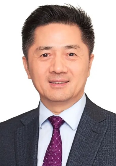 Jia Fan (樊嘉)合作论文数Zhongshan Hospital, Fudan University;Liver Cancer Institute, Fudan University;Shanghai Medical College, Fudan University9Zhang-Fu Yang合作论文数8Wei Gan合作论文数8
 Guoqiang Hua (华国强)合作论文数复旦大学放射医学研究所6Jin-Long Huang合作论文数5Zhang Long合作论文数5XiaoYa Xu (徐小雅)合作论文数4
 Jian Zhou (周健)合作论文数Shanghai Sixth People’s Hospital Affiliated to Shanghai Jiao Tong University School of Medicine4Xinxin Rao合作论文数3Peiyuan Tang合作论文数3Peiyuan Mu合作论文数3Tao Lv合作论文数3Guoxiang Fu合作论文数3
 Guo Mian合作论文数The Second Affiliated Hospital, Harbin Medical University3
 Zhen Zhang (章真)合作论文数Department of Radiotherapy, Shangahi Cancer Center, Fudan University3Shaobo Mo (莫少波)合作论文数College of Chemistry and Molecular Sciences, Wuhan University3Zhisheng Chen合作论文数2Zhou Yi合作论文数2Jie Li (李解)合作论文数2Xiaotong Kong合作论文数Harbin Med Univ, Affiliated Hosp 2, Dept Neurol, Harbin, Heilongjiang, Peoples R China2Ming Bai合作论文数2Jianjun Gao (高建军)合作论文数2Lixia Chen (陈丽霞)合作论文数Guangzhou University of Chinese Medicine2Chunrui Bo合作论文数2Sun Jian合作论文数2Lu Xiao-Yu合作论文数2Yi-Peng Fu合作论文数2Liu Zhaojun合作论文数2Peng Junjie合作论文数2Jianwei Lu (路建伟)合作论文数2Jianjian Wang合作论文数2Huixue Zhang合作论文数2Xingfeng He合作论文数University of Maryland, College Park2
 Ye Luo (罗业)合作论文数School of Software Engineering, Tongji University2Jia Shen合作论文数2Yun Deng (邓云)合作论文数2
 Lihua Wang (王丽华)合作论文数The Second Affiliated Hospital of Harbin Medical University2
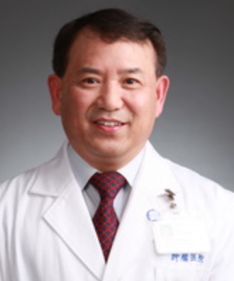 Sanjun Cai (蔡三军)合作论文数Fudan University Shanghai Cancer Center2Chen Hu合作论文数1Xiang Hu (胡祥)合作论文数1




