Objective: Previous studies assessing left atrium (LA) function in patients with atrial fibrillation (AF) have not simultaneously analyzed left ventricle (LV) function.To evaluate differences in LA and LV volume and function in patients with AF during and between AF episodes compared to healthy subjects by using dual-source computed tomography (DSCT).Materials and Methods: Sixty-one consecutive patients with AF and 30 healthy subjects underwent retrospective electrocardiography-gated DSCT.Thirty of 61 AF patients experienced AF during scan acquisition (AF episode subgroup), and 31 were in sinus rhythm (AF non-episode subgroup).LA and LV functions were analyzed with dedicated software and groups were compared with analysis of variance and least significant difference test. Results:The maximal LA volumes in healthy subjects, the AF episode subgroup, and the no AF episode subgroup were 94.4±19.9mL, 121.8±42.4mL, and 101.4±32.9mL, respectively (p=0.005).Left ventricular end-diastolic volumes (LVEDV) in healthy subjects, the AF episode subgroup and the no AF episode subgroup were 160.5±34.4mL, 131.7±37.3mL, and 138.1±27.6 mL, and respectively (p=0.003).Left ventricular end-systolic volumes were 55.6± 19.4 mL, 65.2±32.4mL, and 51.0±14.9mL, respectively (p=0.060).Increased LA volume, decreased LVEDV, and decreased LA and LV systolic function were found during AF episodes. Conclusion:Our data contributes to understanding the mechanism of AF and guides clinical management of patients with AF.
患者女,46岁.活动后胸闷乏力1年,近1个月加重,于2009年12月18日就诊.体检:主动脉瓣听诊区和主动脉瓣第二听诊区可闻及Ⅱ~Ⅲ级收缩期杂音,呈喷射样,向颈部传导.心电图提示左心室高电压,ST-T改变.超声心动图示先天性心脏病,主动脉瓣二瓣化畸形并重度狭窄,升主动脉扩张.X线摄影示两肺纹理重,升主动脉扩张,主动脉结增宽,左心室增大,心胸比为0.63。
Objective To investigate the intra- and interobserver repeatability of coronary artery disease (CAD) diagnosis based on invasive coronary angiography (ICA) and CT coronary angiography (CTCA).Methods Two readers with comparable experience ( over 10 years) independently evaluated ICA results of 42 consecutive patients with a blind method. After 30 days,one of them reviewed the same patients again.Another two comparable-experience (over 10 years) readers evaluated the results of CTCA (prospectively ECG-triggering) from the same 42 patients in the same way.The inter-reader and intra-reader repeatability of ICA and CTCA were analyzed by performing Kappa test and calculating the percentage of the segments with agreement on stenotic degree.Using ICA as reference,the accuracy of CTCA in diagnosing CAD was studied by comparing the area under ROC. Results The Kappa between readers for ICA and CTCA were 0.91 and 0.81.Intra-reader Kappa were 0.92 and 0.83 respectively (x2 =509.4 and 432.5,all P <0.01 ).The percentage of the segments with agreement between readers on the degree of stenosis were 80.8% (494/611) in ICA and 75.2% (469/624) in CTCA ( x2 =2.75,P =0.10),and within the same reader,86.9% (531/611)in ICA and 81.9% (511/624) in CTCA(x2 =3.76,P =0.053).With≥ 50%narrowing as a CAD diagnosis criterion,the agreement rates for two readers were 96.6% (590/611 ) in ICA and 94.4% (589/624) in CTCA( x2 =3.36,P =0.07),and for the same reader,97.4% (595/611) in ICA,95.4% (595/624) in CTCA ( x2 =3.62,P =0.06).Using ICA as reference,two readers of CTCA results achieved a sensitivity and specificity of 84.9% (530/624)and 98.1% (612/624).The area under ROC was 0.94 (95% CI 0.91-0.97).Conclusions Both ICA and CTCA demonstrate good repeatability in diagnosing CAD.The repeatability of ICA is superior to that of CTCA.A certain discrepancy exists in two readings from the same reader or two readers.
 Fangfang Yu合作论文数3Ming-Li Sun合作论文数3
 Shiliang Jiang (蒋世良)合作论文数Fuwai Hospital, Chinese Academy of Medical Sciences3Lei Han合作论文数3
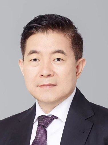 Bin Lv (吕滨)合作论文数Fuwai Hospital, Chinese Academy of Medical Sciences;Peking Union Medical College, Chinese Academy of Medical Sciences3Shiguo Li合作论文数3
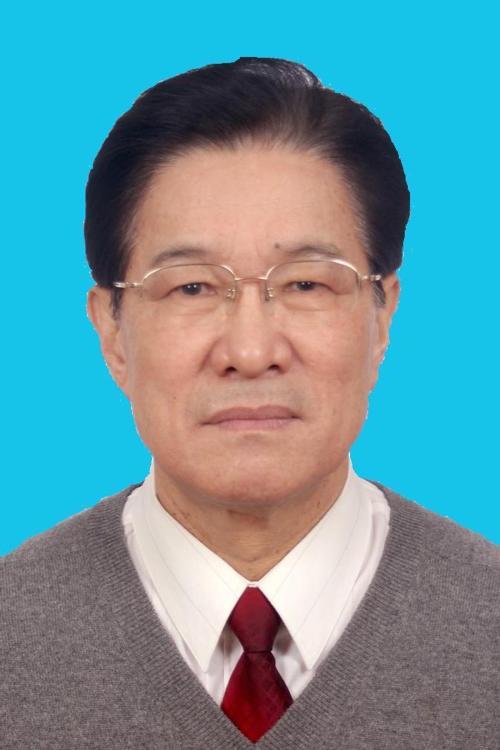 Ruping Dai (戴汝平)合作论文数Fuwai Hospital, Chinese Academy of Medical Sciences3
 Runze Wu合作论文数2
 Zhicheng Jing (荆志成)合作论文数Department of Cardiology, Guangdong Provincial People's Hospital2cuihong zhou (周翠红)合作论文数1Huili Cao合作论文数1JianHua Lv (吕建华)合作论文数1Yang Gao (高扬)合作论文数1
 Felix Meinel合作论文数Institut für Diagnostische und Interventionelle Radiologie, Kinder- und Neuroradiologie, Department für Radiologie, Universitätsmedizin Rostock1Ebersberger Ullrich合作论文数1McQuiston Andrew D合作论文数1
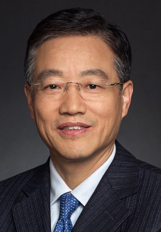 Yuejin Yang (杨跃进)合作论文数Fuwai Hospital, CAMS & PUMC1
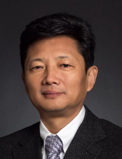 Yongjian Wu (吴永健)合作论文数Fuwai Hospital, CAMS & PUMC1
 U. Joseph Schoepf合作论文数Department of Radiology and Radiological Science, College of Medicine, Medical University of South Carolina1
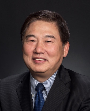 Shubin Qiao (乔树宾)合作论文数Fuwai Hospital, Chinese Academy of Medical Sciences & Peking Union Medical College1
 Zhihui Hou (侯志辉)合作论文数Department of Radiology, Fuwai Hospital, Chinese Academy of Medical Sciences and Peking Union Medical College1




