BACKGROUND:Proficiency in cytopathologic diagnosis depends heavily on extensive hands-on practice and immediate error correction. Traditional teaching models, however, are constrained by limited practice opportunities and delayed feedback, which fails to meet the core skill-development needs of residents. METHODS:In total, 45 pathology residents were enrolled and assigned to two groups. The experimental group (n = 20) adopted a tripartite teacher-artificial intelligence-resident collaborative teaching model, whereas the control group (n = 25) received conventional instruction. Both groups underwent an identical 8-week teaching cycle. RESULTS:The questionnaire results from the experimental group indicated that 19 of 20 residents (9%) deemed the new model highly necessary, and 15 of 20 (75%) believed it significantly improved their diagnostic competence. Semistructured interviews further revealed that the model enhanced diagnostic ability, facilitated personalized learning, and alleviated learning anxiety. For objective metrics, the experimental group demonstrated a significantly higher postintervention concordance rate for gray-zone cell identification (78.65%) compared with both their preintervention baseline (64.38%) and the contemporaneous control group (66.84%; t = 8.962; p < .001). In addition, the experimental group exhibited a markedly faster diagnostic speed (mean ± standard deviation, 3.05 ± 0.52 minutes per case) compared with their preintervention performance (5.92 ± 0.85 minutes per case) and the control group (5.63 ± 0.79 minutes per case; t = 14.821; p < .001). No statistically significant changes were observed in the control group (p > .05). CONCLUSIONS:This study demonstrates that artificial intelligence technology integrated with real-time visual interaction effectively improves the cytopathologic diagnostic skills of residents and merits wider promotion in pathology education.
Aims In recent years, patients with programmed cell death-Ligand 1 (PD-L1)-positive oesophageal squamous cell carcinoma (OSCC) have been able to benefit from immunotherapy. However, method for improving the treatment efficacy of PD-L1-positive patients is a problem that needs further consideration. Studies on the relationship between human epidermal growth factor receptor 2 (HER2) and PD-L1 expression have recently been reported in certain cancers, but the relationship between PD-L1 and HER2 expression in OSCC is still unclear. Methods A total of 263 patients with OSCC were included in the study. PD-L1 protein expression and HER2 protein expression were analysed by immunohistochemistry (IHC), and fluorescence in situ hybridisation (FISH) was performed to assess HER2 gene amplification. The significance of differences between HER2 status, PD-L1 status and clinicopathological parameters was assessed. The relationship between PD-L1 status and HER2 status was examined. Results Of the 263 OSCC cases, the PD-L1-positive expression rates were 39.2% and 77.2% in OSCC for Tumour Proportion Score (TPS) and Combined Positive Score (CPS), respectively, and PD-L1 expression was associated with the degree of tumour differentiation. The HER2 expression was positive in 24% (63/263) of cases based on IHC and FISH. HER2 expression was not significantly associated with clinicopathological characteristics. PD-L1 TPS expression and CPS expression were significantly positively correlated with HER2 expression in OSCC. Conclusions PD-L1 expression was significantly positively correlated with HER2 expression in OSCC. The results provide valuable insight for the future application of HER2-targeted therapy combined with immunotherapy in OSCC.
Background:The prognostic predictors of the synchronous multiple primary lung cancer (SMPLC) still remain unclear, and there is a lack of studies on the prognosis of SMPLC patients excluding those with multifocal ground-glass/lepidic (GG/L) nodules. The aim of this study is to develop an effective model for predicting survival of SMPLC patients. Methods:In this multicenter cohort study, a total of 831 SMPLC patients presenting for lung cancer resection from January 2004 to January 2018 at five institutions were included for developing and validating a nomogram model. Specifically, 499 patients from the Cancer Hospital, Chinese Academy of Medical Sciences, and Beijing Chao-Yang Hospital, Capital Medical University were served as the training cohort. A total of 332 patients from The Third Xiangya Hospital of Central South University, the First Affiliated Hospital of University of Science and Technology of China, and Beijing Liangxiang Hospital were served as the external validation cohort. The nomogram model was compared with the Tumor Node Metastasis (TNM) system for the overall survival. The C-index, net reclassification improvement (NRI), and integrated discrimination improvement (IDI) were used to evaluate the model performance. A user-friendly website for SMPLC survival probability calculation was also provided for a better understanding of prognosis of patients with resected SMPLC. Results:A total of seven independent risk factors were selected by conducting a multivariate analysis on the training set. Further, a nomogram model was developed with these factors. Both the internal and external validations exhibited good discrimination (C-index: internal, 0.827; external, 0.784). The NRI and IDI of this model were 0.33 and 0.21, respectively. The survival rates for 1-year, 3-year, and 5-year were consistent with the actual observed values. A set of cutoff values were determined by grouping the patients into three different groups. For each group, we should expect a significant distinction between survival curves. Conclusions:The novel nomogram model enables accurate survival risk stratification of patients with resected SMPLC and may assist in decision-making that is conducive to patients with SMPLC at high risk.
Background Circumferential resection margin (CRM) is very important in esophageal cancer, but its diagnostic criteria has not been unified. The College of American Pathologists (CAP) and the Royal College of Pathologists (RCP) provide two different criteria. The aim of this study is to evaluate the long-term prognostic significance of CRM status with different CRM criteria in esophageal squamous cell carcinoma (ESCC). Methods Influence of CRM status according to the CAP and RCP criteria on long-term survival of 838 patients with resected pT3 tumors and without neoadjuvant therapy was analyzed. Patients stratified into three groups on the basis of tumor distance from the CRM (CRM > 1 mm, 0-1 mm, and 0 mm) were also analysed. Results Positive CRM was found in 59 (7%) patients according to the CAP criteria and 317 (37.8%) patients according to the RCP criteria. Univariate and multivariate survival analysis showed that CRM status, according to three different criteria, was independent prognostic factor. However, subgroup analysis showed that the prognostic value of CRM status was limited to certain metastatic lymph node load. In pN0 subgroup, patients with CRM > 1 mm had better prognosis than patients with CRM 0-1 mm. Patients with CRM 0 mm had worse outcome than patients with CRM > 0 mm in pN1-2 subgroup. But CRM status had no prognosis value in pN3 subgroup. Conclusions The CRM status is an important prognostic factor in ESCC patients, but this effect was limited to patients without or with less lymph node metastasis (pN0-2). In clinical practice, we recommend the 1 mm-three-tier criteria as it provides more prognostic value than the traditional two-tier criteria.
Background Despite great progress in surgery and other treatments, the prognosis of patients with esophageal squamous cell carcinoma (ESCC) is still very poor. HER2 has strong therapeutic implications in certain cancers, such as breast cancer and gastric cancer. However, literature on the frequency of HER2 expression in ESCC is scarce. In the present study, HER2 protein expression, HER2 gene amplification and the relationship between HER2 status and clinicopathological characteristics were evaluated in a large cohort of Chinese ESCC patients. Methods A total of 857 consecutive ESCC patients who received radical esophagectomy without neoadjuvant therapy between January 2014 and October 2015 were included in this retrospective study. HER2 protein expression was analyzed by immunohistochemistry (IHC), and its correlation with clinicopathological parameters was assessed. In addition, 65 cases, including 13 HER2 overexpression (3+) cases and 52 HER2 equivocal (2+) cases from the 857-case cohort, and another 104 ESCC cases, including 1 HER2 overexpression (3+) case, 3 HER2 equivocal (2+) cases and 100 HER2 negative (1+/0) cases, were selected to construct tissue microarrays (TMAs). Dual-color in situ hybridization (DISH) was performed on the TMAs to assess HER2 gene amplification and the relationship with clinicopathological parameters. Results We found HER2 overexpression (3+) status in 1.5% (13/857) of cases and HER2 equivocal (2+) status in 6.1% (52/857) of cases. HER2 IHC expression was significantly associated with gender ( P = 0.028). However, there were no significant correlations between HER2 IHC expression and age, tumor differentiation, pT stage, pN stage, pM stage and pTNM stage ( P > 0.05). Regarding the 169 cases analyzed by DISH, 14 (of 14, 100%) HER2 overexpression (3+) cases, 10 (of 55, 18.2%) HER2 equivocal (2+) cases, and 0 (of 100, 0%) HER2 negative (1+/0) cases showed HER2 gene amplification. HER2 gene amplification was not significantly associated with clinicopathological characteristics such as age, gender, tumor differentiation, pT stage, pN stage, pM stage and pTNM stage ( P > 0.05). Conclusions Approximately 1.5% of the Chinese ESCC patients had HER2 overexpression based on IHC. IHC and DISH had a high concordance rate. These results provide valuable insight for the future treatment of ESCC.
Esophageal squamous cell carcinoma (ESCC) and leiomyoma are common tumors. The coexistence of these two tumors can be classified into two types: the overlying type and the separate type. The overlying type is rare. We report 12 cases of the overlying type treated by endoscopic submucosal dissection (ESD). They underwent pre-ESD endoscopic examination with white-light imaging, iodine staining, narrow-band imaging, endoscopic ultrasound, and biopsy. The clinical, endoscopic and pathologic characteristics were reviewed. Among the 12 patients, 3 were female and 9 were male. The age range was 49–76 years. They accounted for 4.0% of 300 cases of esophageal leiomyoma and 1.3% of 955 cases of superficial ESCC or high-grade dysplasia treated by endoscopic resection. After endoscopic examination and biopsy, ESCC or high-grade dysplasia, combined with leiomyoma was considered in four cases; leiomyoma was considered but without the squamous lesion (underdiagnosis) in another case; and leiomyoma was mistaken for submucosal ESCC (overdiagnosis) in the other seven cases. ESD specimens showed that nine cases were intramucosal or submucosal ESCC, and three cases were high-grade dysplasia, overlying leiomyoma originating from the muscularis mucosae or muscularis propria. The 12 cases were successfully treated by ESD, with no recurrence during follow-up. We must keep in mind that ESCC or high-grade dysplasia can occur overlying leiomyoma. These cases are rare but should not be neglected, especially in high-risk areas for ESCC. These patients can receive appropriate treatment if overdiagnosis or underdiagnosis can be avoided.
Background: Programmed death ligand 1 (PD-L1) is a ligand for the inhibitory programmed cell death protein 1 (PD-1), which are targeted by several anti-PD-1 and PD-L1 drugs for a variety of human cancers. However, only a few studies have evaluated PD-L1 expression in esophageal squamous cell carcinoma (ESCC) with a large Chinese cohort. Our present study is to evaluate the association of PD-L1 expression with clinicopathological features on ESCC. Methods: Using tissue microarray and immunohistochemistry, PD-L1 expression on tumor cells and tumor-infiltrating immune cells was studied in 378 advanced ESCC patients without neoadjuvant chemoradiotherapy. Its correlation with clinicopathological parameters was analyzed. Results: PD-L1 was expressed on 29.9% (113/378) ESCC tumor cells and 40.2% (152/378) tumor-infiltrating immune cells. PD-L1 expression in tumor cells was significantly correlated with age, degree of differentiation, T stage, N stage and metachronous hematogenous metastasis, and PD-L1 expression in tumor-infiltrating immune cells was significantly associated with N stage (P < 0.05). Patients with PD-L1 expression in tumor cells had poor disease-free survival (Hazard ratio [HR] = 1.436, P = 0.009). There was a positive association between tumor cells and tumor-infiltrating immune cells for PD-L1 expression (r = 0.16, P = 0.002). However, PD-L1 expression in tumor-infiltrating immune cells was not significantly correlated with disease-free survival and overall survival. Conclusions: PD-L1 expression in tumor cells and tumor infiltrating immune cells is not only an indicator for immunotherapy, but also significantly related with age, differentiation, stage, metastasis and disease free survival.
Ras-association (RA) domain family number 6 (RASSF6) is a member of the Ras-association domain protein family. It is epigenetically inactive and negatively regulates the malignant progression of some tumors. However, its precise role in esophageal squamous cell carcinoma (ESCC) has not been reported. In this study, we performed immunohistochemistry (IHC) assay. The results show that RASSF6 is upregulated in ESCC and that the elevated expression level of RASSF6 is associated with lymph node metastasis and poor survival of ESCC patients. Consistent with the clinical observations, the upregulation of RASSF6 greatly promotes ESCC cell proliferation, migration and invasion as well as the cell cycle transition to G1/S phase in vitro. According to models in vivo, the downregulation of RASSF6 considerably inhibits ESCC tumor growth and lung metastasis. Mechanistically, RASSF6 negatively regulates the tumor suppressor tripartite-motif-containing protein 16 (TRIM16) by promoting its ubiquitination-dependent degradation and eventually activates pathways associated with the cell cycle and epithelial-mesenchymal transition (EMT). Together, these results indicate that the RASSF6-TRIM16 axis is a key effector in ESCC progression and that RASSF6 serves as a potential target for the treatment of ESCC.
食管梭形细胞癌是食管鳞状细胞癌的一种特殊亚型,发生率较低,镜下具有双相性的特点,上皮成分常为高中分化的鳞状细胞癌或原位癌,梭形细胞成分常为高级别肉瘤样成分.肿瘤通常呈息肉样的生长方式,分期较早,目前研究普遍认为食管梭形细胞癌侵袭性低,淋巴结转移率低,生存率高,预后较好,但由于食管梭形细胞癌病例发生率较低,目前多为个案报道,关于食管梭形细胞癌的流行病学、临床特征、双相性的组织来源及预后尚不十分清楚.本文综述了食管梭形细胞癌的流行病学、临床特征、分子病理学特征及预后特点.
Clinicopathological parameters are important to predict the prognosis of esophageal squamous cell carcinoma (ESCC),they mainly include TNM stage related indexes of the tumor,tumor length,vessel and nerve invasion, tumor budding, peripheral blood cells, etc. To predict the prognosis of ESCC patients accurately is the prerequisite of precise treatment and the key to improve the patients survival rate and survival quality.
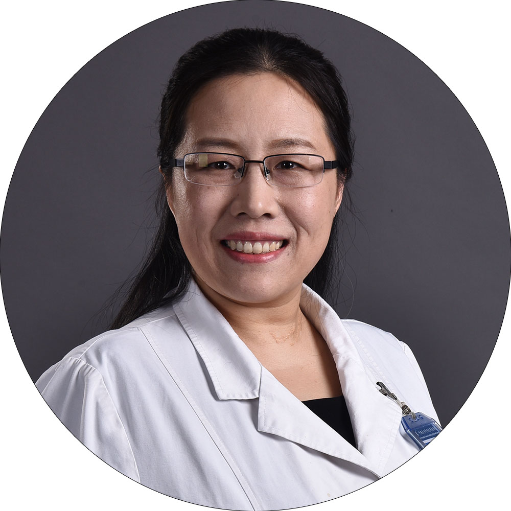 Liyan Xue (薛丽燕)合作论文数国家癌症中心/中国医学科学院肿瘤医院病理科9
 Ning Lu合作论文数4
 Lei Guo (郭磊)合作论文数Peking Union Medical College3Bingzhi Wang合作论文数2
 Jianming Ying (应建明)合作论文数Cancer Hospital Chinese Academy of Medical Sciences2
 Zitong Zhao合作论文数2Xue Xuemin合作论文数2
 Xiuyun Liu (刘秀云)合作论文数Beijing Children's Hospital Affiliated to Capital University of Medical Sciences,Beijing ,China2
 Ning Lv (吕宁)合作论文数Cancer Hospital Chinese Academy of Medical Sciences2Yong Liu (刘勇)合作论文数Cancer Hospital, Chinese Academy of Medical Sciences2
 Yongmei Song (宋咏梅)合作论文数State Key Laboratory of Molecular Oncology, Cancer Hospital, Chinese Academy of Medical Sciences;Peking Union Medical College2Jing Zhou合作论文数1
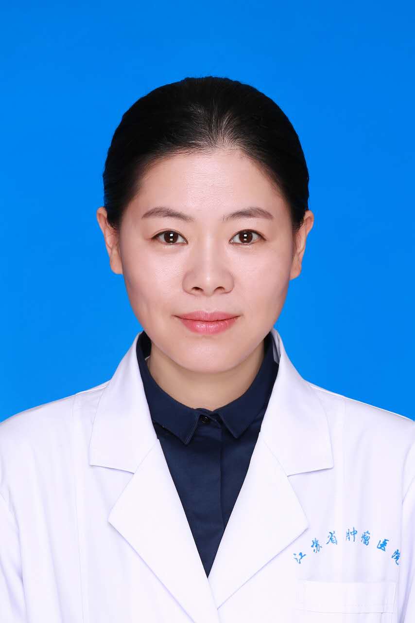 Li Yin (尹丽)合作论文数The Fourth School of Clinical Medicine1Ligong Yuan合作论文数1Yueming Zhang合作论文数1Yong-Jie Lu合作论文数1L. Wang合作论文数1
 Feng Wei合作论文数1Xin Wang合作论文数1Jianjun Qin合作论文数1
 Guangyu Bai合作论文数1Zhongren Zhou合作论文数1Changyuan Guo合作论文数1Hua Fu合作论文数1Ye Tao合作论文数1Wenchao Liu合作论文数1Yangyang Zhao合作论文数1
 Lin Dong合作论文数1Hua Lin合作论文数1Qing Zhao (赵庆)合作论文数1Leilei Zheng合作论文数1Feng Shi合作论文数1Ying Ji合作论文数Department of Thoracic Surgery, National Cancer Center/National Clinical Research Center for Cancer/Cancer Hospital, Chinese Academy of Medical Sciences and Peking Union Medical College, Beijing, China.1Bingning Wang合作论文数1Yunlong Li合作论文数1HongYing Zhao (赵宏颖)合作论文数1
 Xuchen Zhang合作论文数Pathology Faculty, Yale School of Medicine, Yale University1
 Zhen Wang (王真)合作论文数Institute of Medicinal Biotechnology, Chinese Academy of Medical Sciences1Yanling Yuan合作论文数1Li Lin合作论文数1
 Lin Wang (汪麟)合作论文数Cancer Hospital Chinese Academy of Medical Sciences1
 Hui Liu (刘辉)合作论文数School of Traffic and Transportation Engineering, Central South University1Jun Lu合作论文数1
 Bin Hu (胡滨)合作论文数Beijing Chao-Yang Hospital, Capital Medical University1
 Zhouguang Hui (惠周光)合作论文数Cancer Hospital Chinese Academy of Medical Sciences1
 Shuangmei Zou (邹霜梅)合作论文数Cancer Hospital, Chinese Academy of Medical Sciences1
 WenBin Li (李文斌)合作论文数Beijing Shijitan Hospital, Capital Medical University1Bo Zheng (郑波)合作论文数1
 Bin Qiu (邱彬)合作论文数College of Chemistry, Fuzhou University;College of Biological Science and Engineering, Fuzhou University;Key Laboratory for Analytical Science of Food Safety and Biology, Ministry of Education, Fuzhou University1




