Enkurin (ENKUR) is a tumor suppressor in some malignancies. However, its role in endometrial cancer (EC) remains unknown. Here, we firstly observed that reduced ENKUR expression promotes progression and poor prognosis in EC. Moreover,the overexpression of ENKUR suppressed the proliferation, migration, invasion, and intrahepatic dissemination of EC in vitro and in vivo. Repressing ENKUR expression by small-interfering RNA significantly reversed the inhibition of cell proliferation and invasion in vitro. We used co-immunoprecipitation combined with mass spectral analysis to identify the potential interactive proteins of ENKUR. Based on Gene Ontology analysis, we discovered that Wnt/β-catenin (Wnt/CTNNB1) signaling is a ENKUR-modulated key pathway. ENKUR binds to CTNNB1, significantly repressing its protein expression. Furthermore, ENKUR also binds to E3 ligase F-box and WD repeat domain containing 7 (FBXW7), a critical tumor suppressor. Interestingly, the latter binds to CTNNB1 and S502 of CTNNB1 is the key binding site, thereby increasing its protein ubiquitination and degradation. Finally, we confirmed that the predominant ubiquitination sites of CTNNB1 are located at K281 and K394. Transfection of ENKUR-overexpressing EC cells with CTNNB1 reversed the suppressive effects on tumor growth and invasion. ENKUR may be a tumor suppressor via recruiting FBXW7 to directly ubiquitinate and degrade CTNNB1 in EC.
Tumor cell metastasis is the key cause of death in patients with nasopharyngeal carcinoma (NPC). MiR-2110 was cloned and identified in Epstein-Barr virus (EBV)-positive NPC, but its role is unclear in NPC. In this study, we investigated the effect of miR-2110 on NPC metastasis and its related molecular basis. In addition, we also explored whether miR-2110 can be regulated by cinobufotalin (CB) and participate in the inhibition of CB on NPC metastasis. Bioinformatics, RT-PCR, and in situ hybridization were used to observe the expression of miR-2110 in NPC tissues and cells. Scratch, Boyden, and tail vein metastasis model of nude mouse were used to detect the effect of miR-2110 on NPC metastasis. Western blot, Co-IP, luciferase activity, colocalization of micro confocal and ubiquitination assays were used to identify the molecular mechanism of miR-2110 affecting NPC metastasis. Finally, miR-2110 induced by CB participates in CB-stimulated inhibition of NPC metastasis was explored. The data showed that increased miR-2110 significantly suppresses NPC cell migration, invasion, and metastasis. Suppressing miR-2110 markedly restored NPC cell migration and invasion. Mechanistically, miR-2110 directly targeted FGFR1 and reduced its protein expression. Decreased FGFR1 attenuated its recruitment of NEDD4, which downregulated NEDD4-induced phosphatase and tensin homolog (PTEN) ubiquitination and degradation and further increased PTEN protein stability, thereby inactivating PI3K/AKT-stimulated epithelial-mesenchymal transition signaling and ultimately suppressing NPC metastasis. Interestingly, CB, a potential new inhibitory drug for NPC metastasis, significantly induced miR-2110 expression by suppressing PI3K/AKT/c-Jun-mediated transcription inhibition. Suppression of miR-2110 significantly restored cell migration and invasion in CB-treated NPC cells. Finally, a clinical sample assay indicated that reduced miR-2110 was negatively correlated with NPC lymph node metastasis and positively related to NPC patient survival prognosis. In summary, miR-2110 is a metastatic suppressor involving in CB-induced suppression of NPC metastasis.
The small molecule chemical compound cinobufotalin (CB) is reported to be a potential antitumour drug that increases cisplatin (DDP) sensitivity in nasopharyngeal carcinoma. In this study, we first found that CB decreased DDP resistance, migration and invasion in lung adenocarcinoma (LUAD). Mechanistic studies showed that CB induced ENKUR expression by suppressing PI3K/AKT signalling to downregulate c-Jun, a negative transcription factor of ENKUR. Furthermore, ENKUR was shown to function as a tumour suppressor by binding to β-catenin to decrease c-Jun expression, thus suppressing MYH9 transcription. Interestingly, MYH9 is a binding protein of ENKUR. The Enkurin domain of ENKUR binds to MYH9, and the Myosin_tail of MYH9 binds to ENKUR. Downregulation of MYH9 reduced the recruitment of the deubiquitinase USP7, leading to increased c-Myc ubiquitination and degradation, decreased c-Myc nuclear translocation, and inactivation of epithelial-mesenchymal transition (EMT) signalling, thus attenuating DDP resistance. Our data demonstrated that CB is a promising antitumour drug and may be a candidate chemotherapeutic drug for LUAD patients.
ENKUR plays a crucial role in lung and colorectal cancers. Chemically synthesized cinobufotalin (CB) showed its significant anti-cancer effect in nasopharyngeal carcinoma. However, the roles of ENKUR and CB along with their correlation are still unknown in hepatocellular carcinoma (HCC). In this study, ENKUR expression in HCC tissue and cells were detected. The relationship between ENKUR expression and clinical pathology was also assessed. In vivo and in vitro experiments were conducted to explore the effects and molecular basis of ENKUR and CB in HCC. ENKUR expression was correlated with HCC progression and patient prognosis. Furthermore, ENKUR could inhibit tumor proliferation, metastasis, and sorafenib resistance in HCC. Mechanistic studies showed that ENKUR or its Enkurin domain could bind to MYH9 and decrease its expression by binding to β-catenin and inhibiting its nuclear transfer, thus decreasing c-Jun level. Low expression of MYH9 suppressed recruitment of deubiquitination enzyme USP7, promoting degradation of the c-Myc. Therefore, cell cycle and EMT signals were suppressed. CB as a safe and effective anti-cancer compound up-regulates the expression of ENKUR via inhibiting PI3K/AKT/c-Jun-mediated transcription suppression. These findings show that ENKUR induced by CB antagonizes β-catenin/c-Jun/MYH9/USP7 pathway, thus increasing c-Myc ubiquitin degradation and finally suppressing cell cycle and EMT signals.
Clinically, the metastasis of tumor cells is the key factor of death in patients with cancer. In this study, we used a model of metastatic nasopharyngeal carcinoma (NPC) to explore the effects of a new chemical, cinobufagin (CB), combined with cisplatin (DDP). We observed that chemically synthesized CB strongly decreased the metastasis of NPC. Furthermore, a better therapeutic effect was shown when CB was combined with DDP. Molecular analysis revealed that CB induced ENKUR expression by deregulating the PI3K/AKT pathway and suppressing c-Jun, an oncogenic transcriptional factor that binds to the ENKUR promoter and negatively modulated its expression in NPC. ENKUR as a tumor suppressor binds to MYH9 and decreases its expression by recruiting β-catenin via its enkurin domain to prevent its nuclear accumulation, which therefore suppresses c-Jun-induced MYH9 expression. Subsequently, downregulated MYH9 reduces the enlistment of E3 ligase UBE3A and thus decreases the UBE3A-mediated ubiquitination degradation of p53, a key tumor suppressor that decreases epithelial-mesenchymal transition (EMT). Clinical sample analysis demonstrated that the ENKUR expression level was significantly reduced in NPC tissues. Its decreased expression substantially promoted clinical progression and reflected poor prognosis for patients with NPC. This study demonstrated that CB induced ENKUR to repress the β-catenin/c-Jun/MYH9 signal and thus decreased UBE3A-mediated p53 ubiquitination degradation. As a result, the EMT signal was inactivated to suppress NPC metastasis.
Abstract Enkurin, TRPC Channel Interacting Protein (ENKUR) was shown as a suppressor in some tumors. However, the biological role of ENKUR on gastric cancer (GC) and its related molecular mechanisms is not clear. Here, we first observed that ENKUR significantly inhibited cell migration, invasion, and metastasis in GC. The molecular basis showed β‐catenin‐mediated epithelial‐mesenchymal transition (EMT) signaling was inactivated in ENKUR‐overexpressing GC cells. In addition, ENKUR knockdown markedly restored cell migration and invasion. Subsequently, ENKUR bound to MYH9 and decreased its protein expression by recruiting E3 ubiquitin ligase F‐Box And WD Repeat Domain Containing 7 (FBXW7) to form an ubiquitinated degradation complex. The downregulated Myosin Heavy Chain 9 (MYH9) protein weakened the recruitment of the deubiquitinase USP2 and thus promoted the degradation of β‐catenin protein, which finally suppressed EMT signaling. Finally, the oncogenic transcription factor c‐Jun bound to ENKUR promoter and reduced its expression in GC. In clinical samples, decreased ENKUR expression promoted the unfavorable prognosis of GC. Our data proved the vital role of ENKUR on suppressing cell migration, invasion, and metastasis and demonstrated its potential as a therapeutic target for GC.
Background: Clinically, the metastasis of tumor cells is the key factor of death in patients with cancer. Methods: In this study, we used a model of metastatic nasopharyngeal carcinoma (NPC) to explore the effects of a new chemical, cinobufotalin (CB), combined with cisplatin (DDP). Findings: We observed that chemically synthesized CB strongly decreased the metastasis of NPC. Furthermore, a better therapeutic effect was shown when CB was combined with DDP. Molecular analysis revealed that CB induced ENKUR expression by deregulating the PI3K/AKT pathway and suppressing c-Jun, an oncogenic transcriptional factor that binds to the ENKUR promoter. ENKUR as a tumor suppressor binds to MYH9 and decreases its expression by recruiting β-catenin via its enkurin domain to prevent its nuclear accumulation, which therefore suppresses c-Jun-induced MYH9 expression. Subsequently, downregulated MYH9 reduces the enlistment of E3 ligase UBE3A and thus decreases the UBE3A-mediated ubiquitination degradation of p53, a key tumor suppressor that decreases epithelial-mesenchymal transition (EMT). Clinical sample analysis demonstrated that the ENKUR expression level was significantly reduced in NPC tissues. Its decreased expression substantially promoted clinical progression and reflected poor prognosis for patients with NPC. Interpretation: This study demonstrated that CB induced ENKUR to repress the β-catenin/c-Jun/MYH9 signal and thus decreased UBE3A-mediated p53 ubiquitination degradation. As a result, the EMT signal was inactivated to suppress NPC metastasis. Funding: This study was supported by the National Natural Science Foundation of China(81974460, 81572649), the Scientific Research Project of Guangdong Provincial Bureau of Traditional Chinese Medicine (No. 20193010), the People’s Livelihood Science and Technology grant of Guangzhou Municipal Science and Technology Project (No. 201803010023), Nature Science Fund of Guangdong Province (2021A1515012430, 2020A1515010176),Discipline construction project of Guangdong Medical University(4SG21011G).Declaration of Interest: Prof. Fang has a pending patent on Cinobufotalin (No.201910399834.7). All authors declare no conflicts. Ethical Approval: All in vivo experiments were approved by the Animal Care and Use Committee of Southern Medical University.
Gliomas are the most common primary intracranial tumors, which have genetic heterogeneity. It is impossible to accurately evaluate the dynamic evolution of tumor genes and microenvironment only by pathological examination after surgery. As one of the smallest extracellular vesicles secreted by cells, exosomes can be used as potential biomarkers and carriers of intercellular information transmission. It can promote glioma cell proliferation, immune escape and chemoradiotherapy resistance, improve blood oxygen microenvironment, and enhance the ability of invasion and metastasis. The relations of exosomes with glioma occurrence and development are summarized as follows.
The coiled-coil domain containing protein members have been well documented for their roles in many diseases including cancers. However, the function of the coiled-coil domain containing 65 (CCDC65) remains unknown in tumorigenesis including gastric cancer. Methods: CCDC65 expression and its correlation with clinical features and prognosis of gastric cancer were analyzed in tissue. The biological role and molecular basis of CCDC65 were performed via in vitro and in vivo assays and a various of experimental methods including co-immunoprecipitation (Co-IP), GST-pull down and ubiquitination analysis et al. Finally, whether metformin affects the pathogenesis of gastric cancer by regulating CCDC65 and its-mediated signaling was investigated. Results: Here, we found that downregulated CCDC65 level was showed as an unfavourable factor in gastric cancer patients. Subsequently, CCDC65 or its domain (a.a. 130-484) was identified as a significant suppressor in GC growth and metastasis in vitro and in vivo. Molecular basis showed that CCDC65 bound to ENO1, an oncogenic factor has been widely reported to promote the tumor pathogenesis, by its domain (a.a. 130-484) and further promoted ubiquitylation and degradation of ENO1 by recruiting E3 ubiquitin ligase FBXW7. The downregulated ENO1 decreased the binding with AKT1 and further inactivated AKT1, which led to the loss of cell proliferation and EMT signal. Finally, we observed that metformin, a new anti-cancer drug, can significantly induce CCDC65 to suppress ENO1-AKT1 complex-mediated cell proliferation and EMT signals and finally suppresses the malignant phenotypes of gastric cancer cells. Conclusion: These results firstly highlight a critical role of CCDC65 in suppressing ENO1-AKT1 pathway to reduce the progression of gastric cancer and reveals a new molecular mechanism for metformin in suppressing gastric cancer. Our present study provides a new insight into the mechanism and therapy for gastric cancer.
FAM83A is part of an 8-member protein family of unknown function and is reported to be a cancer-promoting and treatment-resistance factor in several cancers. However, its role in hepatocellular carcinoma (HCC) remains unclear. Analysis of the Cancer Genome Atlas (TCGA) showed that FAM83A mRNA expression is upregulated in HCC, as are the protein expression levels in both HCC cell lines and tissues. Clinical data have demonstrated that high FAM83A expression is positively correlated with poor progression-free survival time, thus suggesting its cancer-promoting potential. Functional analyses showed that FAM83A overexpression promoted HCC cell migration and invasion in vitro and suppressed sorafenib sensitivity. Inhibiting FAM83A reversed these results. A pulmonary metastasis model further confirmed that FAM83A promoted HCC cell metastasis in vivo. Mechanistic analyses indicated that FAM83A activated the PI3K/AKT signaling pathway, its downstream c-JUN protein, and epithelial-to-mesenchymal transition (EMT)-related protein levels, including downregulation of E-cadherin and upregulation of Vimentin and N-cadherin. Interestingly, c-JUN induced FAM83A expression by directly binding to its promoter region and thus forming a positive-feedback loop for FAM83A/PI3K/AKT/c-JUN. In conclusion, we demonstrated that FAM83A, as a cancer-metastasis promoter, accelerates migration, invasion and metastasis by activating the PI3K/AKT/c-JUN pathway and inducing its self-expression via feedback, thus forming a FAM83A/PI3K/AKT/c-JUN positive-feedback loop to activate EMT signaling and finally promote HCC migration, invasion and metastasis.
 Weiyi Fang (方唯意)合作论文数Southern Medical University9
 Zhen Liu (刘真)合作论文数School of Basic Medical Sciences, Guangzhou Medical University7
 Yonghao Li合作论文数6
 Jiahao Liu合作论文数6
 Huiling Yang (杨惠玲)合作论文数Zhongshan School of Medicine, Sun Yat-sen University;The First Affiliated Hospital, Sun Yat-sen University6Shuting Deng合作论文数4Xiong Liu (刘雄)合作论文数4
 Yewei Zhang (张业伟)合作论文数The Second Affiliated Hospital of Nanjing Medical University;School of Biomedical Engineering and Informatics, Nanjing Medical University4Yingying Xie合作论文数3Shiyi Fang合作论文数2Liyang Wu合作论文数2Yun Su合作论文数2Zhe Hu合作论文数2Chen Liu合作论文数2Xuemei Peng合作论文数2Beixian Zhou合作论文数2Li Xiaoning合作论文数2Chao Cheng合作论文数2
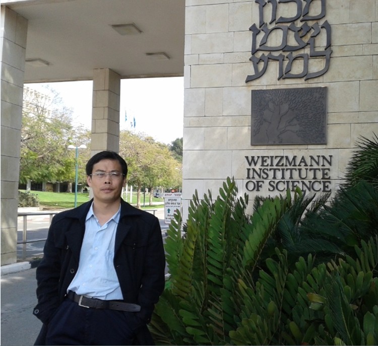 Zhibo Zhu (朱志博)合作论文数南方医科大学药学院2
 Weiwei Yan (严炜炜)合作论文数School of Information Management, Wuhan University2
 Aimin Li (李爱民)合作论文数南方医科大学中西医结合医院2
 Libo Li (李黎波)合作论文数南方医科大学中西医结合医院2
 Peng Shen (沈朋)合作论文数The First Affiliated Hospital, School of Medicine, Zhejiang University2Jianyang Xin合作论文数1Ruiting Xian合作论文数1Tongyuan Deng合作论文数1Mengmin Zhang合作论文数1Lingrong Peng合作论文数1Yinghao Wen合作论文数1Zhan Liu合作论文数1Yu-Ling Deng合作论文数1Shaohua Liu合作论文数1Menglan ZHANG (张梦蓝)合作论文数1Shu Liu合作论文数1Siyuan Han合作论文数1Shi Zuo合作论文数Guizhou Prenatal Diagnosis Center, Affiliated Hospital of Guizhou Medical University1Zibo Tang合作论文数1Xing Deng合作论文数1Wan Zhang合作论文数1
 Yao Tang合作论文数1Wei-Wei Yan合作论文数1Wenmin Li合作论文数1Yun Huang合作论文数1Qian Wang合作论文数1Rong Li合作论文数1Ying Zhu合作论文数1Yanzi Zhang合作论文数1Xiu-qiong WU (吴秀琼)合作论文数1




