PURPOSE:This study aimed to investigate contrast-enhanced ultrasound (CEUS) and clinical features associated with early recurrence (ER) following surgical resection in patients with microvascular invasion (MVI)-negative hepatocellular carcinoma (HCC), and to establish a nomogram-based predictive model. METHODS:A total of 146 pathologically confirmed MVI-negative HCC patients were retrospectively enrolled between August 2021 and June 2024. Univariable and multivariable Cox regression analyses were performed based on CEUS and clinicopathologic variables to identify predictors and develop a nomogram for 2-year recurrence probability. Model performance and discriminative ability were evaluated using the concordance index (C-index), receiver operating characteristic curve, and decision curve analysis (DCA). RESULTS:Multivariable Cox analysis identified smooth morphology, intra-tumoral artery, glypican-3 expression, and albumin-bilirubin (ALBI) grade as independent predictors of ER (all P < 0.05). A nomogram incorporating CEUS and clinicopathologic variables was constructed and demonstrated a favorable C-index of 0.768 (95% confidence interval [CI], 0.711-0.817). The model showed strong discriminative ability, with an area under the curve of 0.870 (95% CI, 0.805-0.952), and DCA indicated good clinical utility. The derived risk score effectively stratified patients into high-risk and low-risk groups. Patients in the low-risk group exhibited significantly better prognosis compared with those in the high-risk group ( P < 0.001). CONCLUSIONS:This CEUS- and clinicopathology-based prognostic model accurately identifies high-risk MVI-negative HCC patients and may provide valuable information for optimizing surveillance and intervention strategies.
Patients with unresectable large HCC may be downstaged through TACE followed by MWA to achieve curative treatment. Controversially, for some patients, single MWA treatment may be sufficient. This study aimed to evaluate the clinical outcomes of unresectable large HCC treated with transarterial chemoembolization (TACE) followed by microwave ablation (MWA) and compare them based on the tumor burden score (TBS), to provide a scientific basis for guiding treatment strategies. This retrospective study included 294 patients with unresectable large HCC (449 tumors) treated with MWA between January 2010 and June 2023 at the General Hospital of the People’s Liberation Army. Propensity score matching (PSM) was used to compare the TACE sequential MWA group (n = 103) with the single MWA group (n = 103). The primary endpoint was progression-free survival (PFS), with secondary endpoints including overall survival (OS) and occurrence of adverse events. Subgroup analyses were conducted by identifying TBS split points using the interaction cubic spline plot method. Among the 294 patients, 76.5
OBJECTIVES:To compare the diagnostic efficacy of 3 imaging modalities-conventional ultrasound (US), contrast-enhanced ultrasound (CEUS), and their combination (US + CEUS)-among physicians with varying experience levels in diagnosing breast intraductal lesions and to elucidate the adjunctive value of CEUS. METHODS:This study included patients who underwent both US and CEUS for breast lesions and were subsequently pathologically confirmed to have intraductal breast lesions between January 2022 and September 2025. Using pathological findings as the gold standard, the efficacy of senior, mid-career, and junior physicians in differentiating benign from malignant intraductal lesions was calculated and compared across the 3 diagnostic modalities. RESULTS:A total of 205 cases were included, comprising 117 benign and 88 malignant lesions. The combined US + CEUS approach demonstrated significantly superior diagnostic efficacy and higher diagnostic confidence scores compared to either CEUS or US alone among physicians at all experience levels. The combined modality yielded optimal efficacy in the senior physician group (p < .001), while the magnitude of improvement was greatest among junior physicians (p < .001), particularly for easily missed lesions like atypical ductal hyperplasia (ADH) and ductal carcinoma in situ (DCIS). For any given examination protocol, diagnostic efficacy exhibited a declining trend with decreasing physician experience (p < .05). CONCLUSION:CEUS serves as a valuable adjunctive tool for diagnosing breast intraductal lesions and is particularly beneficial for junior and mid-career physicians. The combined diagnostic approach enhances overall diagnostic performance.
Objective To explore the clinical value of Automated Breast Ultrasound System (ABUS) combined with two-dimensional ultrasound for intraductal breast lesions, and to provide imaging evidence for preoperative benign and malignant differentiation. Methods Patients with pathologically confirmed intraductal breast lesions hospitalized in our hospital from April 2025 to April 2026 were enrolled. All subjects underwent two-dimensional ultrasound and ABUS examination. Ultrasonographic features were compared between benign and malignant groups. Univariate and multivariate logistic regression were used to screen independent risk factors of malignant lesions and establish a combined diagnostic model. ROC curves were drawn to compare the diagnostic efficiency of different detection schemes. Results A total of 200 patients were enrolled, including 132 benign cases and 68 malignant cases. The malignant group had significantly higher incidences of irregular shape, microcalcification, abundant blood flow, duct dilatation ≥ 5 mm, convergence sign, nipple-oriented lesion extension and marginal spiculation or angulation (P < 0.05). Multivariate logistic regression confirmed seven independent risk factors for malignancy (P < 0.05): irregular lesion morphology, microcalcification, Adler grade II–III blood flow, duct dilatation ≥ 5 mm, convergence sign, lesion extending toward the nipple and spiculation/angulation. The combined model had an AUC of 0.90, superior to standalone two-dimensional ultrasound (AUC = 0.81) and single ABUS (AUC = 0.86), with sensitivity of 90.7% and specificity of 89.2%. Conclusion Combination of ABUS and two-dimensional ultrasound has prominent clinical value for differentiating benign and malignant intraductal breast lesions, which improves preoperative diagnostic accuracy and facilitates clinical decision-making.
Objectives To compare the diagnostic efficacy of 3 imaging modalities—conventional ultrasound (US), contrast‐enhanced ultrasound (CEUS), and their combination (US + CEUS)—among physicians with varying experience levels in diagnosing breast intraductal lesions and to elucidate the adjunctive value of CEUS. Methods This study included patients who underwent both US and CEUS for breast lesions and were subsequently pathologically confirmed to have intraductal breast lesions between January 2022 and September 2025. Using pathological findings as the gold standard, the efficacy of senior, mid‐career, and junior physicians in differentiating benign from malignant intraductal lesions was calculated and compared across the 3 diagnostic modalities. Results A total of 205 cases were included, comprising 117 benign and 88 malignant lesions. The combined US + CEUS approach demonstrated significantly superior diagnostic efficacy and higher diagnostic confidence scores compared to either CEUS or US alone among physicians at all experience levels. The combined modality yielded optimal efficacy in the senior physician group ( p < .001), while the magnitude of improvement was greatest among junior physicians ( p < .001), particularly for easily missed lesions like atypical ductal hyperplasia (ADH) and ductal carcinoma in situ (DCIS). For any given examination protocol, diagnostic efficacy exhibited a declining trend with decreasing physician experience ( p < .05). Conclusion CEUS serves as a valuable adjunctive tool for diagnosing breast intraductal lesions and is particularly beneficial for junior and mid‐career physicians. The combined diagnostic approach enhances overall diagnostic performance.
Glypican 3 (GPC-3) in hepatocellular carcinoma (HCC) is receiving attention as a potential biomarker for treatment options. However, GPC-3 expression can only be assessed through surgical pathology. Therefore, we aim to develop and validate a practical tool based on contrast-enhanced ultrasound (CEUS) and clinical features to preoperatively identify GPC-3 expression in HCC. From August 2021 to July 2024, 197 patients with pathologically confirmed HCC were retrospectively enrolled. The least absolute shrinkage and selection operator (LASSO) and stepwise logistic regression analyses were performed in training cohort to identify predictors and establish a model for predicting GPC-3 expression. The performance of model was evaluated by C-index, area under the receiver operating characteristic curve (ROC), calibration curves, and decision curve analysis (DCA). Our study showed that heterogeneous arterial phase hyperenhancement (APHE), mosaic architecture, alpha-fetoprotein (AFP) ≥ 100 ng/mL, rim enhancement in arterial phase (AP), neutrophil count (Nc) level and age were independently associated with GPC-3 expression. The model demonstrated strong predictive performance and the areas under curve were 0.811 (95
Background Given the current inconsistency in thyroid nodule diagnostic classifications, this study aimed to compare the diagnostic performance between conventional US based and contrast enhanced ultrasound (CEUS) integrated systems, as well as among systems within each category, and to identify the most clinically practical thyroid imaging reporting and data system (TI-RADS). Methods This retrospective study included patients who underwent conventional US, CEUS and fine-needle aspiration (FNA) between January 2023 and January 2025. Nodules were classified using three conventional US-based TI-RADS (ACR-TIRADS, EU-TIRADS, Modified Kwak TI-RADS) and five CEUS-integrated TI-RADS (CEUS1-5) recently reported in the literature, which were modified from US TI-RADS by incorporating CEUS features. Comparisons of diagnostic performance within and between the two categories were conducted using pathology as the reference standard. Results A total of 319 thyroid nodules from 268 patients were included, comprising 120 benign and 199 malignant. CEUS-integrated systems demonstrated superior diagnostic performance compared with conventional TI-RADS, with significantly higher AUC values (0.78–0.88 vs. 0.76–0.77, P < 0.01). Among CEUS-integrated systems, CEUS-1 and CEUS-5 achieved the same highest AUC (0.88) and accuracy (0.80). Using the optimal cut-off values (TR4b), CEUS-5 demonstrated superior Youden index (0.65; 95% CI: 0.57–0.73), specificity (0.93; 95% CI: 0.89–0.97) and positive predictive value (0.95; 95% CI: 0.91–0.98), CEUS-1 offered a balanced performance (sensitivity 0.76, 95% CI: 0.70–0.82; specificity 0.87, 95% CI: 0.80–0.92). CEUS-5 showed 0% malignancy in its lowest risk category and a peak malignancy rate exceeding 95% in its highest risk category, with Spearman correlation analysis revealing a nearly perfect correlation between its risk scores and malignancy ( P < 0.01). Consistency metrics (F1 score, κ, MCC) were also highest for CEUS-1 (0.83, 0.59, 0.61) and CEUS-5 (0.82, 0.60, 0.63), confirming their robust predictive performance. Conclusion CEUS-integrated systems significantly outperform conventional TI-RADS in thyroid nodule risk stratification, with CEUS-1 and CEUS-5 achieving the highest diagnostic performance in terms of AUC. CEUS-5 further demonstrated superior performance across additonal diagnostic metrics, especially Youden index and specificity, representing a more refined and promising tool for clinical implementation.
OBJECTIVE:To evaluate the efficacy and safety of ultrasound-guided thermal ablation (TA)for the treatment of T1N0M0 papillary thyroid carcinoma (PTC) in adolescent patients. METHODS:This retrospective study enrolled adolescent patients with solitary T1N0M0 PTC who underwent TA between January 2015 and January 2025. Patients were stratified into T1a and T1b subgroups based on maximum tumor diameter. The primary outcomes were tumor progression rate and progression-free survival (PFS); secondary outcomes included ablation zone volume dynamics, volume reduction rate, complications, thyroid function, and patient-reported satisfaction. RESULTS:A total of 82 patients were included (56 in T1a group, 26 in T1b group), with a technical success rate of 100%. The overall median follow-up duration was 36 months (interquartile range [IQR]: 24-60 months). Ablation zone volumes progressively decreased and eventually vanished in all cases. The median time to complete disappearance of the ablation zone was significantly longer in the T1b group than in the T1a group (18 months vs. 12 months, p = 0.014). The overall tumor progression rate was 2.4% (2/82), with no significant difference between subgroups (p = 0.579). Progression-free survival (PFS) rates were comparable between T1a and T1b groups (96.5% vs. 94.0%, p = 0.550). No major complications occurred. CONCLUSION:Ultrasound-guided TA is a safe and effective treatment modality for both T1a and T1b subgroups of adolescent patients with solitary T1N0M0 PTC.
Thermal ablation technology is crucial for treating early-stage primary tumors. However, the "water isolation" during clinical surgery is limited by unstable thermal protection caused by fluid loss. More seriously, it is prone to intraoperative bleeding, cancer cell metastasis, and postoperative tissue adhesion. Here we introduce a "one stone, four birds" strategy, which utilizes a self-healing chitosan hydrogel without drug assistance to offer four abilities, including thermal protection, hemostasis, anti-tumor metastasis and anti-tissue adhesion. A rabbit VX2 liver transplantation tumor model is established. The self-healing chitosan hydrogel, crosslink of hydroxyethyl chitosan (HC) and dual aldehyde-PEG (Ald-PEG-Ald) using dynamic Schiff base, adheres to the liver surface and remains in situ upon minimally invasive injection, resulting in outstanding thermal protection compared with saline. The temperature of the isolated tissue near the ablation foci remains below 42 °C. Moreover, the self-healing property of the hydrogel can prevent cell leakage during the procedure, thus avoiding intraoperative bleeding, tumor metastasis and postoperative abdominal adhesion. Therefore, this work identified four efficacies of the self-healing chitosan hydrogel in thermal ablation of metastatic liver tumors, providing an advanced alternative for clinical application.
Purpose:To investigate the feasibility of real-time shear wave elastography (SWE) in evaluating intestinal wall stiffness, and to establish the threshold SWE value of normal intestinal wall and explore the influencing factors of intestinal SWE. Method:659 subjects who underwent intestinal SWE and colonoscopy were retrospectively enrolled. The wall elasticity of colonoscopy-confirmed normal/abnormal intestinal segment was measured by transabdominal SWE. Measurement reliability was evaluated by the intraclass correlation coefficient (ICC). The threshold value of SWE in differentiating normal and abnormal intestine was determined using ROC curve analysis with the largest Youden index, and the diagnostic performance of this threshold was evaluated. We explored the effects of gender, age, depth and type of the targeted intestinal segment on the intestinal wall elasticity by t test and logistic linear regression analysis. Results:The technical success rate of SWE examination is 95.3 % (628/659). The mean SWE value of normal intestinal walls is (5.45 ± 1.34) kPa, which was significantly lower than that of abnormal ones (15.38 kPa±7.22, P < 0.001). Using 8.1 kPa as the threshold, the sensitivity and specificity were 93.5 % and 96.0 % with an AUC of 94.8 %. The overall ICC for SWE measurements was 0.933. Gender (ß=0.278, P = 0.013), depth (ß=0.220, P = 0.043) and type of the targeted segment (ß=0.522, P < 0.001) was associated with the SWE value of intestinal wall, but age was not (ß=0.050, P = 0.484). Conclusions:SWE is feasible in evaluating the stiffness of intestinal wall with high reliability. The SWE threshold value differentiating normal intestinal wall and abnormal intestinal wall is 8.1 kPa.
OBJECTIVES:This study aims to compare and analyze the 3-year efficacy and safety differences of microwave ablation in the treatment of patients with hyperthyroidism with benign nodules/papillary thyroid carcinoma (PTC). METHODS:This retrospective study included 66 patients with hyperthyroidism accompanied by nodules who received microwave ablation treatment from December 2009 to December 2024. A 3-year follow-up was conducted after the operation to monitor indicators such as the complete remission rate of hyperthyroidism and the tumor disappearance rate, and the differences in therapeutic effects between the two groups were analyzed through propensity score matching (PSM). RESULTS:Post-PSM cohort analysis showed that at the 36-month follow-up after surgery, the complete remission rate of hyperthyroidism in the benign nodule group was 93.8%, which was higher than 62.5% in the PTC group (p = 0.083). The tumor disappearance rate in the benign nodule group was 50.0%, which was higher than that in the PTC group at 18.8% (p = 0.0627). During the 3-year follow-up period, no disease progression was observed in either group of patients. No serious complications were reported in either group. CONCLUSION:MWA in the treatment of hyperthyroidism with benign nodules or PTC has certain efficacy and safety. In terms of therapeutic effect, the difference between the benign nodule group and the PTC group was not statistically significant, and further verification with larger sample sizes is still needed.
OBJECTIVE:The objective of this study was to develop a combined model based on radiomics features of Sonazoid contrast-enhanced ultrasound (CEUS) during the Kupffer phase and to evaluate its value in differentiating well-differentiated hepatocellular carcinoma (w-HCC) from atypical benign focal liver lesions (FLLs). METHODS:A total of 116 patients with preoperatively Sonazoid-CEUS confirmed w-HCC or benign FLL were selected from a prospective multiple study on the clinical application of Sonazoid in FLLs conducted from August 2020 to March 2021. According to the randomization principle, the patients were divided into a training cohort and a test cohort in a 7:3 ratio. Seventy-nine patients were used for establishing and training the radiomics model and combined model. In comparison, 37 patients were used for validating and comparing the performance of the models. The diagnostic efficacy of the models for w-HCC and atypical benign FLLs was evaluated using ROCs curves and decision curves. A combined model nomogram was created to assess its value in reducing unnecessary biopsies. RESULTS:Among the patients, there were 55 cases of w-HCC and 61 cases of atypical benign FLLs, including 28 cases of early liver abscess, 16 cases of atypical hepatic hemangioma, 8 cases of hepatocellular dysplastic nodules (DN), and 9 cases of focal nodular hyperplasia (FNH). The radiomics model and combined model we established had AUCs of 0.905 and 0.951, respectively, in the training cohort, and the AUCs of the two models in the test cohort were 0.826 and 0.912, respectively. The combined model outperformed the radiomics feature model significantly. Decision curve analysis demonstrated that the combined model achieved a higher net benefit within a specific threshold probability range (0.25 to 1.00). A nomogram of the combined model was developed. CONCLUSION:The combined model based on the radiomics features of Sonazoid-CEUS in the Kupffer phase showed satisfactory performance in diagnosing w-HCC and atypical benign FLLs. It can assist clinicians in timely detecting malignant FLLs and reducing unnecessary biopsies for benign diseases.
Ultrasound image segmentation of ablation region of liver tumor has recently emerged as a significant tool for assessing tumor ablation surgery outcomes. However, existing segmentation methods are limited to the artifact of ultrasound images caused by hand-held transduces and speckle noise, leading to the background region likely being identified as the ablation region. Therefore, we introduce a MLP-based attentional network, MA-Net, and get accurate segmentation results. We present the hybrid attention cascading module to pay more attention to the ablation region to ensure accurate segmentation. In addition, we present an inverted residual multilayer perceptron module to avoid misrecognizing the ablation region as the background region. We evaluate our method on private and public dataset and achieve state-of-the-art performance.
Objective: The purpose of the study was to develop and validate a radiomics model by using contrast-enhanced ultrasound (CEUS) data for pre-operative differential diagnosis of pancreatic cystic neoplasms (PCNs), especially pancreatic serous cystadenoma (SCA). Methods: Patients with pathologically confirmed PCNs who underwent CEUS examination at Chinese PLA hospital from May 2015 to August 2022 were retrospectively collected. Radiomic features were extracted from the regions of interest, which were obtained based on CEUS images. A support vector machine algorithm was used to construct a radiomics model. Moreover, based on the CEUS image features, the CEUS and the combined models were constructed using logistic regression. The performance and clinical utility of the optimal model were evaluated by area under the receiver operating characteristic curve (AUC), sensitivity, specificity and decision curve analysis. Results: A total of 113 patients were randomly split into the training (n = 79) and test cohorts (n = 34). These patients were pathologically diagnosed with SCA, mucinous cystadenoma, intraductal papillary mucinous neoplasm and solid-pseudopapillary tumor. The radiomics model achieved an AUC of 0.875 and 0.862 in the training and test cohorts, respectively. The sensitivity and specificity of the radiomics model were 81.5% and 86.5% in the training cohort and 81.8% and 91.3% in the test cohort, respectively, which were higher than or comparable with that of the CEUS model and the combined model. Conclusion: The radiomics model based on CEUS images had a favorable differential diagnostic performance in distinguishing SCA from other PCNs, which may be beneficial for the exploration of personalized management strategies.
The aim of this study was to develop a predictive model based on Sonazoid contrast-enhanced ultrasound (SCEUS) and clinical features to discriminate poorly differentiated hepatocellular carcinoma (P-HCC) from intrahepatic cholangiocarcinoma (ICC). Forty-one ICC and forty-nine P-HCC patients were enrolled in this study. The CEUS LI-RADS category was assigned according to CEUS LI-RADS version 2017. Based on SCEUS and clinical features, a predicated model was established. Multivariate logistic regression analysis and LASSO logistic regression were used to identify the most valuable features, 400 times repeated 3-fold cross-validation was performed on the nomogram model and the model performance was determined by its discrimination, calibration, and clinical usefulness. Multivariate logistic regression and LASSO logistic regression indicated that age (> 51 y), viral hepatitis (No), AFP level (≤ 20 µg/L), washout time (≤ 45 s), and enhancement level in the Kupffer phase (Defect) were valuable predictors related to ICC. The area under the receiver operating characteristic (AUC) of the nomogram was 0.930 (95
The aim of this study was to evaluate the potential of Sonazoid contrast-enhanced ultrasound (SNZ-CEUS) as an imaging biomarker for preoperative prediction of microvascular invasion (MVI) in hepatocellular carcinoma (HCC). From August 2020 to March 2021, we conducted a prospective multicenter study on the clinical application of Sonazoid in liver tumor; a MVI prediction model was developed and validated by integrating clinical and imaging variables. Multivariate logistic regression analysis was used to establish the MVI prediction model; three models were developed: a clinical model, a SNZ-CEUS model, and a combined model and conduct external validation. We conducted subgroup analysis to investigate the performance of the SNZ-CEUS model in non-invasive prediction of MVI. Overall, 211 patients were evaluated. All patients were split into derivation (n = 170) and external validation (n = 41) cohorts. Patients who had MVI accounted for 89 of 211 (42.2 • This is the first prospective multicenter study to analyze the possibility of SNZ-CEUS preoperatively predicting MVI. • The model established by combining SNZ-CEUS image features and clinical features has high predictive performance in both derivation cohort and external validation cohort. • The findings can help clinicians predict MVI in HCC patients before surgery and provide a basis for optimizing surgical management and monitoring strategies for HCC patients.
脓毒症是由感染引起的全身炎症反应综合征,常导致多器官功能障碍,病死率高,为临床常见的危重症.急性肾损伤(acute kidney injury,AKI)是一组以肾功能迅速减退或丧失为主要特征的临床综合征,可由许多因素引起,是临床急危重症患者的常见并发症.脓毒症是引起AKI的一大病因[1].多项研究[2-3]表明,脓毒症患者中AKI的发生率高达50%左右.脓毒症并发AKI的患者较单纯脓毒症患者有着更高的病死率,且是否发生AKI及其严重程度是影响脓毒症患者预后的重要因素[4].由于目前血清学检测方法尚不能在AKI发生的初期对其进行诊断,当血清学指标升高时,肾功能已发生不可逆的损伤.患者往往错失最佳的早期干预时期,导致病死率居高不下,给家庭及社会带来沉重的负担.因此,应用更加有效的检测方法提高AKI的早期检出率,完善治疗方案的制定,改善脓毒症患者的预后,成为近年来的研究热点.
Objective:To explore the value of contrast-enhanced ultrasonography (CEUS) quantitative parameters in evaluating renal perfusion and monitoring inflammatory response in sepsis-induced acute renal injury (S-AKI).Methods:Thirty-two healthy Sprague Dawley rats were selected and divided into either a model group or a control group. Sepsis was induced in rats by cecal ligation and puncture. For control rats, the abdominal cavity was only opened and closed, and cecal ligation and puncture were not performed. At 12 h and 24 h after modeling, eight rats from each group were taken to perform contrast-enhanced ultrasonography, and region of interest (ROI) was selected to draw time intensity curve (TIC) and perform quantitative analysis. Blood samples were taken from the inferior vena cava (IVC) to test serum creatinine (SCr), urea, interleukin (IL)-6, tumor necrosis factor-α (TNF-α), and IL-10, and the kidney was taken for pathological examination. Independent sample t test and non-parametric test (Mann-Whitney U) were used to analyze the differences between the two groups at different time points, and Spearman correlation test was used to analyze the correlation between quantitative parameters of CEUS [arrival time (AT), time to peak (TTP), ascending slope (AS), peak intensity (PI), half of descending time (DT/2), descending slope (DS), and area under curve (AUC)] and inflammatory factors.Results:Renal function indexes (Scr and urea) and inflammatory factors (IL-6, TNF-α, and IL-10) in the model group were significantly higher than those of the control group at both time points (P<0.05). At 2-4 s after SonoVue bolus injection, the blood vessels of the kidney were enhanced at first, then the cortex and medulla developed successively, and the medulla and cortex disappeared in turn. Compared with the control group, in the model group at 12 h, the TTP of the renal cortex significantly increased [(10.47±1.25) s vs (8.30±1.53) s, t=-3.105, P=0.008], the DS and PI of the renal cortex significantly decreased [(-0.13±0.02) dB/s vs (-0.17±0.04) dB/s, t=-3.121, P=0.008; (23.90±1.36) dB vs (27.26±1.88) dB, t=4.102, P=0.001], TTP and DT/2 of the medulla significantly increased [(11.66±1.99) s vs (9.00±1.28) s, t=-3.180, P=0.007; (62.49±4.56) s vs (52.15±7.70) s, t=-3.268, P=0.007], and DS of the medulla significantly decreased [(-0.13±0.02) s dB/s vs (-0.17±0.03) dB/s, t=-2.915, P=0.011]. Compared with the control group, in the model group at 24 h, AS and DS of the renal cortex significantly increased [(0.99±0.17) dB/s vs (0.61±0.19) dB/s, t=-4.262, P=0.001; (-0.23±0.03) dB/s vs (-0.15±0.04) dB/s, t=5.138, P<0.001], and DT/2 and AUC of the renal cortex significantly decreased [(42.41±3.03) s vs (61.07±6.52) s, t=7.344, P<0.001; (2477.89±113.37) dB?s vs (3024.93±253.81) dB?s, t=5.566, P<0.001]; AS, DS, and AT of the renal medulla significantly increased [(1.00±0.27) dB/s vs (0.66±0.17) dB/s, t=-3.061, P=0.008; (-0.23±0.06) dB/s vs (-0.15±0.04) dB/s, t=3.108, P=0.008; (4.30±0.34) s vs (3.77±0.29) s, t=-3.349, P=0.005], and DT/2 and AUC of the renal medulla significantly decreased [(42.73±7.02) s vs (59.64±9.23) s, t=4.124, P=0.001; (2335.75±189.77) dB?s vs (2689.72±285.45) dB?s, t=2.921, P=0.011]. The CEUS quantitative parameters PI and AUC in the renal cortex were significantly correlated with serum IL-6, TNF-α, and IL-10 (r=-0.562, -0.398, -0.512, -0.540, -0.638, and -0.430, respectively, P<0.05). Renal PAS staining showed that the kidney of rats in the model group showed unclear outline of renal tubules, dilatation, shedding of brush margin, and flattening and shedding of renal tubular epithelial cells, and part of renal tubules were formed in 24 h in the model group.Conclusion:CEUS can be used to dynamically assess the blood perfusion process of the rat kidney in real time, quantitative evaluate the blood perfusion level, and monitor the inflammatory reaction state of the body.
aim of this study was to evaluate renal perfusion changes in rats with acute kidney injury induced by rhabdomyolysis, using quantitative parameters obtained with contrast-enhanced ultrasound (CEUS). Sprague-Dawley (SD) rats were randomly divided into an experimental group (n = 40) and a control group (n = 20). Each group was further divided into four subgroups (0.5-, 6-, 24-and 72-h groups). Time-intensity curves and related quantitative parameters of the renal cortex and medulla were obtained by CEUS, and the con-trast characteristics analyzed for different time points. In the experimental group, the CEUS quantitative param-eters for the renal medulla of time to peak (TTP), descending time/2 (DT/2) and area under the curve (AUC) increased, whereas ascending slope (AS) and descending slope (DS) decreased. Similarly, renal cortical AS, DS and AUC in the experimental group differed significantly from those in the control group. With respect to the CEUS quantitative parameters for the renal cortex, AUC increased, and AS and DS decreased. These parameters differed significantly between the experimental and control groups. CEUS is sensitive to change in renal perfu-sion in rhabdomyolysis-induced acute kidney injury and, thus, has diagnostic value. (E-mail: lyk301@163. com) (C) 2022 World Federation for Ultrasound in Medicine & Biology. All rights reserved.
Objective:To evaluate the changes of renal perfusion in rats with acute kidney injury induced by rhabdomyolysis using quantitative parameters of contrast-enhanced ultrasound (CEUS).Methods:Sixty Sprague-Dawley rats were randomly divided into a model group and a control group, with 30 rats in each group. The model group was injected with 50% glycerin saline at 10 ml/kg intramuscularly, and the control group was injected with 0.9% saline at 10 ml/kg intramuscularly. At 0.5, 2, 6, 12, and 24 h after injection, six rats in each group were examined by CEUS. Renal cortical and medullary time-intensity curve (TIC) and related quantitative parameters [ascending slope (AS), area under the curve (AUC), descending slope (DS), descending time/2 (DT/2), peak intensity (PI)] were obtained to analyze the CEUS characteristics at different time points. Blood was collected from the inferior vena cava at the end of the examination to detect the serum levels of urea nitrogen, creatinine, cystatin C, and creatine kinase, and pathological sections were prepared for observation after renal tissue was fixed. The t-test or Mann Whitney U test was used to compare the differences in relevant quantitative parameters and serum biochemical indexes between groups, and one-way ANOVA or Kruskal-Wallis H test was used to compare the differences in various indexes at different time points within the groups.Results:Compared with the control group, the levels of serum creatinine, urea nitrogen, and creatine kinase at 0.5, 2, 6, and 12 h and the levels of serum cystatin C at 2 and 6 h were significantly increased in the model group (serum creatine kinase: Z=-2.242, P=0.025; Z=-2.882, P=0.004; Z=-2.882, P=0.004; Z=-2.562, P=0.010; urea nitrogen: t=4.288, P=0.002; t=4.450, P=0.001; t=10.812, P<0.001; t=5.260, P<0.001; creatinine: t=6.327, P=0.001; t=10.577, P<0.001; t=2.612, P=0.035; t=4.694, P=0.001; cystatin C: t=3.460, P=0.009; t=2.391, P=0.038). In the model group, the cortical AUC at 0.5 h was significantly increased [(2636.84±150.99) dB·s vs (2308.20±210.50) dB·s, t=3.107, P=0.011], the cortical DS at 2, 6, and 12 h and cortical AS at 6, 12, and 24 h were significantly decreased [DS: (0.16±0.05) dB/s vs (0.23±0.03) dB/s, t=-3.342, P=0.007; (0.16±0.03) dB/s vs (0.23±0.04) dB/s, t=-3.542, P=0.005; (0.16±0.05) dB/s vs (0.23±0.03) dB/s, t=-3.226, P=0.009; AS: (0.54±0.22) dB/s vs (0.84±0.15) dB/s, t=-2.733, P=0.021; (0.58±0.21) dB/s vs (0.86±0.20) dB/s, t=-2.318, P=0.043; (0.67±0.20) dB/s vs (0.96±0.13) dB/s, t=-2.809, P=0.019], and the cortical DT/2 at 12 h was significantly increased [(55.78±12.28) s vs (40.09±6.29) s, t=2.789, P=0.019]. The medullary AUC at 0.5, 2, 6, and 12 h was significantly increased [(2382.40±189.84) dB·s vs (1910.82±140.13) dB·s, t=5.025, P=0.001; (2637.35±258.55) dB·s vs (1999.88±409.52) dB·s, t=3.217, P=0.009; (2424.63±409.39) dB·s vs (1910.47±263.59) dB·s, t=2.669, P=0.0024; (2353.54±348.2) dB·s vs (1958.43±95.37) dB·s, t=3.014, P=0.013], the medullary DS and AS at 2 and 6 h were significantly decreased [DS: 0.13 (0.07, 0.15) dB/s vs 0.22 (0.19, 0.24) dB/s, Z=-2.892, P=0.004; 0.13 (0.11, 0.16) dB/s vs 0.21(0.20, 0.23) dB/s, Z=-2. 585, P=0.010; AS: (0.44±0.22) dB/s vs (0.77±0.14) dB/s, t=-3.063, P=0.015; (0.41±0.21) dB/s vs (0.80±0.10) dB/s, t=-4.042, P=0.005], and the medullary DT/2 at 2, 6, and 12 h was significantly increased [(61.44±15.52) s vs (38.88±8.80) s, t=3.105, P=0.011; (54.60±12.47) s vs (37.59±8.71) s, t=2.724, P=0.021; (56.08±14.57) s vs (38.42±6.00) s, t=3.003, P=0.013]. The pathological examination showed that the glomerular and tubular structures were normal in the control group. In the model group, the glomerular structure did not change significantly, the brush border of some tubules was sloughed off, the epithelial cells were flattened, the dilation of the tubular lumen was obvious starting at 0.5 h, with the distal tubules being the most prominent one, tubular cast appeared at 2 h, and the pathological changes were aggravated with the prolongation of time, being the most severe at 24 h.Conclusion:Contrast-enhanced ultrasound can sensitively reflect the dynamic changes of renal perfusion in rats with acute kidney injury induced by rhabdomyolysis, and thus has certain diagnostic value.
 Ping Liang (梁萍)合作论文数School of Medicine, Nankai University;Chinese PLA General Hospital9
 Xiaoling Yu (于晓玲)合作论文数School of Medicine, Tongji University9
 Fangyi Liu (刘方义)合作论文数Chinese PLA General Hospital7
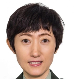 Jie Yu (于杰)合作论文数Chinese PLA General Hospital7
 QiuYang Li (李秋洋)合作论文数6
 Yukun Luo (罗渝昆)合作论文数School of Medicine, Nankai University;Chinese PLA General Hospital6
 ZhiGang Cheng (程智刚)合作论文数中南大学湘雅医院 中南大学第一临床学院5Zhi-Yu Han合作论文数5Erpeng Qi合作论文数4Yiqiong Zhang合作论文数4Zhu LianHua合作论文数4Shuilian Tan合作论文数4Jianing Zhu合作论文数4JunDong Yao (姚俊东)合作论文数4Jian-Ping Dou合作论文数4Ping Zhao (赵平)合作论文数College of Life Sciences, South-Central Minzu University3
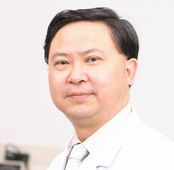 Shichun Lu (卢实春)合作论文数School of Medicine, Nankai University3
 Kaiyan Li (李开艳)合作论文数Tongji Hospital, Tongji Medical College of Huazhong University of Science and Technology3Xiang Jing (经翔)合作论文数3
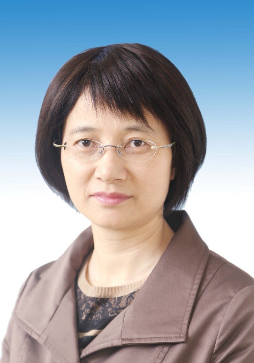 Hong Ding (丁红)合作论文数Huashan Hospital, Fudan University3Yunlin Li合作论文数2Chuan Pang合作论文数2Guang-Zhi He合作论文数2XueJuan Dong (董雪娟)合作论文数2YiRu Wang (王一茹)合作论文数2Xin Li合作论文数2
 Jie Tang (唐杰)合作论文数Chinese PLA General Hospital2
 Tianan Jiang (蒋天安)合作论文数浙江大学医学院附属第一医院超声科2
 Wei Wu (吴薇)合作论文数Department of Ultrasound, Key Laboratory of Carcinogenesis and Translational Research (Ministry of Education), Peking University Cancer Hospital and Institute2
 Kai Li (李凯)合作论文数State Key Laboratory of Rare Earth Resource Utllization, Changchun Institute of Applied Chemistry, Chinese Academy of Sciences2
 Wen Cheng (程文)合作论文数Harbin Medical University2
 Qing Song (宋青)合作论文数Chinese PLA General Hospital2Xiaoyan Xie合作论文数Department of Medical Ultrasonics, The First Affiliated Hospital, Sun Yat-Sen University, China2
 Yaqiong Zhu合作论文数1Yuan-Yuan Yan合作论文数1Yu Jie合作论文数1Zhen Wang合作论文数1Jing Xiao合作论文数1Lifei Huang合作论文数1Manying LI (李蔓英)合作论文数1Shuling Chen (陈淑玲)合作论文数1
 Chuyue Zhang合作论文数Key Lab Bioact Mat,Minist Educ, Nankai Univ1
 Wensheng Xie (谢文升)合作论文数College of Life Science and Technology, Beijing University of Chemical Technology1Ya Sun合作论文数1
 Yang Liu (刘旸)合作论文数The First Affiliated Hospital of Guangzhou Medical University1
 Ying Zhang合作论文数1
 Li Lu (陆立)合作论文数School of Medicine, Sun Yat-sen University1
 Guofeng Li合作论文数1




